Surgery

Self-adhesive mesh in open inguinal hernia repair, how does it compare to conventional suture fixation?

Authors
Ali W & Nugud O
Article Citation and PDF Link
BJMP 2019;12(3):a023

Introduction

Lichtenstein tension-free mesh repair has been the standard practice in open inguinal hernia repair for many years. The procedure involves suture fixation of the mesh via an anterior approach to the inguinal canal. It is hypothesised that this invasive fixation contributes to the development chronic postoperative inguinal pain (CPIP), a condition which can cause significant morbidity.

A sound repair should restore the groin anatomy whilst minimising recurrence and not adversely affecting the patient quality of life. Considering the large number of these operations performed each year, reducing complications such as chronic postoperative pain will have a significant impact on healthcare resources.

The introduction of anatomical self-adhesive meshes such as Parietx ProGripTM addresses this concern in theory by obviating the need for mesh fixation. This mesh is a macro porous polyester mesh that utilizes polylactic acid micro grips (PLA) to aid placement within 60 seconds1 without the need for additional fixation. The manufacturer does suggest, however, that additional fixation is left to the discretion of the operating surgeon.

We conducted a review of the literature to evaluate the reported outcomes of using this mesh in open inguinal hernia repair.

Methods

We conducted a PUBMED/MEDLINE search using the search words “Self-adhesive mesh”, “Lichtenstein repair”, “Open inguinal hernia repairand “Self-gripping mesh” .We looked primarily at the outcomes of postoperative pain and recurrence. The result highlighted five well-structured meta-analyses and several RCTs and retrospective reviews.

Results

In a retrospective review of 211 patients who underwent open inguinal hernia repair with self-adhesive mesh, Tarchi P et al reported a recurrence rate of 0.5% at 1 year and 2.4% at 2 years. They incidence of chronic pain was less than 3%. There were no cases of seroma, testicular complications or mesh infection at 1, 2 and 3-year follow-up. The report highlighted the shorter operative duration with no effect on recurrence rates as a point in favour of self-adhesive mesh. The authors acknowledged the limitation of the study design and the need for randomised trials to address the issue.8 A few other small non-randomised trials draw similar conlusions.9

A randomised blinded trial from the Danish Multicentre DANGRIP Study Group allocated 163 vs 171 patients to self-adhesive and suture fixation respectively. There were no significant differences between the groups in postoperative complications (33.7 versus 40.4 %; P = 0·215), rate of recurrent hernia within 1 year (1.2 % in both groups) or quality of life. The 12 month prevalence of moderate or severe symptoms was 17.4 and 20.2% respectively (P = 0.573).

The study concluded that the avoidance of suture fixation using a self-gripping mesh was not accompanied by a reduction in chronic symptoms after inguinal hernia repair. 5

The FinnMesh trial is a randomised multicentre trial from Finland that Compared glue fixation, self-gripping mesh, and suture fixation of mesh. 625 patients were randomised to cyanoacrylate glue (Histoacryl, n = 216), self-gripping mesh (Parietex ProGrip, n = 202), or conventional non absorbable sutures (Prolene 2-0, n = 207) There was no significant differences postoperatively in pain response or need for analgesics between the study groups at 1 year follow up. The mean operative duration was lower in the self-adhesive mesh group.6

The HIPPO trial is a randomised double-blinded trial of 165 patients. The reported hernia recurrence rate after 24 months was 2.4% for the ProGrip mesh and 1.8% for the sutured mesh (P = 0.213).

The incidence of CPIP was 7.3% at 3 months declining to 4.6% at 24 months and did not differ between both groups. 7 The mean duration of surgery was significant shorter with the ProGrip mesh (44 vs 53 minutes, P < 0.001).

In a systematic review of 7 studies comparing self-gripping versus sutured mesh for inguinal hernia repair totalling 1353 patients, Zhang C et al found no difference in recurrence (risk difference -0.02 [95% confidence interval -0.07 to 0.03], P = 0.40) or chronic pain (risk difference -0.00 [95% confidence interval -0.01 to 0.01], P = 0.57). 2 This review found no difference in wound infection, hematoma, and seroma formation. Self-adhesive mesh was again associated with a shorter mean operative duration. In its conclusion, the authors surmised that both mesh types are comparable in outcome but further long term analysis might be needed.

Pandanaboyana S published a meta-analysis of 5 RCTs and 1170 patients, that also found no significant difference in recurrence or chronic pain. Wound infection was lower in the self-gripping mesh group compared to sutured mesh but this was not statistically significant (risk ratio (RR) 0.57, 95% confidence interval 0.30-1.06, P = 0.08). The duration of operation was significantly shorter with self-gripping mesh compared to sutured mesh with a mean difference of -5.48 min [-9.31, -1.64] Z = 2.80 (P = 0.005).3

In another meta-analysis, Li J et al included 5 RCTs and 2 prospective comparative studies and 1353 patients. Statistically, there was no difference in the incidence of chronic pain [odds ratio = 0.74, 95% confidence interval (CI) (0.51-1.08)]. There was no statistical difference in the incidence of acute postoperative pain [odds ratio = 1.32, 95% CI (0.68-2.55)], hematoma or seroma [odds ratio = 0.89, 95% CI (0. 56-1.41)], wound infection [risk difference = -0.01, 95% CI (-0.02 to 0.01)], and recurrence [risk difference = 0.00, 95% CI (-0.01 to 0.01)]. The self-gripping mesh group was associated with a shorter operating time (1-9 minutes).10

In Ismail A et al’s meta-analysis of 12 randomized controlled trials and 5 cohort studies, 3722 patients were included in the final analysis. The two groups, using self-gripping mesh or sutured mesh fixation, did not differ significantly in terms of recurrence rate (odds ratio = 0.66, 95% confidence interval 0.18-2.44; P = 0.54) or postoperative chronic groin pain (odds ratio = 0.75, 95% confidence interval 0.54-1.05; P = 0.09). The operative time was less in the self-gripping mesh group (mean difference = -7.85, 95% confidence interval -9.94 to -5.76; P < .0001). There were comparable risks between self-gripping mesh and sutured mesh fixation groups in terms of postoperative infection (odds ratio = 0.81, 95% confidence interval 0.53-1.23; P = 0.32), postoperative hematoma (odds ratio = 0.97, 95% confidence interval 0.7-1.36; P = 0.9), and urinary retention (odds ratio = 0.66, 95% confidence interval 0.18-2.44; P =0.54).11

A more recent meta-analysis including 10 RCTs and 2541 patients also draws similar conclusions, with no significant difference in the incidence of chronic pain (odds ratio = 0.93; 95% confidence interval, 0.74-1.18), recurrence (odds ratio = 1.34; 95% confidence interval, 0.82-2.19), or foreign body sensation (odds ratio = 0.82; 95% confidence interval, 0.65-1.03).4 The mean operating time was significantly shorter (odds ratio = -7.58; 95% confidence interval, -9.58 to -5.58) in the self-gripping mesh group which is consistent with the reported literature.

Discussion

Open inguinal hernia repair is a routinely performed operation and chronic postoperative inguinal pain is a significant cause of morbidity that can impact negatively on patients’ quality of life. Eliminating the need for suture fixation seems theoretically a step in the right direction.

The published literature, however, seems to arrive at similar conclusions. Whilst using self-adhesive mesh results in a shorter operative duration and seemingly does not affect the outcome negatively otherwise, there is no evidence that it reduces postoperative chronic pain and therefore should not be advocated on this merit. A shortened operative time coupled with a non-inferior outcome does seem like a more reasonable evidence-based argument for its proponents.

The decision of which mesh fixation technique to use can be left to the discretion of the operating surgeon. Further long-term follow up data is required to arrive at more definitive conclusions as the mean follow up duration in the reviewed studies ranged from 4 months to 3 years. The cost implications involved in the choice of mesh used should also be taken into account in future studies.

Acknowledgements / Conflicts / Author Details
Competing Interests: 
None declared
Details of Authors: 
WADAH ALI, MBBS MRCS(Glas) CABHS, General Surgery Registrar, Health Education, England North East. O NUGUD, Consultant General and Laparoscopic Surgeon, England.
Corresponding Author Details: 
waddahabdelazim@hotmail.com
Corresponding Author Email: 
waddahabdelazim@hotmail.com
References
References: 
  1. Philippe Chastan, MD (2006). Tension-Free Open Inguinal Hernia Repair Using an Innovative Self-Gripping Semi-Resorbable Mesh. Journal of Minimal Access Surgery; pp 139-43.
  2. Zhang C1, Li F, Zhang H, Zhong W, Shi D, Zhao Y. Self-gripping versus sutured mesh for inguinal hernia repair: a systematic review and meta-analysis of current literature. J Surg Res. 2013 Dec; 185(2):653-60.
  3. Pandanaboyana S, Mittapalli D, Rao A, Prasad R, Ahmad N. . Meta-analysis of self-gripping mesh (Progrip) versus sutured mesh in open inguinal hernia repair. Surgeon. 2014 Apr; 12(2):87-93.
  4. Molegraaf M, Kaufmann R, Lange J. Comparison of self-gripping mesh and sutured mesh in open inguinal hernia repair: A meta-analysis of long-term results. Surgery. 2018 Feb; 163(2):351-360.
  5. Jorgensen L N, Sommer T,   Assaadzadeh S,   Strand L,  Dorfelt  A, Hensler M, Rosenberg J. Randomized clinical trial of self‐gripping mesh versus sutured mesh for Lichtenstein hernia repair. Br J Surg. 2013 Mar; 100(4):474-81.
  6. Rönkä K1, Vironen J, Kössi J, Hulmi T, Silvasti S, Hakala T, Ilves I, Song I, Hertsi M, Juvonen P, Paajanen H. Randomized Multicenter Trial Comparing Glue Fixation, Self-gripping Mesh, and Suture Fixation of Mesh in Lichtenstein Hernia Repair (FinnMesh Study). Ann Surg. 2015 Nov;262(5):714-9.
  7. Molegraaf MJ, Grotenhuis B, Torensma B, de Ridder V, Lange JF, Swank DJ. The HIPPO Trial, a Randomized Double-blind Trial Comparing Self-gripping Parietex Progrip Meshand Sutured Parietex Mesh in Lichtenstein Hernioplasty: A Long-term Follow-up Study. Ann Surg. 2017 Dec;266(6):939-945.
  8. Tarchi P, Cosola D, Germani P, Troian M, De Manzini N. Self-adhesive mesh for Lichtenstein inguinal hernia repair. Experience of a single centre. Minerva Chir. 2014 Jun; 69(3):167-76.
  9. Yilmaz A, Yener O, Kaynak B, Yiğitbaşi R, Demir M, Burcu B, Aksoy F. Self-gripping Covidien™ ProGrip™ mesh versus polypropylene mesh in open inguinal hernia repair: multicentre short term results. Prague Med Rep. 2013; 114(4):231-8.
  10. Li J1, Ji Z, Li Y. The comparison of self-gripping mesh and sutured mesh in open inguinal hernia repair: the results of meta-analysis. Ann Surg. 2014 Jun; 259(6):1080-5. 
  11. Ismail A1, Abushouk AI2, Elmaraezy A1, Abdelkarim AH3, Shehata M3, Abozaid M4, Ahmed H3, Negida A3. Self-gripping versus sutured mesh fixation methods for open inguinal hernia repair: A systematic review of clinical trials and observational studies. Surgery. 2017 Jul; 162(1):18-36. 

Updated ‘two-week wait’ referral guidelines for suspected colorectal cancer have increased referral volumes without improving cancer detection rates

Authors
Christopher P Jones, Rebecca C Fallaize & Robert J Longman
Article Citation and PDF Link
BJMP 2019;12(2):a012
Abstract / Summary
Abstract: 

Introduction: In November 2015, the National Institute for Clinical Health and Care Excellence updated its two-week wait (2WW) referral guidelines for suspected colorectal malignancy. This study measured the effect of the change in 2WW referral guidelines on: (i) the volume of 2WW referrals, (ii) the rates of detection of cancer in those patients referred to the 2WW service, and (iii) adherence to the referral guidelines.
Methods and materials: A retrospective case note review of all colorectal cancer 2WW referrals during two periods (July to August 2015 and July to August 2016), to a large inner-city teaching hospital, was undertaken. Cancer detection rates were calculated based on diagnosis obtained from review of patient clinical records and were cross-referenced against the regional cancer registry database. 
Results: There was a significant increase in the numbers of patients referred to the colorectal 2WW service in the period following the change in guidelines (193 vs. 268, p<0.01). There was no significant change in the rate of colorectal cancer detection between the two periods observed (8.3% vs. 7.5%, p=0.75), although adherence to the referral guidelines increased (72% vs 89%, p<0.01).
Conclusion: Changes to the 2WW referral criteria have led to an increased number of patients being referred, but have not resulted in a change in the rate of colorectal cancer detection. Further work should seek to assess the impact on survival rates from colorectal cancer, and to contextualise these findings with wider trends in non-2WW routes to diagnosis.

Abbreviations: 
2WW - two week wait
Keywords: 
Colorectal cancer, Two-week wait

Introduction

Colorectal cancer is the fourth most common cancer in the United Kingdom (UK), and accounts for 10% of all cancer deaths.1 The symptoms of colorectal cancer are often non-specific and in its early stages there may be no symptoms at all. Survival is directly linked to stage of disease at diagnosis – five-year survival falls from 98% for stage I disease down to 40% for stage IV disease.2

Thirty percent of all colorectal cancers are diagnosed via the ‘Two-Week Wait’ (2WW) referral route in the UK. The remainder are diagnosed following emergency presentation (24%), non-2WW GP referral (24%), bowel cancer screening (9%) or by other pathways (13%).3

The TWW referrals for patients with suspected cancer were introduced in 2000 by the NHS Cancer Plan,4 and built on the earlier recommendations of the Calman-Hine report into commissioning cancer services.5 These improvements sought to address the United Kingdom’s relatively low cancer survival rates compared to the rest of Europe, and to address the delays in diagnosis and treatment that some patients were encountering. In order to standardise cancer care nationally, 2WW referral guidelines were introduced by the Department of Health in 2000.6 These guidelines were reviewed and updated in 2005 by the National Institute for Clinical Health and Care Excellence (NICE).7

In November 2015, NICE updated all its 2WW referral guidelines, including those for suspected colorectal malignancy.8 The recommendations were developed following a systematic review of the literature which recommended referral for patients with symptoms deemed to have a positive predictive value for colorectal cancer of 3% or more. This was a reduction from the previous guidelines, which used a positive predictive value of greater than 5%.9 The original (2005) and updated (2015) NICE colorectal 2WW referral guidelines are outlined in Table 1.

This study measured the effect of the change in colorectal 2WW referral guidelines on the following outcomes:

  • Volume of referrals to the colorectal 2WW clinic
  • Rate of detection of colorectal cancer
  • Rate of detection of non-colorectal cancer
  • Adherence to the 2WW referral guidelines

Table 1. Summary of the 2005 and 2015 NICE Two-Week Wait referral guidelines for suspected colorectal cancer 7,8

2005 Criteria 2015 Criteria
Age >40 with rectal bleeding and a change in bowel habit for >6 weeks Age >40 with unexplained weight loss and abdominal pain
Age >60 with rectal bleeding without a change in bowel habit for >6 weeks Age >50 with unexplained rectal bleeding
Age >60 with change in bowel habit without rectal bleeding for >6 weeks Age >60 with change in bowel habit or iron-deficiency anaemia
Right lower abdominal mass consistent with involvement of the large bowel Positive faecal occult blood test
Palpable rectal mass (intra-luminal) Palpable rectal or abdominal mass
Unexplained iron deficiency anaemia in: non-menstruating Women with an Hb <10g/100mL
men with an Hb <11g/100mL
Age <50 with rectal bleeding and one of: abdominal pain
change in bowel habit
weight loss
iron-deficiency anaemia

Methods and materials

We undertook a retrospective analysis of referrals to the colorectal 2WW service at a large inner city teaching hospital (Bristol Royal Infirmary, UK). All the patients referred in two-month periods before (July to August 2015) and after (July to August 2016) were included in the study. The referral guidelines changes were identified and their clinical notes were reviewed. The specific variables recorded for each referral included: age, gender, presenting symptoms and signs and subsequent diagnosis. All records were cross-referenced against the regional cancer registry.

Differences between the two groups were assessed for statistical significance using Chi-Squared and unpaired T-tests. Count data was assessed for significance using the Poisson Means test at a 95% confidence interval. Statistical tests were calculated using the MEDCALC statistical software.

Results

A total of 193 and 268 patients were referred in each of the two study periods. The data collection was complete for all patients. The demographics, referral data, and cancer detection rates are summarised in Table 2.

There was a significant increase in the volume of patients referred via the 2WW pathway following the change in the guidelines (193 vs. 268, p<0.01). There was no significant change in the rate of colorectal cancers detected (8.3% vs. 7.5%, p=0.75).

There was no significant difference in the rate of detection of any cancer (including colorectal cancer) following the 2WW referral (11.4% vs 10.8%, p=0.83). The non-colorectal cancers detected (15 in total) were predominantly metastatic cancers; from lung, ovarian, or prostatic primary malignancies. There was no significant difference in the detection rate of non-colorectal cancers (3.1% vs. 3.4%, p=0.85).

The rate of compliance to the referral guidelines was significantly higher following the update in referral guidelines (72% vs 89%, p<0.01).

In the second study period (July - August 2016), there was a sub-group of 31 patients whose referrals met the new (2015) referral guidelines, but who would not meet the previous (2005) referral guidelines. The mean age in this group was 58.5 and none of these patients had a cancer detected following the 2WW referral.

Table 2. Summary of the results

  Jul-Aug 2015 Jul-Aug 2016 p value
Patients referred 193 268 <0.01 a
Cancers detected
(% of total)
22
11.4%
29
10.8%
0.74 a
0.83 b
Colorectal cancers
(% of total)
16
8.3%
20
7.5%
0.58 a
0.75 b
Non-Colorectal cancers
(% of total)
6
3.1%
9
3.4%
0.61 a
0.85 b
% of referrals compliant with the guidelines (at that time) 72% 89% <0.01 b
Mean age in years
(Median age, range)
68.2
(69, 24-92)
67.9
(69, 22-93)
0.81 c
Sex ratio (M : F) 43 : 56 46 : 53  
Frequency of referral signs/symptoms (%)
Change in bowel habit 60 63 0.51 b
Rectal bleeding 33 39 0.18 b
Abdominal pain 37 33 0.37 b
Unexplained weight loss 22 20 0.60 b
Iron deficiency anaemia 27 22 0.21 b

Statistical test used: a Poisson Means Test, b Chi squared test, c Unpaired t-test

Discussion

This study has shown that the volume of patients being referred to the colorectal 2WW service has significantly increased in a large inner city unit following the update to referral guidelines in 2015. A significantly greater proportion of referrals are compliant with the new guidelines compared with the previous guidelines. Despite this, we found no significant change in the rate of colorectal cancer detection. Our colorectal cancer detection rates following 2WW referral are similar to the published data series (6-14%).10,11,12

The factors contributing to the increased referral rate includes removal of time constraints and referral for symptoms not previously included within the guidelines (e.g. abdominal pain, unexplained weight loss). The updated guidelines are subsequently less specific and use signs and symptoms with a lower positive predictive value for colorectal cancer than previously.8

In their costing statement for the new guidelines, NICE acknowledge that the updated guidelines are likely to increase referral volumes. The justification given is that “benefits are anticipated from earlier diagnosis of cancer”.9 This study challenges that supposition – no cancers were detected in the latter group of 31 patients whose referrals met the new guidelines, but would not have met the old referral guidelines.

Studies prior to the update in guidelines have also challenged the view that 2WW referrals lead to earlier detection of cancer. When compared with ‘non-2WW’ outpatient referrals, patients referred via a 2WW pathway had no significant difference in the stage of disease at diagnosis,13,14 nor any significant difference in the related outcomes such as 2-year survival,15,16 5-year survival,15,17 or proportion undergoing curative surgery.14,15

Bowel cancer screening remains the only method with a strong evidence base for detecting colorectal cancers at an earlier stage.18 Cancers detected in this manner are disproportionately lower in stage,19 and are associated with a significant reduction in mortality.20 This study did not assess the impact of screening on cancer detection rates via the 2WW referral process, although the logical effect of increased detection of cancers via screening would be a proportional fall in cancers detected by other routes, including the 2WW pathway.

The findings of this study appear to challenge the anticipated benefits of the new 2WW referral guidelines. A group of patients were identified whose referrals only met the 2015 guidelines; these referrals would have been deemed inappropriate by the 2005 guidelines. This group of patients were generally younger and none went on to a cancer diagnosis. If other units (or multi-centre studies) corroborate these findings then this should prompt urgent review of the 2WW guidelines with regards to cancer stage at diagnosis and longer term outcomes.

Conclusion

The updated 2WW referral guidelines for suspected colorectal cancers have increased the volume of patients being seen via the 2WW service without increasing cancer detection rates. This is anticipated to have secondary effects on waiting times for routine and endoscopic services; this has not been evaluated in this study. Further research is needed to contextualise all of these findings with cancer detection rates via screening and other non-2WW routes to diagnosis.

Acknowledgements / Conflicts / Author Details
Acknowledgement: 
Miss Hannah Marder of United Hospitals Bristol Cancer Services for her kind clerical assistance
Competing Interests: 
None declared
Details of Authors: 
CHRISTOPHER JONES, MBBS MRCS, Yeovil Hospital, Higher Kingston, Yeovil, Somerset, BA21 4AT. REBECCA FALLAIZE, MBBS FRCS PGCMedEd, Bristol Royal Infirmary, University Hospitals Bristol NHS Foundation Trust, Bristol, BS2 8HW, UK. ROBERT LONGMAN, BSc MBChB PhD FRCS, Bristol Royal Infirmary, University Hospitals Bristol NHS Foundation Trust, Bristol, BS2 8HW, UK.
Corresponding Author Details: 
REBECCA FALLAIZE, Bristol Royal Infirmary, University Hospitals Bristol NHS Foundation Trust, Bristol, BS2 8HW, UK.
Corresponding Author Email: 
rcullwick@gmail.com
References
References: 
  1. Office for National Statistics. Cancer Registration Statistics, England: 2015. https://www.ons.gov.uk/peoplepopulationandcommunity/healthandsocialcare/conditionsanddiseases/bulletins/cancerregistrationstatisticsengland/2015(Cited 1 November 2017).
  2. Office for National Statistics.  Cancer survival by stage at diagnosis for England: Adults diagnosed 2012-2014 and followed up to 2015. https://www.ons.gov.uk/peoplepopulationandcommunity/healthandsocialcare/conditionsanddiseases/bulletins/cancersurvivalbystageatdiagnosisforenglandexperimentalstatistics/adultsdiagnosed20122013and2014andfollowedupto2015(Cited 1 November 2017).
  3. National Bowel Cancer Audit. Annual Report 2017. https://www.nboca.org.uk/reports (Cited 17 December 2017)
  4. UK Department of Health. The NHS Cancer Plan: A plan for investment, A plan for reform. London: Stationery Office; 2000.
  5. UK Department of Health. Expert Advisory Group on Cancer: A policy framework for commissioning cancer services. London: Stationery Office; 1995.
  6. UK Department of Health. HSC 2000/013: Referral guidelines for suspected cancer. London: Stationery Office; 2000.
  7. National Institute for Health and Care Excellence. (2005) Clinical Guideline 27 Referral for suspected cancer. http://www.nice.org.uk/guidance (Cited 1 November 2017).
  8. National Institute for Health and Care Excellence. (2015) Suspected cancer: recognition and referral [NG12]. http://www.nice.org.uk/guidance (Cited 1 November 2017).
  9. National Institute for Health and Care Excellence. Costing Statement: Implementing the (2015) NICE guideline on suspected cancer: recognition and referral [NG12]. http://www.nice.org.uk/guidance (Cited 1 November 2017).
  10. Thorne K, Hutchings H, Elwyn G. The effects of the Two-Week Rule on NHS colorectal cancer diagnostic services: A systematic literature review. BMC Health Serv Res 2006; 6: 43.
  11. Bhangu A, Khan M, Roberts L et al. Detection and survival of colorectal cancer from a 2 week wait service. Surgeon 2011; 9(2): 78-82.
  12. Patel RK, Sayers AE, Seedat S et al. The 2-week wait service: a UK tertiary colorectal centre's experience in the early identification of colorectal cancer. Eur J Gastroenterol Hepatol 2014; 26: 1408-14.
  13. Aslam M, Chaudhri S, Singh B, Jameson J. The “two-week wait” referral pathway is not associated with improved survival for patients with colorectal cancer.  Int J Surg 2017; 43: 181-5.
  14. Bevis P, Donaldson OW, Longman RJ et al. The association between referral source and stage of disease in patients with colorectal cancer. Colorectal Dis 2008; 10(1): 58-62.
  15. Thornton L, Reader H, Stojkovic S et al. Has the “Fast-Track’ referral system affected the route of presentation and/or clinical outcomes in patients with colorectal cancer? World J Surg Onc 2016; 14: 158.
  16. Walsh SR, Gilson NL, Brown K, Novell JR. Trends in colorectal cancer survival following the 2 week rule. Colorectal Dis 2007; 9(3): 207-9.
  17. Schneider C, Bevis P, Longman RJ et al. The association between referral source and outcome in patients with colorectal cancer. Surgeon 2013; 11(3): 141-6.
  18. Bretthauer M. Evidence for colorectal cancer screening. Best Prac Res Clin Gastroenterol 2010; 24(4): 417-25.
  19. McClements PL, Madurasinghe V, Thomson CS et al. Impact of the UK colorectal cancer screening pilot studies on incidence, stage distribution and mortality trends. Cancer Epidemiol 2012; 36(4): e232-42.
  20. Atkin WS, Edwards R, Kralj-Hans I et al. Once-only flexible sigmoidoscopy screening in prevention of colorectal cancer: a multicentre randomised controlled trial. Lancet 2010; 375(9726): 1624-33.

Denosumab-associated Osteonecrosis of the Jaw; A Case Series and Literature Review

Authors
Elena Kyriakidou,Mohamed Badr, Simon Atkins and Sheelah Harrison
Article Citation and PDF Link
BJMP 2016;9(4):a930
Abstract / Summary
Abstract: 

Introduction: Medication-related osteonecrosis of the jaw (MRONJ) is a severely debilitating condition of multifactorial pathogenesis. It primarily involves patients receiving intravenous bisphosphonates (BPs) and most recently the new antiresorptive drug, denosumab, for the treatment of skeletal-related malignancies. There is no curative treatment and no consensus exists regarding the clinical management of such patients. This review aims to share our current clinical experience at Sheffield Teaching Hospitals’ Trust and raise awareness of the increase in severity of ONJ in patients receiving denosumab.
Patients and Methods: Four new cases with clinical diagnosis of MRONJ were presented to Sheffield Teaching Hospitals' Trust. MRONJ was attributed to denosumab therapy, as all patients were treated solely with denosumab for skeletal-related malignancies.
Results: All cases appear to have a more aggressive mode of ONJ compared to that seen with IV and/or oral BPs so far. The cause of MRONJ was observed in the presence of periodontal disease alone and following dental extractions. Progression of the disease occurred considerably faster with the development of widespread suppuration and tooth mobility within weeks. Imaging revealed rather extensive areas of bony destruction, sometimes with associated periosteal reaction in keeping with a chronic bony infection.
Conclusion: It is imperative for all dental and medical teams involved in treating these patients to understand the side effects of RANKL inhibitors on bone metabolism and how it affects treatment. Helping patients to understand the chronicity and potential progression of the disease is essential to a satisfactory outcome.

Abbreviations: 
MRONJ- Medication-related osteonecrosis of the jaw; BPs- Bisphosphonates; SREs- skeletal-related events; IV- intravenous; RANKL-Receptor activator of nuclear factor kappa-B ligand; MM- Multiple myeloma; ONJ- Osteonecrosis.
Keywords: 
Osteonecrosis of the jaw, bisphosphonates, denosumab

Introduction

Metastatic bone disease is a relatively common event in the advanced stages of many malignancies.1 Bone-modifying agents decrease the incidence of skeletal-related events (SREs) such as spinal cord compression and bone fracture, as well as the need for skeletal radiotherapy or surgery.2

Bone modifying agents such as intravenous bisphosphonates (IV BPs) (e.g. pamidronate and zoledronic acid) and denosumab are approved for prevention of SREs. IV BPs are primarily used and effective in the treatment and management of cancer related conditions such as multiple myeloma (MM), and breast cancer with skeletal metastases, because they reduce bone pain, hypercalcemia, and the risk of pathologic fractures.3

Denosumab, a receptor activator of nuclear factor kappa-B ligand (RANKL) inhibitor, represents a breakthrough in the treatment of osteoporosis, MM, and bone metastases. The Food and Drug Administration (FDA) approved it in 2010 for the prevention of SREs in patients with bone metastases and in 2011 for the prevention of endocrine-therapy induced bone loss in patients taking aromatase inhibitors for breast cancer and in patients with non-metastatic prostate cancer.

Three international, randomised, double-blind, double-dummy phase III studies have evaluated denosumab versus zoledronic acid for the treatment of SREs in breast and prostate cancers, and in combined solid tumours and MM. Denosumab’s superior efficacy over zoledronic acid was demonstrated in the studies of patients with advanced breast or prostate cancer, as well as in a pre-specified integrated analysis of all patients enrolled across the three studies.4

In the 2014 position paper of the American Association of Oral and Maxillofacial Surgeons (AAOMS), the nomenclature “bisphosphonate-related osteonecrosis of the jaw” changed to “medication related osteonecrosis of the jaw” (MRONJ). MRONJ is defined as cases in which all of the following 3 characteristics are present5:

  • current or previous treatment with antiresorptive or antiangiogenic agents
  • exposed bone or bone that can be probed through an intraoral or extra-oral fistula in the maxillofacial region that has persisted for longer than 8 weeks
  • no history of radiation therapy to the jaws or obvious metastatic disease to the jaws

Other terminologies used previously include “denosumab related osteonecrosis of the jaw” (DRONJ), and “antiresorptive agent-induced ONJ” (ARONJ).

The aetiopathogenesis of MRONJ related to denosumab therapy remains enigmatic, and hypotheses have focused on reduced bony turnover, infection, toxicity of the soft tissue, and antiangiogenesis. The epidemiology also remains unclear, and reported incidence varies widely.6 Overall, it is estimated that bone necrosis can develop in about 0.7-1.9% of patients with malignancy who are given high-potency IV BPs (such as zoledronic acid), and in 0.01–0.1% of those with osteoporosis who take low-potency oral BPs (such as alendronate). Data relevant to denosumab given subcutaneously in patients with metastatic cancer and osteoporosis seem to replicate those when IV high-potency BPs are administered.7 The risk of osteonecrosis of the jaw (ONJ) is higher in patients exposed to concomitant antiagiogenic medication. The individuals’ risk of ONJ is further determined by factors such as the potency of agent, cumulative dosage or duration of antiresorptive treatment, route of administration, comorbidities and local factors such as periodontal disease.8,9 Oral hygiene plays a significant role with evidence supporting a strong correlation between bacteria associated with periodontal disease and MRONJ.10

MRONJ typically manifests as painful and often infected areas of necrotic bone, which subsequently may lead to severe chronic pain and facial disfigurement. This adversely affects the ability to eat, speak and lowers the quality of life. Adverse events related to RANKL inhibitors are usually considered to be infrequent and low in occurrence. Unfortunately from our recent clinical experience at Sheffield Teaching Hospitals' Trust, there have been several new cases presented in a very short period of time. In this paper we present a case series of MRONJ related to denosumab therapy since adverse events of denosumab in the mandible or maxilla have received relatively little attention.

The aim of this article is to highlight the elevated risk of MRONJ in patients receiving denosumab treatment and educate all health care providers involved in the management of such patients. Furthermore, the mechanisms of denosumab, comparison with bisphosphonates and the reported management strategies are reviewed.

Mechanism of Denosumab

Denosumab is an antiresorptive agent that exists as a human IgG2 monoclonal antibody and inhibits the binding of the receptor activator of nuclear factor kappa-B ligand (RANKL) to RANK (Receptor Activator of Nuclear Factor kappa-B). The binding normally signals the proliferation of osteoclasts, as RANK is expressed on the surface of osteoclasts and their precursors, whereas its ligand, RANKL, is a membrane bound protein expressed by bone marrow stromal cells, osteoblasts and T-lymphocytes. The activation of RANK is integral to the function of osteoclasts. Osteoprotegerin binds to membrane bound RANKL on osteoblast which in turns decreases the osteoclastic activity and in theory negatively effects bone turnover. Denosumab acts similarly to osteoprotegerin but has a higher affinity for RANKL.11-13

Denosumab follows nonlinear, dose-dependent pharmacokinetics. The bioavailability of one subcutaneous denosumab injection is 61% and serum concentrations are detected within 1 hour. Maximum serum concentrations occur in 5-21 days and cessation of osteoclast activity occurs within six hours of the subcutaneous injection. The normal function is restored approximately six to nine months later, whilst bone turnover returns to normal shortly after this.14 Based upon monoclonal antibody pharmacokinetics, denosumab is most likely cleared by the reticuloendothelial system with minimal renal filtration and excretion thus avoiding nephrotoxicity. Its elimination half-life is 32 days, and it does not incorporate into bone.15

It is currently marketed as Prolia® and Xgeva®, approved by FDA. Prolia® is administered subcutaneously every six months and has shown to reduce the incidence of new vertebral, non-vertebral, and hip fractures in osteoporotic patients.16,17 Xgeva® is also effective in reducing SRE related to metastatic bone disease from solid tumours when administered intravenously on a monthly basis.17,18

RANKL Inhibitors and BPs Pharmacokinetics

There are fundamental differences between denosumab and BPs with regard to their mode of action. Denosumab is an antibody and acts extracellularly whereas BPs act intracellularly. As such, BPs must be present in the circulation and available for reuptake into bone for prolonged periods to function.19 There is not any evidence of drug recycling with RANKL inhibitors, and therefore it is suggested that their adverse effects can be reversible with discontinuation, in fact leading to a transient rebound phenomenon, which can be restored, with subsequent treatment.14,20 On the other hand, recycling of BPs in the circulation system has been proposed as a reason for the long duration of action even after cessation which can be up to 12 years.

The US FDA-approved manufacturer’s package insert for both zolendronate and pamidronate states that “there are no data available to suggest whether discontinuation of bisphosphonate treatment reduces the risk of ONJ in patients who require dental procedures during therapy and that clinical judgment of the treating physician should guide the management plan of each patient based on individual benefit/ risk assessment”. The package insert for denosumab does not address the issue of treatment continuation in patients who develop MRONJ to date.

Denosumab is a circulating protein capable of distributing throughout extravascular space. It is expected to reach all sites within bone including intracortical sites unlike with BPs. BPs have strong affinity for hydroxyapatite and bone mineral which limits their even distribution throughout the skeleton, particularly to sites deep within the bone.19,21 This can explain the more profound inhibition of bone remodelling with denosumab than that seen with BPs.

Case Series

Case 1

A 55 year-old lady referred to a dedicated Oral Surgery nerve injury clinic for an opinion and management of her left sided inferior alveolar nerve (IAN) paraesthesia. The patient presented with a history of numbness in the left sided inferior alveolar nerve distribution following removal of the left mandibular second premolar (LL5) in July 2014. She was asymptomatic until she had the LL5 removed and since had suffered with constant pain and numbness. A year later, she had removal of the left mandibular first molar (LL6) and gave a history of recurrent infections and excruciating pain in her mandible over the past two months. On presentation she had an obvious submental swelling and left sided IAN anaesthesia.

Medically she was diagnosed with breast cancer in 2011, for which she underwent wide local excision followed by chemotherapy. She then was placed on unknown clinical trial that we identified at the time to be denosumab trial, following liaison with the Oncology team. She is currently receiving intravenous denosumab every three months.

Clinical examination revealed a grossly mobile anterior mandible with widespread bony necrosis and associated osteomyelitis. Sensory testing revealed complete anaesthesia in the left sided IAN distribution secondary to MRONJ.

An OPG (Orthopantogram) and CBCT (Cone-Beam Computerised Tomography) revealed an extensive patchy area of ill-defined bone loss in the anterior mandible extending posteriorly to the premolar/molar areas bilaterally (Fig 1).

Figure 1 A) OPG showing non-healing sockets in the left mandible with extensive bony destruction together with periosteal reaction extending to the right mandible as shown by the arrows.

Rather interestingly, the bony destruction was evident bilaterally with the patient only having had extraction of teeth in the left mandible (Fig 1). This could be the case of spontaneous ONJ in the right mandible or an extensive ONJ arising from simple extractions on the left side.

Figure 2 3D reconstruction of the CBCT image demonstrating extensive bony destruction involving the lower border of anterior mandible in keeping with a spreading chronic bony infection and clinical presentation of submental swelling as showing by arrows.

Case 2

A 66-year-old female referred by her general medical practitioner (GMP) with a 3-month history of delayed healing following a tooth extraction in the left posterior mandible. She had moderate to severe discomfort and reported multiple previous infections and purulent discharge from the area, which treated with multiple courses of antibiotics. In addition, she reported discomfort from the root treated right mandibular first and second premolar teeth (LR4 and LR5).

Medically she was diagnosed with breast cancer over 10 years ago for which she underwent resection followed by chemotherapy. Three years ago, she diagnosed with metastatic deposits and therefore has been receiving intravenous denosumab every six weeks since then. Other medications include steroids, chemotherapy agents, antihypertensives and analgesics. She did not receive any radiotherapy or BPs treatment in the past.

Clinical presentation revealed a heavily restored dentition with chronic generalised periodontal disease. There was evidence of widespread bone loss clinically and radiographically. The slow healing socket in the left mandible was visible but did not have any exposed bone (Fig 3). The lower right first and second premolar teeth (LR4 and LR5) were clinically and radiographically sound.

Figure 3. Non-healing socket in the left posterior mandible with no evidence of exposed bone or suppuration as showing by white arrow. Gingiva recession (black arrows) is evident in the LL6 and LL5 teeth in keeping with chronic periodontal disease.

Figure 4 Coronal sections of CBCT A and B showing multiple lytic areas within the inferior cortex of the mandible and incomplete healing of the extraction sockets.

On follow-up appointments, the patient suffered multiple repeated infections in the right and left posterior mandible and due to deteriorating periodontal disease, the LR4, LR5, LR6 were extracted by her own general dental practitioner (GDP) due to severe mobility. All three extraction sockets failed to heal (Fig 5) leading to an extensive area of exposed bone in the right mandible, extending from the lower right first premolar (LR4) to lower left first molar (LL6) region. Conservative management was embarked which included antibiotics, chlorhexidine mouthwash and routine oral hygiene appointments. Selective sharp bone trimming and three sequestrectomies were undertaken. At the same time, liaison with the patient’s oncologist resulted in cessation of the denosumab therapy and complete resolution of her oral symptoms.

Figure 5 Clinical picture of exposed necrotic bone (white arrows) following simple extractions of periodontally involved teeth.

Case 3

A 76-year-old lady referred to the Oral Surgery department by her GDP with a 3-month history of a non-healing lower left first premolar (LL4) socket. The patient was treated with two courses of antibiotics prior to referral which provided only temporary relief to her symptoms.

Medically she was diagnosed with breast cancer 10 years ago and recently commenced intravenous denosumab for metastatic disease. She also receives hormone therapy and palliative radiotherapy to the spine.

On clinical examination, there was a partially healed LL4 socket with a rather granulomatous appearance. There was no clinical evidence of suppuration or bony exposure. Radiographs confirmed the absence of bony infill in the socket. Local debridement and biopsy of the granulomatous tissue was performed to exclude any metastatic disease. Biopsy report confirmed the presence of inflammation tissue.

Figure 6 CBCT scan; A and B sagittal views, C axial view and D 3D reconstruction. Extensive periosteal reaction extending from the midline of the mandible to the left molar region is evident in keeping with chronic osteomyelitis secondary to MRONJ.

Liaison with the microbiologist suggested a long-term antibiotic course to arrest osteomyelitis. Further liaison with the oncology team, resulted in denosumab being stopped for 4 months. On subsequent review appointments, patient’s symptoms improved however, there is now an area of exposed bone in the LL4 region as shown in Fig 7.

Figure 7 Clinical photo illustrating exposed bone (white arrow) in the LL4 region without evidence of local infection.

Case 4

A 65-year-old lady referred to the Oral Surgery department by her GDP with a history of a sore upper mouth and jaw underneath the dentures which is unable to wear.

Medically she was diagnosed with disseminated breast malignancy including bone metastases 3 years ago and for that, she is on exemestane and IV Denosumab monthly.

Clinical examination revealed multiple draining sinuses in the anterior maxilla. There was a partially healed LL4 socket with a rather granulomatous appearance and tenderness on palpation. There was neither discharge from the area nor any exposed bone. Radiographs confirmed the absence of bony infill in the LL4 socket. Local debridement and biopsy of the granulomatous tissue was performed to exclude any potential malignancy and it was confirmed as inflammation tissue.

Figure 8 CBCT scan; A axial view, B and C 3D reconstruction. A 25mm fragment of right anterior maxilla is beginning to sequestrate. This extends from the anterior margin of the right maxillary sinus approximately to the position of the upper left lateral incisor, crossing the midline. The sequestrated fragment involves the lateral margin of the nasal cavity. There is bilateral moderate mucosal thickening in the maxillary sinuses. Extensive periosteal reaction extending from the midline of the mandible to the left molar region is evident in keeping with chronic osteomyelitis secondary to MRONJ.

Table 1 Summary of cases

Cases Indications Duration (months) Clinical Findings
Case 1 Metastatic deposits from primary breast malignancy 48

Anaesthesia in the distribution of the left inferior alveolar nerve
Osteomyelitis
Excruciating pain

Case 2 Metastatic deposits from primary breast malignancy 36 Chronic generalised adult periodontal disease
Non-healing extraction sockets
Exposed bone persisted for longer than 8 weeks
Severe pain
Case 3 Metastatic deposits from primary breast malignancy and myeloma 24 Non-healing extraction socket with granulomatous tissue
Severe pain
Case 4 Disseminated breast malignancy including bone metastases 30 Multiple draining sinuses in anterior maxilla
Non-healing extraction socket with granulomatous tissue
Severe pain

Discussion

ONJ associated with antiresorptive therapy deserves distinction from other causes and diseases/medications associated with the development of osteonecrosis of the jaw. AAOMS recently published stage specific treatment recommendation for MORNJ.22 The various stages and suggested stage-specific treatment strategies are not evidence-based, and in particular, stage 0 disease is not universally accepted. AAOMS recommendations echoed those stated in previous years for BRONJ, namely supporting conservative therapy, with aggressive surgery offered only to symptomatic patients. In contrast, the MRONJ guideline report from the German Dental and the German Oral and Maxillofacial Associations refrains from recommending therapy at least for certain stages of the disease. This might be attributed to the pitfalls of current MRONJ criteria. Furthermore, due to poor guidelines specifically related to RANKL inhibitors, no agreement exists on a universally acceptable therapy strategy of such cases.

Management strategies are largely based on expert opinion rather than experimental data. It includes prevention, conservative and surgical modalities. Prevention of the condition is the gold standard. It is highly recommended all patients have a comprehensive dental examination and preventive dentistry (pre-emptive extraction of unsalvageable teeth and optimised periodontal health) before commencing antiresorptive therapy.23,24 Oral hygiene should be kept meticulous during the course of therapy as periodontal disease and associated bacteria claim to be implicated in this condition and also observed in these cases.

The success rate of conservative treatment regimens range from less than 20% 25,26 to above 50%27,28 although some cases become chronic and develop complications.29

Microbial cultures from areas of exposed bone are not always helpful since normal oral microbes are isolated. However, when there is extensive soft tissue involvement, microbial cultures may help to define comorbid oral infections, which may guide the selection of an appropriate antibiotic regimen.30

Regardless of the stage of disease, areas of necrotic bone that are a source of chronic soft tissue irritation and loose bony sequestra should be removed or recontoured so that soft tissue healing can be optimised. This is in line with our clinical experience. The extraction of symptomatic teeth within exposed, necrotic bone should be considered as it appears unlikely that extraction will worsen the established necrotic process. Otherwise, surgical resection of necrotic bone should generally be reserved for refractory or advanced cases.31 Resection may occasionally result in even larger areas of exposed and painful infected bone.32

A recently published MISSION study7 reported that the AAOMS system misclassified/ underestimated the severity of the disease at a rate of about 1 in 3, in particular in patients suffering from MRONJ stage 1 and 2. The authors conclude that these findings may explain why the treatment of stage 3 ONJ, namely surgery with success rate over 85%33,34, has been deemed to be more predictable and therefore yields more favourable outcomes than the treatment of stages 1 and 2.35

Denosumab is characterised by reversibility of its effect after treatment discontinuation, in contrast with bisphosphonates. This is in line with our findings since cessation of denosumab in two cases helped to improve their symptoms significantly.

MRONJ has been reported to occur after a mean administration period of 39.3 months and 35 infusions in oncology patients.23 It is interesting that all published cases of denosumab-related ONJ occurred early after commencement of therapy, independent of the number of previous administrations.36,37 In our experience, all patients developed MRONJ within the first 3 months of teeth extractions; well ahead of the reported period and number of administrations of denosumab.

Furthermore, all four cases have had extensive lytic lesions developed following removal of a single tooth. The common radiographic findings in all cases include:

  • non-healing extraction socket
  • areas of focal and diffuse sclerosis
  • thickened lamina dura
  • early sequestrum formation
  • reactive periosteal bone
  • osteolysis of cortical and spongious bone

These findings, although common in MRONJ cases, have had extensive bony involvement and rapid progression of ONJ, demonstrating a far more aggressive nature of the disease compared to that seen with BPs.

In our experience, not all patients are adequately informed of the risks and adverse events of denosumab therapy. This highlights the importance of educating patients and inter-professional communication regarding the prevention and best management of MRONJ cases. In one of the cases, the lack of patient education concerning denosumab side effects and the failure of inter-professional communication had a detrimental effect on the patient’s overall management and subsequently patient’s oral health.

Table 2 Important Points

  • All patients prior to start of any antiresorptive medication should have a dental check-up and receive dental or surgical treatment beforehand to avoid the possibility of complications associated with antiresorptive medications
  • Strongly recommend regular dental check-ups to prevent  
    • Periodontal disease – dental caries – surgical treatment
  • Avoid surgical treatment where possible
  • Use of chlorhexidine mouthwash
  • Liaison between professions
  • Patients should be advised to contact their Doctor/Dentist/Oral surgeon immediately if notice following symptoms:
    • Feeling of numbness, heaviness or other unusual sensation in the jaw
    • Pain in the jaw / toothache
    • Delayed healing to the gums, especially after dental work
    • Bad taste / infection
    • Swelling of the jaw
    • Loose teeth
    • Exposed bone
    • Pus like discharge from the affected area

Conclusion

We present our experience with denosumab-related ONJ from Sheffield Teaching Hospital’s NHS Trust. This case series should contribute to the existing sparse clinical literature on this topic. The pathogenesis, treatment and outcome of ONJ are complex and multifactorial. Patients treated with denosumab may be more prone to developing ONJ even without a precipitating dental event. ONJ may have a more aggressive profile and develop significantly earlier in patients receiving denosumab. Prevention of ONJ still remains the most important goal, and this is most directly accomplished by avoiding invasive dental procedures and establishing inter-professional communication.

Acknowledgements / Conflicts / Author Details
Acknowledgement: 
Radiographic reports used is courtesy of Mr Martin Payne, Consultant in Dental Radiology and Assessment and Casualty, Charles Clifford Dental Hospital, Wellesley Road, Sheffield S10 2SZ.
Competing Interests: 
None declared
Details of Authors: 
ELENA KYRIAKIDOU BDS MFDS RCSEd MClinDent MOral Surg RCS Eng University of Sheffield, School of Clinical Dentistry, 19 Claremont Crescent, Sheffield S10 2TA, UK. MOHAMED BADR BDS MSc PhD University of Sheffield, School of Clinical Dentistry, 19 Claremont Crescent, Sheffield S10 2TA, UK. SIMON ATKINS BDS MFDS RCSEd FDSRCS PhD University of Sheffield, School of Clinical Dentistry, 19 Claremont Crescent, Sheffield S10 2TA, UK. SHEELAH HARRISON BDS, FDSRCS, PhD University of Sheffield, School of Clinical Dentistry, 19 Claremont Crescent, Sheffield S10 2TA, UK.
Corresponding Author Details: 
ELENA KYRIAKIDOU University of Sheffield, School of Clinical Dentistry, 19 Claremont Crescent, Sheffield S10 2TA, UK.
Corresponding Author Email: 
e.kyriakidou@sheffield.ac.uk
References
References: 
  1. Hofbauer LC, Rachner TD, Coleman RE, Jakob F. Endocrine aspects of bone metastases. The Lancet Diabetes & Endocrinology. 2014;2(6):500-12.
  2. Gralow JR, Biermann JS, Farooki A, Fornier MN, Gagel RF, Kumar RN, et al. NCCN Task Force Report: Bone Health in Cancer Care. Journal of the National Comprehensive Cancer Network : JNCCN. 2009;7 Suppl 3:S1-32; quiz S3-5.
  3. Lipton A, Uzzo R, Amato RJ, Ellis GK, Hakimian B, Roodman GD, et al. The Science and Practice of Bone Health in Oncology: Managing Bone Loss and Metastasis in Patients With Solid Tumors. Journal of the National Comprehensive Cancer Network : JNCCN. 2009;7(Suppl 7):S1-S30.
  4. Brown-Glaberman U, Stopeck AT. Impact of denosumab on bone mass in cancer patients. Clinical Pharmacology : Advances and Applications. 2013;5:117-29.
  5. Ruggiero. American Association of Oral and Maxillofacial Surgeons Position Paper on Medication-Related Osteonecrosis of the Jaw-2014 Update (vol 72, pg 1938, 2014). J Oral Maxil Surg. 2015;73(9):1879-.
  6. Borumandi F, Aghaloo T, Cascarini L, Gaggl A, Fasanmade K. Anti-resorptive Drugs and their Impact on Maxillofacial Bone among Cancer Patients. Anti-cancer agents in medicinal chemistry. 2015;15(6):736-43.
  7. Fedele S, Bedogni G, Scoletta M, Favia G, Colella G, Agrillo A, et al. Up to a quarter of patients with osteonecrosis of the jaw associated with antiresorptive agents remain undiagnosed. British Journal of Oral and Maxillofacial Surgery. 2015;53(1):13-7.
  8. Kulkarni R, Cymerman JA, Pick A, Patel, Sutton, Abdel G, et al. Antiresorptive related osteonecrosis of the Jaw Bone (ARONJ): A single Maxillofacial Unit Case series and Analysis. British Journal of Oral and Maxillofacial Surgery. 2014;52(8):e74.
  9. Barasch A, Cunha-Cruz J, Curro FA, Hujoel P, Sung AH, Vena D, et al. Risk factors for osteonecrosis of the jaws: a case-control study from the CONDOR dental PBRN. Journal of dental research. 2011;90(4):439-44.
  10. Sim I-W, Sanders KM, Seymour J, Ebeling PR. Declining Incidence of Antiresorptive Drug-Associated Osteonecrosis of the Jaw (ARONJ) in Patients with Cancer: The Importance of Oral Hygiene.  Osteoporosis: Risk Factors and Complications of Therapy. p. PP22-3-PP-3.
  11. Kearns AE, Khosla S, Kostenuik PJ. Receptor Activator of Nuclear Factor κB Ligand and Osteoprotegerin Regulation of Bone Remodeling in Health and Disease. Endocrine Reviews. 2008;29(2):155-92.
  12. Van den Wyngaert T, Wouters K, Huizing MT, Vermorken JB. RANK ligand inhibition in bone metastatic cancer and risk of osteonecrosis of the jaw (ONJ): non bis in idem? Supportive care in cancer : official journal of the Multinational Association of Supportive Care in Cancer. 2011;19(12):2035-40.
  13. Campisi G, Fedele S, Fusco V, Pizzo G, Di Fede O, Bedogni A. Epidemiology, clinical manifestations, risk reduction and treatment strategies of jaw osteonecrosis in cancer patients exposed to antiresorptive agents. Future oncology. 2014;10(2):257-75.
  14. O'Halloran M, Boyd NM, Smith A. Denosumab and osteonecrosis of the jaws – the pharmacology, pathogenesis and a report of two cases. Australian Dental Journal. 2014;59(4):516-9.
  15. Narayanan P. Denosumab: A comprehensive review. South Asian Journal of Cancer. 2013;2(4):272-7.
  16. Boonen S, Adachi JD, Man Z, Cummings SR, Lippuner K, Torring O, et al. Treatment with denosumab reduces the incidence of new vertebral and hip fractures in postmenopausal women at high risk. The Journal of clinical endocrinology and metabolism. 2011;96(6):1727-36.
  17. Epstein MS, Ephros HD, Epstein JB. Review of current literature and implications of RANKL inhibitors for oral health care providers. Or Surg or Med or Pa. 2013;116(6):E437-E42.
  18. FDA Approval for Denosumab. Available from: http://www.cancer.gov/about-cancer/treatment/drugs/fda-denosumab.
  19. Roelofs AJ, Coxon FP, Ebetino FH, Lundy MW, Henneman ZJ, Nancollas GH, et al. Fluorescent Risedronate Analogues Reveal Bisphosphonate Uptake by Bone Marrow Monocytes and Localization Around Osteocytes In Vivo. Journal of Bone and Mineral Research. 2010;25(3):606-16.
  20. Heymann D. Anti-RANKL therapy for bone tumours: Basic, pre-clinical and clinical evidences. Journal of Bone Oncology. 2012;1(1):2-11.
  21. Russell RGG, Watts NB, Ebetino FH, Rogers MJ. Mechanisms of action of bisphosphonates: similarities and differences and their potential influence on clinical efficacy. Osteoporos Int. 2008;19(6):733-59.
  22. Ruggiero SL, Dodson TB, Fantasia J, Goodday R, Aghaloo T, Mehrotra B, et al. American Association of Oral and Maxillofacial Surgeons Position Paper on Medication-Related Osteonecrosis of the Jaw-2014 Update. J Oral Maxil Surg. 2014;72(10):1938-56.
  23. Khan AA, Morrison A, Hanley DA, Felsenberg D, McCauley LK, O'Ryan F, et al. Diagnosis and management of osteonecrosis of the jaw: a systematic review and international consensus. Journal of bone and mineral research : the official journal of the American Society for Bone and Mineral Research. 2015;30(1):3-23.
  24. Epstein JB, Guneri P, Barasch A. Appropriate and necessary oral care for people with cancer: guidance to obtain the right oral and dental care at the right time. Supportive care in cancer : official journal of the Multinational Association of Supportive Care in Cancer. 2014;22(7):1981-8.
  25. O'Ryan FS, Khoury S, Liao W, Han MM, Hui RL, Baer D, et al. Intravenous Bisphosphonate-Related Osteonecrosis of the Jaw: Bone Scintigraphy as an Early Indicator. J Oral Maxil Surg. 2009;67(7):1363-72.
  26. Watters AL, Hansen HJ, Williams T, Chou JF, Riedel E, Halpern J, et al. Intravenous bisphosphonate–related osteonecrosis of the jaw: Long-term follow-up of 109 patients. Oral Surgery, Oral Medicine, Oral Pathology and Oral Radiology. 2013;115(2):192-200.
  27. Badros A, Terpos E, Katodritou E, Goloubeva O, Kastritis E, Verrou E, et al. Natural History of Osteonecrosis of the Jaw in Patients With Multiple Myeloma. Journal of Clinical Oncology. 2008;26(36):5904-9.
  28. Van den Wyngaert T, Claeys T, Huizing MT, Vermorken JB, Fossion E. Initial experience with conservative treatment in cancer patients with osteonecrosis of the jaw (ONJ) and predictors of outcome. Annals of Oncology. 2009;20(2):331-6.
  29. Saad F, Brown JE, Van Poznak C, Ibrahim T, Stemmer SM, Stopeck AT, et al. Incidence, risk factors, and outcomes of osteonecrosis of the jaw: integrated analysis from three blinded active-controlled phase III trials in cancer patients with bone metastases. Annals of Oncology. 2011.
  30. Allen MR, Ruggiero SL. A review of pharmaceutical agents and oral bone health: how osteonecrosis of the jaw has affected the field. The International journal of oral & maxillofacial implants. 2014;29(1):e45-57.
  31. Scoletta M, Arduino PG, Dalmasso P, Broccoletti R, Mozzati M. Treatment outcomes in patients with bisphosphonate-related osteonecrosis of the jaws: a prospective study. Oral surgery, oral medicine, oral pathology, oral radiology, and endodontics. 2010;110(1):46-53.
  32. Ruggiero SL, Dodson TB, Fantasia J, Goodday R, Aghaloo T, Mehrotra B, et al. American Association of Oral and Maxillofacial Surgeons Position Paper on Medication-Related Osteonecrosis of the Jaw—2014 Update. J Oral Maxil Surg.72(10):1938-56.
  33. Voss PJ, Joshi Oshero J, Kovalova-Müller A, Veigel Merino EA, Sauerbier S, Al-Jamali J, et al. Surgical treatment of bisphosphonate-associated osteonecrosis of the jaw: Technical report and follow up of 21 patients. Journal of Cranio-Maxillofacial Surgery. 2012;40(8):719-25.
  34. Pautke C, Bauer F, Otto S, Tischer T, Steiner T, Weitz J, et al. Fluorescence-Guided Bone Resection in Bisphosphonate-Related Osteonecrosis of the Jaws: First Clinical Results of a Prospective Pilot Study. J Oral Maxil Surg. 2011;69(1):84-91.
  35. Graziani F, Vescovi P, Campisi G, Favia G, Gabriele M, Gaeta GM, et al. Resective surgical approach shows a high performance in the management of advanced cases of bisphosphonate-related osteonecrosis of the jaws: a retrospective survey of 347 cases. Journal of oral and maxillofacial surgery : official journal of the American Association of Oral and Maxillofacial Surgeons. 2012;70(11):2501-7.
  36. Otto S, Baumann S, Ehrenfeld M, Pautke C. Successful surgical management of osteonecrosis of the jaw due to RANK-ligand inhibitor treatment using fluorescence guided bone resection. Journal of Cranio-Maxillofacial Surgery. 2013;41(7):694-8.
  37. Diz P, López-Cedrún JL, Arenaz J, Scully C. Denosumab-related osteonecrosis of the jaw. Journal of the American Dental Association. 2012;143(9):981-4.

Tuberculous Abscess of the Anterior Abdominal Wall: An Unusual Site of Presentation

Authors
Awad Ali Mohamed Alawad and Mohamed Daffalla Gismalla
Article Citation and PDF Link
BJMP 2016;9(4):a927
Abstract / Summary
Abstract: 

The skeletal muscles are rarely affected by tuberculosis (TB) because they are not a favourable site for the survival and multiplication of Mycobacterium tuberculosis. A case of tuberculous abscess in rectus abdominis muscle is described in a 20-year-old apparently healthy female with no past history of TB. The diagnosis was made by ultrasound-guided fine-needle aspiration and cytological examination which revealed caseating granuloma with central necrosis, lymphocytes, and giant cells, consistent with TB. After four weeks of antituberculous treatment, she responded well and the abscess regressed considerably. In most cases, the muscle involvement is secondary and caused by haematogenous route or direct inoculation from a tuberculous abdominal lymph node or extension from underlying tubercular synovitis and osteomyelitis. The objective of this study was to alert clinicians and radiologists of the possibility of TB when considering the differential diagnosis of any lesion even in any unlikely anatomical area, especially in those regions where TB is endemic.

Keywords: 
Skeletal muscle tuberculosis, Intramuscular abscess, antituberculous drugs therapy

Introduction

The skeletal type of muscles are rarely affected by tuberculosis (TB) because they are not a preferred site for the survival and multiplication of Mycobacterium tuberculosis.1 Even in patients with widespread involvement of the disease, TB rarely involves muscles. Petter et al recorded only one case of primary skeletal muscles TB in over 8,000 cases of all types of TB, with an incidence of 0.015%.2 Few cases of tubercular myositis have been described in literature until now, mostly in adults. This, together with the decline in TB in general, makes it unlikely that one would immediately consider TB as the cause of a rectus sheath abscess.

There are only limited cases reports of isolated tubercular involvement of the anterior abdominal wall even though TB is rampant in developing countries, and with the rapid spread of acquired immune deficiency syndrome (AIDS) it has made inroads into the developed nations as well.3 We are presenting a case of primary tuberculous abdominal wall abscess, with no evidence of pulmonary, skeletal or gastrointestinal TB in an immunocompetent patient. This case report should serve as a reminder that TB, in all of its various manifestations, remains very much among us.


Case report

A 20-year-old female presented to the outpatient department of surgery, with a complaint of a progressive swelling in the left lower abdomen for the last three months. There was no history of preceding trauma, fever, cough, malaise or pain. There was no history of contact with any case of TB. On examination, there was a swelling in the left iliac fossa, measuring 8x8cm in size, non-tender with smooth and ill-defined margins, and normal overlying skin. The swelling was firm in consistency and moved with respiration. Examination of the cardiovascular and respiratory systems were within normal limits.

Laboratory investigation revealed: haemoglobin 11.5 g/dl; total leukocyte count 8510/cumm with a differential count of 54% neutrophils, 42% lymphocytes and 4% eosinophils; erythrocyte sedimentation ratio 70 mm; and enzyme-linked immunosorbent assay (ELISA) for human immunodeficiency virus (HIV) negative. The chest radiograph was unremarkable. Other biochemical blood investigations, including liver and kidney function tests, were within normal limits. Ultrasonography of the abdomen revealed a 6.5x8.5cm left iliac fossa cystic mass with a liquefied necrotic centre in the anterior abdominal wall (Fig. 1). Computerized Tomography (CT) scan of the abdomen showed an abscess in the left antero-lateral portion of the abdominal wall limited to the muscle layer (Fig. 2). Ultrasound-guided fine-needle aspiration and cytological examination revealed caseating granuloma with central necrosis, lymphocytes, and giant cells, consistent with TB (Fig. 3). The patient was diagnosed to have tuberculous abscess of the anterior abdominal wall and antituberculous treatment was started following internal medicine consultation. She improved rapidly over the next few days. After four weeks of antituberculous treatment, she responded well to the treatment and the abscess regressed considerably. The patient did not require any surgical intervention.


Figure 1.
Ultrasonography of the abdomen revealed a left iliac fossa cystic mass.


Figure 2.
Focal cystic collection seen in the anterior abdominal wall of the left iliac fossa with mild peripheral enhancement (white arrow)


Figure 3.
Photomicrograph revealed caseating granuloma with central necrosis, lymphocytes, and giant cells, consistent with tuberculosis.

Discussion

TB of the anterior abdominal wall is a rare entity and only isolated cases are reported in the literature. Possible explanation for the rarity of muscle involvement in TB includes high lactic acid content, lack of reticulo-endothelial tissue in muscle, lack of lymphatic tissue, the abundant blood supply, and the highly-differentiated state of muscle tissue.4 Although none of them seems to be an adequate explanation, all theories (except the first one) have been criticized.2

Two forms of skeletal muscle involvement are recognized.5 In the first type the tuberculous process spreads into the muscle through direct extension from a neighbouring structure e.g. bone, joint, tendon, or lymph node. In the second type the spread is haematogenous. Our patient is of interest because she seems to have a primary tubercular anterior abdominal muscular lesion with no evidence of immunoincompetence.

A tuberculous focus in the muscle usually manifests as progressive swelling and pain. The infection is usually restricted to one muscle.6 There may be a frank tuberculous abscess (as seen in our case) or a nodular sclerosis followed by calcification. Ultrasonography usually shows a cystic mass of mixed echogenicity with irregular walls and a liquefied, necrotic centre. Computed scan of the abdomen usually shows a well-defined abscess in the abdominal wall.7, 8 Ultrasonography or CT-guided aspiration followed by cytological examination usually reveals tuberculous granulomas with areas of caseous necrosis.

Management of this entity is mainly in the form of antituberculous drugs. Surgical intervention in the form of sonography, CT-guided aspiration or open drainage is usually reserved to patients where medical treatment has failed.3 Our patient responded well to medical treatment.

Although localized swelling in the rectus abdominis muscle is commonly due to necrotizing fasciitis, rectus sheath haematoma or tumours (benign / desmoid / malignant), a rare possibility of TB should also be considered. The prognosis is good in tuberculous myositis with appropriate chemotherapy.

Conclusion

This case alerts clinicians and radiologists of the possibility of TB when considering the differential diagnosis of any lesion even in any unlikely anatomical area, especially in those regions where TB is endemic.

Acknowledgements / Conflicts / Author Details
Competing Interests: 
None declared
Details of Authors: 
AWAD ALI MOHAMED ALAWAD, Assistant Professor, Department of Surgery, University of Medical sciences and Technology, Sudan. MOHAMED DAFFALLA GISMALLA, Assistant Professor, Department of Surgery, University of Gezira, Sudan.
Corresponding Author Details: 
Awad Ali Mohamed Ahmed Alawad, Department of Surgery, University of Medical Sciences and Technology, Sudan.
Corresponding Author Email: 
awadali82@hotmail.com
References
References: 
  1. Sabat D, Kumar V. Primary tuberculous abscess of rectus femoris muscle: a case report. J Infect Dev Ctries. 2009;3(6):476-8.
  2. Malhotra MK. Cold abscess of the anterior abdominal wall: an unusual primary presentation. Niger J Surg. 2012 Jan;18(1):22-3.
  3. Rebai W, Chebbi F, Bouassida M, Azouz H, Ksantini R, Makni A, et al. [Tuberculosis of the abdominal wall: a rare localization]. Med Trop (Mars). 2010 Feb;70(1):6.
  4. Desai N, Patil S, Thakur BS, Das HS, Manjunath SM, Sawant P. Abdominal wall abscess secondary to subcapsular tubercular liver abscess. Indian J Gastroenterol. 2003 Sep-Oct;22(5):190-1.
  5. Saika A, Kanno T, Ishiguchi T, Oka H, Shiotani A, Yamanishi T, et al. [A case of the tuberculous abscess in the abdominal wall]. Kansenshogaku Zasshi. 1997 Jun;71(6):540-4.
  6. Abid M, Ben Amar M, Abdenadher M, Kacem AH, Mzali R, Mohamed IB. [Isolated abscess of the thoracic and abdominal wall: an exceptional form of tuberculosis]. Rev Mal Respir. 2010;27(1):72-5.
  7. Yilmaz T, Sever A, Gur S, Killi RM, Elmas N. CT findings of abdominal tuberculosis in 12 patients. Comput Med Imaging Graph. 2002 Sep-Oct;26(5):321-5.
  8. Sinan T, Sheikh M, Ramadan S, Sahwney S, Behbehani A. CT features in abdominal tuberculosis: 20 years experience. BMC Med Imaging. 2002 Nov 12;2(1):3.

Colonic Metastasis from a Breast Carcinoma, an Unusual Colonoscopic Finding

Authors
Wadah Ali, Zakir K Mohamed and D Thekkinkattil
Article Citation and PDF Link
BJMP 2016;9(1):a901
Abstract / Summary
Abstract: 

Breast cancer is a leading cause of cancer deaths in females in the UK. Distant metastases are the commonest cause of death and the lung, liver and bones are the most common sites. Metastases to the gastrointestinal (GI) tract are rare with colonic metastases even rarer and as such may pose a diagnostic challenge.  They are much less common than primary intestinal tumours. Here, we report an interesting case of a patient who presented with colonic metastasis over six years following treatment of a breast carcinoma.

Keywords: 
Breast cancer, colon metastasis, colonoscopy

CASE REPORT

A 61-year-old lady underwent a modified radical mastectomy and axillary clearance in 2008 for a carcinoma of the left breast. Histopathology examination revealed two tumours within the left breast; a 16mm Grade 2 lobular carcinoma with probable vascular invasion and a 9mm Grade 1 infiltrating ductal carcinoma with no vascular invasion. She had clear resection margins. 21 out of 34 removed lymph nodes were positive for metastatic deposits. The tumour was oestrogen receptor positive and HER2 negative. She was staged as T1 N3a Mx and the tumour had a Nottingham Prognostic Index of 5.32. Metastatic workup revealed no distant metastasis.

Postoperatively, she required aspiration of a seroma but her recovery was otherwise satisfactory. She received adjuvant chemotherapy in the form of three cycles of Fluorouracil, Epirubicin and Cyclophosphamide and 3 cycles of Docetaxel. In addition, she had postoperative radiotherapy to the chest wall and supraclavicular fossa (40 Gy in 15 Fractions over 3 weeks) and hormonal therapy with Letrozole 2.5mg once daily.

The patient opted to undergo a prophylactic right mastectomy in 2010. She was regular in follow up and appeared to be free of disease recurrence for 6 years.

Her past surgical history included abdominal hysterectomy and bilateral salpingo-ophorectomy for fibroid disease as well as varicose vein stripping. She is a non-smoker and doesn’t consume alcohol. She had a family history of colon and cervical cancer in her uncle and sister respectively.

The patient visited the surgical outpatient clinic complaining of abdominal cramps, altered bowel habits and fatigue of a few months duration. There was no associated rectal bleeding, haematemesis, melaena, weight loss or urinary symptoms. Physical examination was unremarkable but she was noted to have gradually worsening renal function. Her symptoms were at first attributed to side effects of intravenous antibiotic treatment, which she received during an admission for cellulitis. She had already undergone an upper GI endoscopy which showed oesophagitis and ulceration; biopsies were within normal limits. She received treatment with proton pump inhibitors but her symptoms persisted.

A non-contrast abdominal CT scan was done, on account of her poor renal function, which showed bilateral hydronephrosis and thickening of the postero-superior aspect of the bladder wall. Considering the limitations of the non-contrast study, there were no other abnormalities. A colonoscopy was also done to investigate her altered bowel habit and it revealed a benign-looking stricture in the sigmoid about 25cm from the anal verge which was easily bypassed by the scope.


Figure 1.
Benign stricture on flexible sigmoidoscopy

Biopsies of the sigmoid stricture showed an infiltrate of small to medium sized tumour cells in the submucosa, which had an Indian file pattern. They were positive for AE1/AE3 (pancytokeratins) and negative for CD68. They were positive for CK7 and negative for CK20, strongly positive for oestrogen receptors and HER2 negative. Taken in conjunction with the patient’s past history of an invasive lobular carcinoma of the breast, the appearance was consistent with a metastatic lobular carcinoma.


Figure 2.
Clusters and cords of cells with positive cytoplasm for the cytokeratin immunostain CK7. Although the classical ‘Indian filing’ of lobular carcinoma is not well seen, the image clearly demonstrates that the large bowel glands are negative (normally CK20+, CK7-) and that the infiltrate is beneath the glandular mucosa (i.e. not originating from dysplastic glands within the mucosa and raising the possibility of infiltration from outside the bowel wall). The magnification is x200. Lobular carcinoma is usually CK7 +, CK20 -, ER +.


Figure 3.
The same cells with their nuclei staining positively with an immunostain to oestrogen receptors. There are a few short chains of ‘Indian filing’ with the cells appearing rather rectangular in shape with straight margins. You can make out slight ‘moulding’ of the nuclei as they press against one another. The magnification is x 400.


Figure 4.
Haematoxylin and Eosin section at 400 magnification. This shows a diffuse infiltrate of single cells with eccentric nuclei.

The patient required a right nephrostomy and a cystoscopy with left double J ureteric stent insertion to address her hydronephrosis and deteriorating renal function before undergoing restaging of her disease.

DISCUSSION

In patients with history of breast cancer, isolated GI metastases are less common than benign disease processes or second primaries of the GI tract.1.2 In a retrospective review, 73 out of 12001 cases of breast cancer had gastrointestinal metastases, out of which 24 were to the colorectum3 and invasive lobular carcinoma was the commonest histological subtype. 3.4 However, sixteen percent of patients with breast cancer have GI metastases at postmortem examination1.

There might be a long interval of time between the diagnosis of breast cancer and development of gastrointestinal metastasis which together with their rare occurrence and nonspecific clinical and radiological manifestations adds to the diagnostic challenge. The median interval between the diagnosis and the development of GI metastasis was reported to be 6 years (range 0.25 to 12.5 years) by Schwarz et al 5with 25 years being the longest reported in the literature.6 Because of this long interval the history of a primary breast cancer can be missed. This also highlights the importance of long term follow up and maintaining an index of suspicion when these patients develop GI symptoms.

In our case, the interval between the diagnosis of breast cancer and colonic metastasis was 81 months. Her GI symptoms were initially attributed to side effects of antibiotic treatment for cellulitis and dyspepsia before investigating her with a colonoscopy. Even at colonoscopy the appearance was that of a smooth benign-looking stricture which did not seem to harbour any sinister pathology

Histological examination is probably the most reliable tool to make a diagnosis and it is prudent in such cases to compare the specimen with the original breast tumour. In this case, there were two primary tumours; an invasive ductal carcinoma as well as a lobular carcinoma but the metastatic disease favoured the lobular component, which is consistent with other published reports in the literature. The reasons why metastases favour lobular carcinoma are poorly understood. One explanation is the loss of E-cadherin expression, a molecule involved in cellular adhesion, in invasive lobular carcinoma7. A similar case in which the primary was a mixed ductal and lobular type with lobular subtype colonic metastasis was reported by Uygun et al.8 Immunohistochemistry can also help in establishing a diagnosis. Metastatic breast cancers tend to be positive for Oestrogen or Progesterone receptors as well as Gross Cystic Disease Fluid Protein-15.9, 10 It is, however, worth noting that primary colonic cancers can be oestrogen receptor positive in 30 to 70% of cases.11

Accurate histopathological diagnosis probably saved our patient an unnecessary surgical treatment for a primary colonic neoplasm as the main focus of her treatment should be systemic therapy for metastatic breast cancer.

CONCLUSION

GI tract metastases from breast cancer are a rare occurrence. The patients may present after a long interval from the original diagnosis and the clinical and radiological features are nonspecific with the diagnosis often established on histological examination. Moreover, the history of breast cancer may not be elicited in all cases and these patients may present to a gastroenterologist or colorectal surgeon rather than a breast surgeon or oncologist. Therefore, remaining vigilant to this possibility is advised in any patient with a history of breast cancer who presents with unexplained GI symptoms.

Acknowledgements / Conflicts / Author Details
Competing Interests: 
None declared
Details of Authors: 
WADAH ALI MBBS MRCS(Glasg) CABHS, General Surgery Registrar, Pilgrim Hospital, Boston, Lincolnshire. ZAKIR K MOHAMED MRCSEd MSc FRCSEd FRCSEng, Consultant Colorectal Surgeon, Pilgrim Hospital, Boston, Lincolnshire. DINESH THEKKINKATTIL MS MD FRCS, Consultant Breast and Oncoplastic Surgeon, Pilgrim Hospital, Boston, Lincolnshire.
Corresponding Author Details: 
WADAH ALI MBBS MRCS(Glasg) CABHS: General Surgery Registrar, Pilgrim Hospital, Boston, Lincolnshire, United Kingdom.
Corresponding Author Email: 
Wadah.Ali@ULH.nhs.uk
References
References: 
  1. Cifuentes N, Pickren JW: Metastases from carcinoma of mammary gland: an autopsy study. J Surg Oncol 1979, 11:193–205.
  2. Yokota T, Kunii Y, Kagami M, Yamada Y, Takahashi M, Kikuchi S, et al: Metastatic breast carcinoma masquerading as primary colon cancer.  Am J Gastroenterol 2000, 95:3014–3016.
  3. McLemore EC, Pockaj BA, Reynolds C, Gray RJ, Hernandez JL, Grant CS, Donohue JH:Breas cancer: presentation and intervention in women with gastrointestinal metastasis and carcinomatosis. Ann Surg Oncol 2005, 12:886-894. 
  4. Taal BG, den Hartog Jager FC, Steinmetz R, Peterse H: The spectrum of gastrointestinal metastases of breast carcinoma: II. The colon and rectum. Gastrointest Endosc 1992, 38:136-141
  5. Schwarz RE, Klimstra DS, Turnbull AD: Metastatic breast cancer masquerading as gastrointestinal primary. Am J Gastroenterol 1998, 93:111–114. 
  6. Winston CB, Hadar O, Teitcher JB, Caravelli JF, Sklarin NT, Panicek DM, Liberman L: Metastatic lobular carcinoma of the breast: patterns of spread in the chest, abdomen, and pelvis on CT.  AJR Am J Roentgenol 2000, 175:795-800.
  7. Sastre-Grau X, Jouve M, Asselain B et al:  Infiltrating lobular carcinoma of the breast. Clinicopathologic analysis of 975 cases with reference to data on conservative therapy and metastatic patterns. Cancer 1996, 77: 113–120.
  8. Uygun K, Kocak Z, Altaner S, Cicin I, Tokatli F, Uzal C: Colonic Metastasis from Carcinoma of the Breast that Mimics a Primary Intestinal Cancer. Yonsei Medical Journal  2006, 47(4):578-582.
  9. Monteagudo C, Merino MJ, LaPorte N, Neumann RD: Value of gross cystic disease fluid protein-15 in distinguishing metastatic breast carcinomas among poorly diffentiated neoplasms involving the ovary. Hum Pathol 1991, 22:368–372.
  10. Bracali G, Caracino AM, Rossodivita F, Bianchi C, Loli MG, Bracali M: Estrogen and progesterone receptors in human colorectal tumour cells (study of 70 cases). Int J Biol Markers 1988, 3:41–48.
  11. Sastre-Grau X, Jouve M, Asselain B et al:  Infiltrating lobular carcinoma of the breast. Clinicopathologic analysis of 975 cases with reference to data on conservative therapy and metastatic patterns. Cancer 1996, 77: 113–120.


Current Management of Achalasia – A Review

Authors
Hanna Winter, Rajeev Shukla, Mohamed Elshaer and Amjid Ali Riaz
Article Citation and PDF Link
BJMP 2015;8(2):a810
Abstract / Summary
Abstract: 

Introduction: Achalasia is a rare oesophageal motility disorder characterised by oesophageal aperistalsis and incomplete relaxation on swallowing of the lower oesophageal sphincter. This review aims to identify and critique literature detailing the available management options for these patients and provide an up to date account of current thoughts and controversies in the treatment of achalasia.

Methods: An extensive literature search was performed for articles and reviews published on the management of achalasia, using Ovid MEDLINE, Cochrane library and PubMed search databases.

Results: The management of achalasia is controversial. Simple options such as pharmacological treatments and Botulinum toxin A injections do not provide sufficient relief of symptoms but may serve to treat those not suitable for surgery or dilatation. However, in those who are deemed suitable, the literature suggests that the optimum treatment is laparoscopic transabdominal Heller myotomy which has demonstrated the best long term results with few complications or perforations.

Conclusion: It is not possible to treat the underlying cause of achalasia but only to improve symptoms. Whilst the literature may suggest that the Heller myotomy is the best method to achieve this, it is clear that the outcomes are dependent on surgeon or physician technique and experience. It is important therefore that these patients are treated in a specialist centre with experience with such procedures. Recent advances in surgical and endoscopic technologies, with robotic Heller myotomy and per-oral endoscopic myotomy, provide promising progress for the treatment for achalasia

Keywords: 
Achalasia, manometry

INTRODUCTION

Achalasia is a rare oesophageal motility disorder, typically presenting with symptoms of dysphagia, regurgitation of food and retrosternal chest pain made worse on eating. The annual incidence in the UK, Ireland and USA is between 0.5 to 1.2 per 100,0001 and seems to affect both sexes and all races equally.

The aetiology of achalasia remains largely unknown. However, suggested influences include a genetic predisposition, infection and autoimmunity2,3. The changes responsible for achalasia include a combination of both poor oesophageal contractility and impairment of relaxation of the lower oesophageal sphincter resulting in oesophageal distension and symptoms described above. Reaching a diagnosis relies on oesophageal manometry in addition to barium swallow and oesophagogastroduodenoscopy (OGD).

The condition was first described by a British physician in 1674, Sir Thomas Willis, and treated with dilatation using a sponge attached to a whale bone4. It was not until many years later in 1913 that a German surgeon, Heller, performed the first cardiomyotomy5. The optimal treatment for achalasia remains controversial with treatment largely dependent on the preference of the physician. Cases are few and far between and therefore large studies reviewing the optimal treatments are limited.

This review aims to identify and collaborate relevant literature detailing the management options available to treat achalasia.

METHODS

An extensive literature search was performed using Ovid MEDLINE, Cochrane library and PubMed databases for relevant articles relating to medical, endoscopic and surgical management of patients with achalasia. Keywords including achalasia, Heller’s myotomy and balloon dilatation were used and relevant articles included.

MANAGEMENT

Diagnosis

All patients presenting with dysphagia should initially be investigated with OGD to exclude a mitotic lesion. OGD has little value however in diagnosing achalasia but remains an essential component of the investigation of the upper gastrointestinal tract. The gold standard for diagnosing achalasia is oesophageal manometry6,7. This typically shows a high resting pressure in the lower oesophageal sphincter which fails to relax on swallowing with associated impaired oesophageal contractility. A barium swallow may show very little in early disease, but in more advanced disease may demonstrate a ‘bird’s beak’ appearance or a sigmoid oesophagus, distension due to longstanding obstruction at the gastro-oesophageal junction (GOJ)8.

Achalasia Subtypes

Whilst the diagnosis of achalasia is dependent upon the above, high resolution manometry can further classify achalasia into three subtypes dependent on the pattern of oesophageal peristaltic abnormalities and oesophageal pressure dynamics (Figure 1). The three subtypes differ in responsiveness to treatment and as such, can be used to guide the most appropriate treatment and counsel patients appropriately.

Figure 1: The Chicago classification for achalasia subtypes9

Type I (classic) Achalasia with minimal oesophageal pressurisation
Type II Achalasia with oesophageal compression
Type III Achalasia with oesophageal spasm

Treatment

The treatment for achalasia is aimed entirely at symptom control. The underlying pathological processes which lead to myenteric plexus neurodegeneration are not fully understood and as such, cannot as yet be prevented or reversed. Current treatment options exist therefore to reduce the contractility of the lower oesophageal sphincter and hence improve the obstruction to passage of food and symptoms of dysphagia.

Various options exist for this, including pharmacological therapies which are available in the form of nitrates, calcium channel blockers, anticholinergic agents and beta agonists. Endoscopic therapy is a preferable alternative, with pneumatic balloon dilatation or intrasphincteric Botulinum toxin injection being the most commonly used techniques. The ultimate and generally accepted optimal treatment, however, is the surgical Heller’s myotomy (Figure 2).

Figure 2: Treatment options available for the management of achalasia

Pharmacological options Oral nitrates (GTN, Isosorbide dinitrate)
Calcium channel blockers (Nifedipine, verapamil)
Anticholinergics
Opioids (loperamide)
Phosphodiesterase inhibitors
Β2 agonists
Nitric oxide agonists
Endoscopic techniques Pneumatic balloon dilatation
Botulinum toxin injections
Peroral endoscopic myotomy (POEM)
Surgical options Heller’s cardiomyotomy (transabdominal or transthoracic / open or laparoscopic)

Medical

Pharmacological therapies as treatment for achalasia have been largely superseded by improvements in both endoscopic and surgical techniques. However, their potential role still exists in those with early disease, in elderly patients unsuitable for surgery or dilatation and in whom Botulinum toxin injections have failed. They may also have potential use in patients awaiting surgery for interim symptom control10,11,12. Most trials reviewing the effect of drug therapy for achalasia are limited by small numbers and short follow up so long-term benefits remain poorly understood13.

As with all achalasia treatments, the aim of drug therapy is to relax the lower oesophageal sphincter. Nitrates have been used as vasodilators within cardiovascular disease since the 1970s. Within the smooth muscle of the gastrointestinal tract, they behave similarly by increasing the production of cyclic GMP and in turn, causing dephosphorylation of the myosin light chain and subsequent inhibition of smooth muscle contraction. It is with this concept in mind that medical treatment with nitrates can cause relaxation of the lower oesophageal sphincter. There are only two randomised controlled trials which have reviewed the effect of nitrates on patients with achalasia and compared them to alternative treatment modalities14,15. However, as a Cochrane review has established, the results of these studies cannot be reliably interpreted due to both the methodology and the limitations with regards to follow up13. Regardless, nitrates are not without side effects and can cause headaches and changes in blood pressure. In view of this, their routine use is not recommended.

Calcium channel blockers, including Nifedipine, are more commonly used and are given sublingual 15-30 minutes before meals16. These limit the intracellular uptake of calcium and hence reduce the contractility of muscle cells. Reports of success as high as 65-80% have been documented17,18,19. However, up to 30% experience significant side effects.

Additional agents that have been described include β2- agonists, anticholinergics and phosphodiesterase inhibitors, the latter of which induces nitric oxide release and thereby relaxation of lower oesophageal sphincter muscle but can also result in significant side effects, including angina, and so routine use is again not advised20,21. It is for these reasons, that progress has been encouraged elsewhere with developments in both endoscopic and surgical techniques for the treatment of achalasia.

Endoscopic

Endoscopic treatments are again aimed at reducing the contractility of the lower oesophageal sphincter and several options exist for this. Injection of Botulinum toxin A is the most commonly performed and has fewer associated side effects and complications than its alternatives, hence is often used as first line treatment and especially in patients not suitable for surgical intervention. Alternative options include pneumatic balloon dilatation and more recently, per-oral endoscopic myotomy (POEM).

Botulinum toxin A is used as an intrasphincteric injection and exerts its action by inhibiting the release of acetylcholine, necessary for muscular contractions. This in turn lowers the tone and pressure of the lower oesophageal sphincter. 80-100 units of Botulinum toxin A are injected in divided doses in all four quadrants at the level of the squamocolumnar junction via endoscopic guidance. Patients recover quickly and can go home the same day22, typically seeing improvements in symptoms between days 1-323. Results are variable. Certainly the side effects are minimal and it appears to be a safe procedure without the risk of perforation seen with other techniques24,25. Short term improvement in symptoms is described as high as 85%. However, over time this is seen to decrease significantly to only 30% at one year. Most will require further injections or alternative treatments such as pneumatic balloon dilatation or surgical myotomy24.

Pneumatic balloon dilatation includes inflating a 30mm balloon at the level of the GOJ26,27. This process fractures the muscular fibers of the lower oesophageal sphincter hence disrupting the sphincter mechanism. It can be performed under fluoroscopic or endoscopic guidance dependent on operator experience and preference. The major risk is oesophageal perforation, which in experienced hands occurs in 1.9% (range 1-16)28. In addition, gastro-oesophageal reflux post procedure can be troublesome, affecting 4-16% of patients29.

A Cochrane review compared outcomes with Botulinum toxin injections and pneumatic balloon dilatation30. Whilst little difference in short term improvement was noted, longer term remission rates were considerably higher in those treated with balloon dilatation. However, even with balloon dilatation, up to a quarter require further treatments at five years31,32.

An emerging endoscopic technique is the peroral endoscopic myotomy (POEM). This is performed by incising the mucosa endoscopically, dissecting and developing a plane in the submucosal layer and performing a myotomy inferiorly to beneath the gastro-oesophageal junction. The mucosa is thereafter closed with staples. Studies have shown it to be both safe and effective with short term results demonstrating similar relief in dysphagia and improvements in Eckardt scores as patients undergoing laparoscopic myotomy33,34. The added benefit of POEM is the potential for faster return to normal activities34 and with preserving the need for surgery, dissection at the hiatus can be avoided which may reduce symptoms of post-operative reflux. However, it is technically challenging and studies demonstrating long term outcomes are not yet available.

Surgical

The surgical treatment for achalasia involves performing a myotomy at the level of the gastro-oesophageal junction. There has been controversy regarding the most appropriate method of achieving this and experience includes open versus laparoscopic, transthoracic versus transabdominal. Further controversy exists in the importance of performing simultaneous antireflux surgery.

With the development of laparoscopic abdominal surgery, there is little doubt that this has lowered the complications and improved patient recovery and inpatient hospital stay35,36,37. Not only is the approach to the GOJ easier via the abdomen, also single lung ventilation is not required and so pulmonary complications are fewer.

Surgical myotomy offers superior long-term relief of achalasia-related symptoms compared to medical and endoscopic alternatives, alleviating dysphagia in 88%-94% at ten years following surgery36,38. Improvements have also been demonstrated in patient satisfaction and quality of life post operatively39. Performing a complete myotomy is essential to outcome and prevention of recurrent symptoms, hence accuracy and precision is paramount40. Where this is concerned, robotic surgery is becoming more accessible and early results would suggest improvements over conventional laparosopic surgery41.

The risk of perforation is small with laparoscopic myotomy42 and even smaller with robotic surgery. The main complication associated with performing a myotomy is symptomatic reflux. Controversy exists regarding simultaneous anti-reflux procedure and some would argue that in the absence of posterior dissection at the level of the GOJ, there is not the need43. A meta-analysis performed by Lyass et al reviewed patients undergoing surgery for achalasia44. The authors concluded that the rates of reflux post operatively were no different between those who had anti-reflux procedures and those who did not. Ultimately, the decision to proceed with anti-reflux surgery will vary surgeon to surgeon. However, what is generally accepted is that a complete 360 degree Nissen’s fundoplication is not required, and may serve only to give the patient ongoing symptoms of dysphagia. Therefore, Toupet (posterior 270 degrees) or Dor (anterior 180 degrees) fundoplication are more commonly used, the latter providing cover to the myotomy and thus potentially protecting any unidentified mucosal breach45.

Surveillance

Studies have demonstrated that patients with a diagnosis of achalasia have an increased risk of squamous cell carcinoma of the oesophagus46. For this reason, guidelines developed by the American Society for Gastrointestinal Endoscopy suggest surveillance oesophagogastroduodenoscopy every 1-3 years for 15-20 years47.

CONCLUSIONS

Achalasia is a difficult condition to diagnose and treat. All treatments are aimed at disrupting the lower oesophageal sphincter mechanism and none are without risk or complication. Treatment modalities vary in their short and long term success rates. Pharmacological treatments are of limited value and Botulinum toxin injections have limited long term results but both may play a role in patients who cannot tolerate more invasive procedures48. The main debate has historically lain between advocating the use of endoscopic dilatation versus laparoscopic Heller myotomy.

Studies looking at endoscopic dilatation versus myotomy have comparable initial symptomatic relief. Direct comparison between the long term outcomes does, however, favour laparoscopic myotomy49,50. Traditionally, endoscopic dilatation has been the first line treatment, with surgery reserved for those in whom dilatation has failed51. However, subsequent intervention is common and there are many studies examining outcomes of second treatment with either surgery or dilatation. In cases where initial treatment has failed and recurrent symptoms of dysphagia present, dilatation has been shown to be more effective in those who have had surgery rather than those who have had previous dilatations or Botulinum toxin injections52,53. Importantly, there is not a greater risk of perforation in these patients than in those who have not undergone myotomy54.

Performing a surgical myotomy after previous treatment with dilatation or injection may complicate the surgery slightly and has been shown to increase complications and failure of myotomy55,56, providing an argument for surgery as first line treatment. That said, surgery is still recommended in these patients as the most successful option57.

Ultimately, the optimal treatment will vary dependent on physician or surgeon technique and experience. Cases are limited and so it is recommended that these patients are treated in a specialist Upper GI unit where all options are presented to the patient and the risks and benefits of each counselled appropriately. It is an exciting time for achalasia as new treatment options including POEM come to light and robotic surgery becomes more available.

Acknowledgements / Conflicts / Author Details
Competing Interests: 
None declared
Details of Authors: 
Prof.A A RIAZ, Hunterian Professor and Consultant Upper GI, West Hertfordshire Hospitals NHS Trust, Hertfordshire, WD18 0HB, UK. HANNA WINTER, Surgical Registrar, West Hertfordshire Hospitals NHS Trust, Hertfordshire, WD18 0HB, UK. RAJEEV SHUKLA, Surgical Registrar, West Hertfordshire Hospitals NHS Trust, Hertfordshire, WD18 0HB, UK. MOHAMED ELSHAER, and Surgical Registrars, West Hertfordshire Hospitals NHS Trust, Hertfordshire, WD18 0HB, UK.
Corresponding Author Details: 
Professor A A Riaz, Hunterian Professor and Consultant Upper GI, Laparoscopic and General Surgeon, Department of Surgery, West Hertfordshire Hospitals NHS Trust, Vicarage Road, Hertfordshire, WD18 0HB.
Corresponding Author Email: 
mrariaz@hotmail.com
References
References: 
  1. O’Neill OM, Johnston BT, Coleman HG. Achalasia: A review of clinical diagnosis, epidemiology, treatment and outcomes. World J Gastroenterol. 2013. 21;19(35):5806-5812. 
  2. Hirano I. Pathophysiology of achalasia and diffuse esophageal spasm. GI Motility Online. 2006; doi: 10.1038/gimo22.
  3. Hirano I. Pathophysiology of achalasia and diffuse esophageal spasm. Table 1 - Classification of secondary causes of achalasia. GI Motility Online. 2006. Available from: http://www.nature.com/gimo/contents/pt1/fig_tab/gimo22_T1.html. 
  4. Willis T. Pharmaceutice Rationalis Sive Diatribe de Medicamentorum Operationibus in Human Corpore. London, England: Hagae Comitis; 1674. 
  5. Heller E. Extramukose Kardioplastik beim chronischen Kardiospasmus mi Dilatation des Oesophagus. Mitt Grenzgeh Med Chir. 1914; 27:141-9.
  6. Smout AJ. Advances in esophageal motor disorders. Curr Opin Gastroenterol. 2008 Jul;24(4):485-9. 
  7. Pohl D, Tutuian R. Achalasia: an overview of diagnosis and treatment. J Gastrointestin Liver Dis. 2007 Sep;16(3):297-303. 
  8. Richter JE. Achalasia. In: Richter JE, Castell DO, editors. The esophagus. 4th ed. New York: Lippincott, Williams & Wilkings, 2004. p. 221-61.
  9. Pandolfino JE, Kwiatek MA, Nealis T, Bulsiewicz W, Post J, Kahrilas P. Achalasia: A New Clinically Relevant Classification by High-Resolution Manometry. 2008;135(5):1526-33.
  10. Vaezi MF, Richter JE. Current therapies for achalasia. Comparison efficacy. J Clin Gastoenterol 1998; 27: 21-35.
  11. Bassotti G, Annese V. Review article: pharmacological options in achalasia. Aliment Pharmacol Ther. 1999; 13(11):1391-6.
  12. Gelfond M, Rozen P, Gilat T. Isosorbide dinitrate and nifedipine treatment of achalasia: a clinical, manometric and radionuclide evaluation. Gastroenterology. 1982 Nov;83(5):963-9.
  13. Wen AAW, Gardener E, Wang Y. Nitrates for achalasia. Cochrane Database of Systematic Reviews 2005, Issue 3. Art. No.: CD002299. DOI: 10.1002/14651858.CD002299.pub2.
  14. Gelfond M, Rozen P, Gilat T. Isosorbide dinitrate and nifedipine treatment of achalasia: a clinical, manometric and radionuclide evaluation. Gastroenterology. 1982;83(5):963-9.
  15. Wong RKH, Maydonovitch C, Garcia JE, Johnson LF, Castell DO. The effect of terbutaline sulphate, nitroglycerin, and aminophylline on lower esophageal sphincter pressure and radionuclide esophageal emptying in patients with achalasia. Journal of Clinical Gastroenterology. 1987;9(4):386-9.
  16. Short TP, Thomas E. An overview of the role of calcium antagonists in the treatment of achalasia and diffuse esophageal spasm. Drugs 1992; 43: 177-84.
  17. Traube M, Hongo M, McCallum RW. Effects of nifedipine on esophageal smooth muscle function in normals and in patients with esophageal motility disorders. Gastroenterology 1983; 84: 1336A.
  18. Coccia G, Bortolotti M, Michetti P, Dodero M. Prospective clinical and manometric study comparing dilation and sublingual nifedipine in the treatment of esophageal achalasia.Gut 1991; 32: 604-6.
  19. Traube M, Dubovik S, Lange RC, McCallum RW. The role of nifedipine therapy in achalasia: results of a randomized, doubleblind, placebo-controlled study. Am J Gastroenterol. 1989;84:1259-62.
  20. Bortolotti M, Mari C, Lopilato C, Porrazzo G, Miglioli M. Effects of sildenafil on the oesophageal motility of patients with idiopathic achalasia. Gastroenterology. 2000;118:253-7.
  21. Lake JM, Wong RKH. Review article: the management of achalasia- a comparison of different treatment modalities. Aliment Pharmacol Ther. 2006;24:909-18.) 
  22. Storr M, Born P, Frimberger E, Weigert N, Rösch T, Meining A, et al. Treatment of achalasia: the short-term response to botulinum toxin injection seems to be independent of any kind of pretreatment. BMC Gastroenterol. 2002;2:19.
  23. Dughera L, Chiaverina M, Cacciotella L, Cisaro F. Management of achalasia. Clin Exp Gastroenterol. 2011;4:33-41.
  24. Cuilliere C, Ducrotte P, Zerbib F, Metman EH, de Looze D, Guillemot F, et al. Achalasia: outcome of patients treated with intrasphincteric injection of botulinum toxin. Gut 1997;41:87-92.
  25. Pehlivanov N, Pasricha PJ. Achalasia: botox, dilatation or laparoscopic surgery in 2006. Neurogastroenterol Motil. 2006;18(9):799-804.
  26. Annese V, Basciani M, Perri F, Lombardi G, Frusciante V, Simone P, Andriulli A, Vantrappen G. Controlled trial of botunilnum toxin injection versus placebo and pneumatic dilation in achalasia. Gastroenterology. 1996;111(6):1418-24.
  27. Ghoshal UC, Chaudhuri S, Pal BB, Dhar K, Ray G, Banerjee PK. Randomized controlled trial of intrasphincteric botulinum toxin A injection versus balloon dilatation in treatment of achalasia cardia. Dis Esophagus. 2001;14(3-4):227-31.
  28. Richer JE. Update on the management of achalasia: balloons, surgery and drugs. Expert Rev Gastroenterol Hepatol 2008;2:435–45.
  29. Gideon RM, Catel DO, Yarze J. Prospective randomized comparison of pneumatic dilatation technique in patients with idiopathic achalasia. Dig Dis Sci 1999;44:1853-1857. 
  30. Leyden JE, Moss AC, MacMathuna  P. Endoscopic pneumatic dilation versus botulinum toxin injection in the management of primary achalasia, Cochrane Database Syst Rev. 2006 Oct 18;(4):CD005046.
  31. Bravi I, Nicita MT, Duca P, Grigolon A, Cantù P, Caparello C, et al. A pneumatic dilation strategy in achalasia: prospective outcome and effects on oesophageal motor function in the long term. Aliment Pharmacol Ther. 2010;31(6):658-65.
  32. Hulselmans M, Vanuytsel T, Degreef T, Sifrim D, Coosemans W, Lerut T, et al. Long-term outcome of pneumatic dilation in the treatment of achalasia. Clin Gastroenterol Hepatol. 2010 Jan;8(1):30-5.
  33. Von Rentein D, Fuchs KH, Fockens P, Bauerfeind P, Vassiliou MC, Werner YB, Fried G, Breithaupt W, Heinrich H, Bredenoord AJ, Kersten JF, Verlaan T, Trevisonno M, Rosch T. Peroral endoscopic myotomy for the treatment of achalasia: an international prospective multicentre study. Gastroenterology. 2013;145(2):309-11.
  34. Ujiki MB, Yetasook AK, Zapf M, Linn JG, Carbray JM, Denham W. Peroral endoscopic myotomy: A short-term comparison with the standard laparoscopic approach. Surgery. 2013;154(4):893-7.
  35. Dang Y, Mercer D. Treatment of esophageal achalasia with Heller myotomy: retrospective evaluation of patient satisfaction and disease-specific quality of life. Can J Surg. 2006 Aug;49(4):267-71.
  36. Jeansonne LO, White BC, Pilger KE, Shane MD, Zagorski S, Davis SS, et al. Ten-year follow-up of laparoscopic Heller myotomy for achalasia shows durability. Surg Endosc. 2007 Sep;21(9):1498-502.
  37. Ali A, Pellegrini CA. Laparoscopic myotomy: technique and efficacy in treating achalasia. Gastrointest Endosc. Clin N Am 2001;11:347-58.
  38. Dang Y, Mercer D. Treatment of esophageal achalasia with Heller myotomy: retrospective evaluation of patient satisfaction and disease-specific quality of life. Can J Surg. 2006 Aug;49(4):267-71.
  39. Youssef Y, Richards WO, Sharp K, Holzman M, Sekhar N, Kaiser J, et al. Relief of dysphagia after laparoscopic Heller myotomy improves long-term quality of life. J Gastrointest Surg. 2007 Mar;11(3):309-13.
  40. Litle VR. Laparoscopic Heller Myotomy for Achalasia: A review of the controversies. Ann Thorac Surg. 2008 Feb;85(2):S743-6.
  41. Melvin WS, Dundon JM, Talamini M, Horgan S. Computer-enhanced robotic telesurgery minimizes esophageal perforation during Heller myotomy. Surgery. 2005;138(4):553-8.
  42. Suárez J, Mearin F, Boque R, Zanón V, Armengol JR, Pradell J, et al. Laparoscopic myotomy vs endoscopic dilation in the treatment of achalasia. Surg Endosc. 2002 Jan;16(1):75-7.
  43. Andreollo NA, Earlam RJ. Heller’s myotomy for achalasia: is an added anti-reflux procedure necessary? Br J Surg. 1987;74:765-9.
  44. Lyass S, Thoman D, Steiner JP, Phillips E. Current status of an antireflux procedure in laparoscopic Heller myotomy. Surg Endosc. 2003;17(4):554-8.
  45. Torquati A, Lufti R, Khaitan L, Sharp KW, Richards WO. Heller myotomy vs Heller myotomy plus Dor fundoplication: cost-utility analysis of a randomized trial. Surg Endosc. 2006;20(3):389-93.
  46. Peracchia A, Segalin A, Bardini R, Ruol A, Bonavina L, Baessato M. Esophageal carcinoma and achalasia: prevalence, incidence and results of treatment. Hepatogastroenterology. 1991;38(6):514-6.
  47. American Society for Gastrointestinal Endoscopy. ASGE guideline: the role of endoscopy in the surveillance of premalignant conditions of the upper GI tract. 2006. 63(4):570-80.
  48. Moawad FJ, Wong RKh. Modern management of achalasia. Curr Opin Gastroenterol. 2010 Jul;26(4):384-8.
  49. Kostic S, Kjellin A, Ruth M, Lönroth H, Johnsson E, Andersson M,  et al. Pneumatic dilatation or laparoscopic cardiomyotomy in the management of newly diagnosed idiopathic achalasia. Results of a randomized controlled trial. World J Surg. 2007 Mar;31(3):470-8.
  50. Wang L, Li YM, Li L, Yu CH. A systematic review and meta-analysis of the Chinese literature for the treatment of achalasia. World J Gastroenterol. 2008 Oct 14;14(38):5900-6
  51. Leconte M, Douard R, Gaudric M, Dousset B. Surgical management of primary esophageal motility disorders. J Chir (Paris). 2008 Sep-Oct;145(5):428-36.
  52. Portale G, Costantini M, Rizzetto C, Guirroli E, Ceolin M, Salvador R, et al. Long-term outcome of laparoscopic Heller-Dor surgery for esophageal achalasia: possible detrimental role of previous endoscopic treatment. J Gastrointest Surg. 2005 Dec;9(9):1332-9.
  53. Lopushinsky SR, Urbach DR. Pneumatic dilatation and surgical myotomy for achalasia. JAMA. 2006 Nov 8;296(18):2227-33.
  54. Guardino JM, Vela MF, Connor JT, Richter JE. Pneumatic dilation for the treatment of achalasia in untreated patients and patients with failed Heller myotomy. J Clin Gastroenterol. 2004 Nov-Dec;38(10):855-60.
  55. Leonard DS, Broe P. Oesophageal achalasia: an argument for primary surgical management. Surgeon. 2009 Apr;7(2):101-13.  
  56. Smith CD, Stival A, Howell DL, Swafford V. Endoscopic therapy for achalasia before Heller myotomy results in worse outcomes than Heller myotomy alone. Ann Surg. 2006 May;243(5):579-84; discussion 584-6.
  57. Rosemurgy AS, Morton CA, Rosas M, Albrink M, Ross SB. A single institution's experience with more than 500 laparoscopic Heller myotomies for achalasia. J Am Coll Surg. 2010 May;210(5):637-45, 645-7.

Current management of oesophageal cancer

Authors
Naufal Rashid, Mohamed Elshaer, Michael Kosmin and Amjid Riaz.
Article Citation and PDF Link
BJMP 2015;8(1):a804
Abstract / Summary
Abstract: 

Background: Oesophageal cancer is the eighth most common cancer and it’s the sixth leading cause of death in the world. The five years overall survival is reported to be between 15-20%. The aim of this review is to highlight the current trends of management of oesophageal cancer.

Methods: A literature search of PubMed/MEDLINE, EMBASE and Cochrane Library and Central Register of Controlled Trials (CENTRAL) databases up to November 2014 was conducted.

Results: Oesophageal cancer accounts for almost 3% of all cancers and is the ninth most common malignancy in the UK. Diagnosis is usually made by oesophago-gastro-duodenoscopy where multiple biopsy samples must be taken from any mucosal abnormality to exclude early tumours. The management of oesophageal cancers requires a multi-disciplinary team approach involving surgeons, oncologists, radiologists, pathologists, specialist nurses, dietitians and specialists from other specialties if required.

Conclusions: Treatment of oesophageal cancer is still a challenge however recent advances in surgery, endoscopic treatments and new therapeutic agents will hopefully improve prognosis.

Keywords: 
Oesophageal cancer, staging, Transhiatal oesophagectomy, Ivor-Lewis oesophagectomy, chemotherapy.

Introduction

Oesophageal cancer (OC) is the eighth most common cancer affecting an estimated 481,000 people worldwide with a rapidly rising incidence. Due to the poor prognosis of patients with these cancers it is the sixth leading cause of cancer related mortality with 406,000 deaths.1,2 Although the overall 5-year survival has increased from 4% in the 1970s3 to currently ranging between 15 to 20%4, it remains a challenge to treat as clinical presentation is often late and diagnosis is made at advanced stages. Incidence and mortality rates for OCs are two fold higher in males compared to females, however this ratio rises to up to 5-10:1 for oesophageal adenocarcinomas. Cohort studies have shown that the incidence of OC increases with age; the average of onset is between 65 to 70 years. 14 This article seeks to discuss the epidemiology, diagnosis and staging, prevention and current trends in the management of OC.

Methods

We searched PubMed/MEDLINE, EMBASE and Cochrane Library and Central Register of Controlled Trials (CENTRAL) databases up to November 2014. Our search strategy used a combination of MeSH, textwords, and appropriate words variants of “oesophagus”, “cancer”, “epidemiology”, “adenocarcinoma”, or “squamous cell carcinoma”, and “staging”, “transhiatal oesophagectomy”, “transthoracic oesophagectomy”, “chemotherapy”, “radiotherapy”. This was supplemented with selected systematic reviews, evidence based guidelines and consensus statements.

Epidemiology

There have been major changes in the epidemiology of OC over the last thirty years. The two key histological types of OC are adenocarcinoma and squamous cell carcinoma (SCC) and they differ significantly in their fundamental patterns of incidence and aetiological factors. Oesophageal SCC comprises the majority of cases worldwide and represents 90% of all OCs in most Eastern countries. However the incidence of adenocarcinoma has risen rapidly over the last three decades and it is now the predominant histological type in Western Europe, USA and Australia, particularly amongst white males.5, 6 There are other rare histological types, which include lymphoma, leiomyosarcoma, melanoma, rhabdomyosarcoma and small cell carcinoma.7 OC accounts for almost 3% of all cancers in the UK and is the ninth most common malignancy in the UK. There were 8,173 new cases in 2008; incidence rates have increased over the last thirty years in the UK and are now one of the highest in Europe.8 Incidence rates of OC differ markedly by geographical locations and between ethnic groups; overall, rates are twice as high in less developed regions compared with more-developed regions and the highest rates occur in Asia. In this region, especially in Iran, Turkey, Kazakhstan and China, a very high incidence of oesophageal SCC exists with greater than 100 cases per 100,000 population annually. A similar trend is also seen in South Africa.9-12 In contrast, the rate of rise in incidence of oesophageal adenocarcinoma in more-developed countries has exceeded that of oesophageal SCC, which has remained the same or decreased. Oesophageal adenocarcinoma now comprises approximately 50% of all OCs in these countries. 13

Who gets oesophageal cancer?

The aetiology of OC is multifactorial, with interactions between environmental risk exposures and genetic factors. These can be divided between the two different histological types of OC.

Pathology of oesophageal tumours

Oesophageal tumours are classified as epithelial and non epithelial. Epithelial tumours include papilloma, intraepithelial neoplasia, carcinoma and carcinoid tumours. Non epithelial tumours include leiomyoma, lipoma and gastrointestinal stromal tumours (Table 1).

Table 1: WHO histological classification of oesophageal tumours

Epithelial Non Epithelial
Squamous cell papilloma Intraepithelial neoplasia
· Squamous
· Glandular (adenoma)
Carcinoma
· Squamous cell carcinoma
· Verrucous (squamous) carcinoma
· Basaloid squamous cell carcinoma
· Spindle cell (squamous) carcinoma
· Adenocarcinoma
· Adenosquamous carcinoma
· Mucoepidermoid carcinoma
· Adenoid cystic carcinoma
· Small cell carcinoma
· Undifferentiated carcinoma
· Others
Carcinoid tumour
Leiomyoma Lipoma
Granular cell tumour
Gastrointestinal stromal tumour
· benign
· uncertain malignant potential
· malignant
Leiomyosarcoma
Rhabdomyosarcoma
Kaposi sarcoma
Malignant melanoma
Others
Secondary tumours

Oesophageal adenocarcinoma

Established risk factors for oesophageal adenocarcinoma (Fig. 1) include gastro-oesophageal reflux disease, Barrett’s oesophagus, obesity, male sex, tobacco smoking and a low intake of fruit and vegetables.15, 16 There is evidence to suggest that previous infection with Helicobacter Pylori and the use of non-steroidal anti-inflammatory drugs may decrease the risk of OC. 17

Fig. 1: Adenocarcinoma of the oesophagus (from Lewin et al. Gastrointestinal pathology and its clinical implications)

Barrett’s oesophagus (Fig. 2) occurs when there is metaplastic change in the lining of the oesophagus from normal stratified squamous mucosa to single layered columnar glandular mucosa with variable degrees of goblet cell differentiation.18 This transition usually occurs in the context of chronic gastro-oesophageal reflux disease, which causes exposure of the epithelium to refluxate. Gastro-oesophageal reflux disease is a major contributory factor and 5% of people with reflux disease develop Barrett’s oesophagus. The estimated prevalence of Barrett’s oesophagus is just under 2% amongst adults in the West and the annual incidence is approximately 0.1%. However, there is evidence to suggest that the rate of diagnosis is increasing by 2% annually.19 There has been a rise in the incidence of gastro-oesophageal reflux disease, which may be explained by a number of factors. The rise in the prevalence of obesity, specifically central and intra-abdominal obesity has been found to have a link with oesophageal adenocarcinoma. This can be explained by the fact that an increase in adiposity will cause a rise in intra-abdominal pressure thereby increasing reflux that may be asymptomatic. However, studies also suggest that obesity is a strong independent risk factor for oesophageal adenocarcinoma regardless of gastro-oesophageal reflux symptoms implying an underlying link. 20, 21 Another factor that may contribute to the rise in reflux disease is the increased use of drugs that relax the lower oesophageal sphincter. There is evidence to suggest that individuals with previous H. Pylori infections are less likely to develop oesophageal adenocarcinoma.22 This might be explained by the gastric atrophy that results from this infection, which will reduce the acidity and quantity of gastric secretions and thus decreasing the chances of gastro-oesophageal reflux. However, the prevalence of H. Pylori infections is decreasing in the Western population, which may contribute to the rising incidence of oesophageal adenocarcinoma. Gastro-oesophageal junction (GOJ) adenocarcinoma was classified by Siewert and Stein into three types. Type I arises from 1 to 5 cm proximal to the GOJ (tumours of the distal oesophagus), type II arises from 1 cm proximal to 2 cm distal to the GOJ (true cardiacarcinoma), and type III arises from 2 to 5 cm distal to the GOJ (subcardial gastric carcinoma). 61

Fig. 2: Barrett’s oesophagus (adapted from WHO classification of oesophageal tumours)

Oesophageal squamous cell carcinoma

The major risk factors for the development of oesophageal SCC (Fig. 3,4) are tobacco use and alcohol consumption; particularly a combination of both.23, 24 Nitrosamine exposure in tobacco smoking and the alcohol metabolite aldehyde, which is a known carcinogen, are probably the underlying reasons for these two risk factors. The high incidence of oesophageal SCC in Northern China, Iran and areas of Southern Africa may be related to a diet rich in nitrosamines and deficient in trace elements and vitamins A & C.

Other risk factors for oesophageal SCC in the Western world include low socioeconomic status, poor oral hygiene, achalasia, history of thoracic radiation, caustic injury, hereditary tylosis and Plummer-Vinson Syndrome.25

Fig. 3: Squamous cell carcinoma of the oesophagus

Fig. 4: Microscopic picture of squamous cell carcinoma (adapted from WHO classification of oesophageal tumours)

How does oesophageal cancer present clinically?

Patients presenting with symptoms of OC almost invariably have advanced disease. The most common presenting symptom is progressive dysphagia with 74% of patients reporting difficulty swallowing.26 This is graded according to the following: 27

  • Grade 1: Able to swallow most foods
  • Grade 2: Able to swallow soft foods only
  • Grade 3: Able to swallow liquids only
  • Grade 4: Unable to swallow anything

17% of patients will also report pain on swallowing food and liquids (odynophagia). 26 Typically, patients with oesophageal adenocarcinoma will be white males with a background of gastro-oesophageal reflux disease who have developed dysphagia. On the other hand, patients with oesophageal SCC will present with dysphagia, associated with weight loss and a history of smoking and increased alcohol intake may exist.

Other less common symptoms include dyspnoea, cough, hoarseness, acute haemorrhage and pain which may be retrosternal, back or right upper abdominal. These will usually represent the existence of metastatic disease.

Physical examination is often normal; positive clinical findings may include cachexia, lymphadenopathy and hepatomegaly in the presence of metastases.

How is oesophageal cancer diagnosed?

It is essential to have a low threshold if cancers are to be detected at an early, treatable stage. National Institute for Health and Clinical Excellence (NICE) guidelines state that a patient presenting with symptoms suggestive of upper gastrointestinal cancer should be referred to a team specialising in the management of these cancers. Specifically; patients of any age presenting with dyspepsia in association with alarm symptoms should be urgently referred for endoscopy or to a specialist. The classical ‘alarm’ symptoms associated with OC includes dysphagia, vomiting, anorexia, weight loss and symptoms associated with gastro-intestinal blood loss. Patients aged 55 or more with persistent, recent onset, and unexplained dyspepsia should be referred urgently for an endoscopy.

Diagnosis is usually made by oesophago-gastro-duodenoscopy where multiple biopsy samples must be taken from any mucosal abnormality to exclude early tumours. Suspicious lesions including oesophageal strictures may require repeated biopsies if initial results are negative.

Once diagnosis is made patients should be urgently referred to an Upper Gastro-intestinal team at a specialist centre for investigations to stage disease and further management.

Staging oesophageal cancers

It is essential to accurately stage disease to exclude patients with widespread metastatic disease for whom surgery will not be curative and to identify subgroups of patients who will require neo-adjuvant therapies. Whilst it is difficult to completely eliminate the possibility of ‘open and shut’ cases where tumours are found to be inoperable at the time of surgery; it is important to develop a staging strategy with investigations and procedures that help to minimise this risk. The TNM (Tumour, Node, Metastasis) staging system is used to classify the depth of tumour invasion into or through the oesophageal wall, the status of regional lymph nodes and metastases to distant sites. The TNM7 categories are shown in Tables 2 and the current stage groupings is shown in Table 3. 28

Table 2: TNM7 staging system

Primary Tumour (T)
TX Primary tumour cannot be assessed
T0 No evidence of primary tumour
Tis High-grade dysplasia
T1 Tumour invades lamina propria, muscularis mucosae, or submocusa
T1a Tumour invades lamina propria, muscularis mucosae
T1b Tumour invades submucosa
T2 Tumour invades muscularis propria
T3 Tumour invades adventitia
T4 Tumour invades adjacent strictures
T4a Resectable tumour invading pleura, pericardium, or diaphragm
T4b Unresectable tumour invading other adjacent structures, such as aorta, vertebral body, trachea etc.
Regional Lymph Nodes (N)
NX Regional lymph nodes cannot be assessed
N0 No regional lymph node metastasis
N1 Metastases in 1-2 regional lymph nodes
N2 Metastases in 3-6 regional lymph nodes
N3 Metastases in ≥ 7 regional lymph nodes
Distant Metastasis (M)
M0 No distant metastasis
M1 Distant metastasis

Table 3: Stage classification for oesophageal cancer in the 2010 TNM7 staging system

Squamous-cell carcinoma
Stage Tumour Node Metastasis Grade Tumour location
0 Tis (HGD) N0 M0 1, X Any
IA T1 N0 M0 1, X Any
IB T1 N0 M0 2-3 Any
T2-3 N0 M0 1, X Lower, X
IIA T2-3 N0 M0 1, X Upper, middle
T2-3 N0 M0 2-3 Lower, X
IIB T2-3 N0 M0 2-3 Upper, middle
T1-2 N1 M0 Any Any
IIIA T1-2 N2 M0 Any Any
T3 N1 M0 Any Any
T4a N0 M0 Any Any
IIIB T3 N2 M0 Any Any
IIIC T4a N1-2 M0 Any Any
T4b Any M0 Any Any
Any N3 M0 Any Any
IV Any Any M1 Any Any
Adenocarcinoma  
Stage Tumour Node Metastasis Grade  
0 Tis (HGD) N0 M0 1, X  
IA T1 N0 M0 1-2, X  
IB T1 N0 M0 3  
T2 N0 M0 1-2, X  
IIA T2 N0 M0 3  
IIB T3 N0 M0 Any  
T1-2 N1 M0 Any  
IIIA T1-2 N2 M0 Any  
T3 N1 M0 Any  
T4a N0 M0 Any  
IIIB T3 N2 M0 Any  
IIIC T4a N1-2 M0 Any  
T4b Any M0 Any  
Any N3 M0 Any  
IV Any Any M1 Any  

Initial staging assessment includes Computed Tomography (CT) (Fig. 5) of the thorax, abdomen and pelvis and its major role will be in evaluating the T stage to detect local tumour invasion into adjacent structures and determining the presence or absence of metastatic disease. However CT will not be able to determine the depth of tumour invasion. Endoscopic ultrasound (EUS) (Fig. 6) is the main modality used to stage the primary tumour and primarily aids in distinguishing T1 lesions from T2-4 lesions. This method has an accuracy ranging from between 73% to 89% in tumour staging.29 Accurately distinguishing tumour stage will affect treatment as T1 lesions may be treated with endoscopic therapy or with oesophagectomy whereas T2-4 lesions may require neo-adjuvant chemo-radiotherapy prior to surgery. EUS is also used for evaluation of regional lymph nodes however although sensitivity is approximately 80%, the specificity is lower at approximately 70%. 30 It is best to perform a EUS-guided lymph node biopsy for confirmation of involvement.

Fig. 5: CT scan shows irregular wall thickening of the esophagus and enlarged metastatic lymph node.

Fig. 6: Endoscopic ultrasound (EUS) of oesophagus showing T3 tumour

FDG-PET (18F-fluoroudeoxyglucose PET) (Fig. 7) is a key modality for the detection of distant metastatic disease in OC.31, 32 PET may reveal previously occult distant metastases in nodal and non-nodal sites with a sensitivity of 67% and high specificity of 97%. 33 It can also reveal metastases to bone, which may not be detected using conventional methods. An investigation has shown PET to be the only modality that predicted intended curative resection and it may also be used to prevent unnecessary surgical explorations.34 The use of PET has been shown in a study to change the management of patients from curative to palliative due to detection of previously unknown metastases.35 It has also been used in a prospective study to assess response early after neo-adjuvant chemotherapy to determine the need for urgent surgery or further chemotherapy. The usage of CT and PET in combination has become increasingly available and is useful in selective cases.36

Fig. 7: FDG PET/CT image demonstrating increased uptake at the distal oesophagus and coeliac lymph node in oesophageal cancer case

Minimally invasive surgery is also used as a method to stage OC in many specialist centres.37 A staging laparoscopy can visualise the primary tumour, identify metastases such as hepatic and regional nodal and can accurately detect intraperitoneal dissemination of disease, which may not have been appreciated on other radiological staging investigations. Samples of peritoneal ascites or washings for cytology can also be obtained at this stage if present.

Endoscopic mucosal resection (EMR) can provide accurate histological staging for high grade dysplasia and intramucosal carcinomas. 38 In many cases EMR alone can be a therapeutic intervention depending on the depth of invasion on the specimen.

Treatment

The management of OCs requires a multi-disciplinary team approach involving surgeons, oncologists, radiologists, pathologists, specialist nurses, dietitians and specialists from other specialties if required. Patients considered for surgery or chemo-radiotherapy will require a fitness assessment. In addition to pulmonary function tests, ECG and echocardiogram, cardio-pulmonary exercise testing (CPEX/CPET) is now being increasingly used to assess fitness for major surgery.

OCs can be managed with surgery, chemotherapy or radiotherapy, a combination of the three or palliation in many cases. Disease that is locally advanced without signs of distant metastases is treated with an intention to cure and this will involve a multimodal approach. Metastatic, disseminated and recurrent disease will be treated with palliative intent with chemotherapy to increase survival or measures such as radiotherapy or stent placement for symptomatic relief.

Surgical

Surgical resection can be part of a multimodal approach or alone and is the main option for curative treatment. There are a number of surgical procedures that can be used however it is important to ensure removal of macroscopic and microscopic tumour in association with dissection of lymph nodes with either method as these are vital prognostic factors following surgery.

Open oesophagectomy (OO):

Options for resection include trans-hiatal oesophagectomy and transthoracic approaches and the choice of approach will depend on the location of the tumour, access to lymph nodes and surgeon preference. An Ivor Lewis oesophagectomy (also known as Lewis-Tanner oesophagectomy) involves abdominal mobilization of the stomach and right thoracic approach for resection of the oesophagus. The three-stage modified McKeown oesophagectomy involves a laparotomy, right thoracotomy and neck anastomosis. A resection margin 8-10 cm proximally and 7 cm distally is recommended to achieve an R0 resection (recommendation class IIB, level of evidence C). The next step is to construct a conduit for the anastomosis and this can be achieved by using a gastric tube, large or small bowel. A gastric tube is preferred due to the following factors; ease of use, tension free and longest term conduit survival (recommendation class IIA, level of evidence C). The anastomosis can be performed in the chest or the neck. This relies on multiple factors such as ease of the anastomosis, tension on the repair, ability to diagnose and treat complications and the oncological status. Circular staplers or hand sewn technique usually used with no significant differences in the outcomes. A drainage procedure such as pyloroplasty is recommended to avoid delayed gastric emptying (recommendation class I, level of evidence B). 62

Radical oesophagectomy using either approach has a perioperative mortality of 5-10% and morbidity of 30-40%. 39 Lymph node dissection plays an important role owing to the extensive submucosal lymphatic drainage of the oesophagus. This has meant that nearly 80% of patients undergoing surgery have positive lymph nodes and prognostically this is of importance.40, 41 However, there has been controversy with regards to the extent of lymph node dissection required. For optimal staging 10 lymph nodes for T1 and 20-30 lymph nodes for T2 and T3 tumours should be harvested. 62 In order to perform a curative resection for carcinoma of the middle and lower third of the oesophagus it is recommended to dissect the abdominal and mediastinal lymph nodes. Three-field lymphadenectomy in the abdomen, chest and neck, is performed in Japan for oesophageal SCC.42 Proponents of radical lymphadenectomy argue that it does allow optimal staging, improves loco-regional disease free survival improving the quality of life for these patients.

Minimally invasive oesophagectomy (MIO):

Minimally invasive approaches, which involve laparoscopic mobilisation of the stomach, thoracoscopic mobilisation of the oesophagus and hybrid or robotic approaches, are increasing in many specialist centres. Benefits of this approach include shorter recovery times, decreased post-operative pain and reduced cardiopulmonary complications without jeopardising the oncological outcomes. Luketich et al. reported a mortality rate of 1.7%, leak 5% and empyema 6% following MIO. 63Several randomised controlled trials (RCTs) and comparative studies were conducted to investigate the efficacy and outcomes of MIO. A study by Li et al was conducted on 407 patients underwent MIO and OO found that the overall incidence of complications was lower in the MIO patients. The incidence of pulmonary complications was 20.7% in contrast to 39.7% in the OO group. However, there was no difference in the overall survival among the groups. Another comparative retrospective study by Mu et al. didn’t reveal any difference in the morbidity, anastomotic leak rate, pulmonary complications and length of stay between the approaches and the authors concluded that both techniques are equivalent. 63, 64

Neo-adjuvant chemotherapy

This aims to improve operability; achieving this by shrinking the tumour prior to surgery, down-staging the disease as well as treating occult metastatic disease. Response to treatment can be assessed prior to surgery with repeat radiological investigations. It is now common for patients in the UK with locally advanced disease to undergo neo-adjuvant chemotherapy followed by resection. This is based on the results of a multi-centre study conducted by the Medical Research Council (OEO2), which showed a 9% improvement in two-year survival in patients given 2 cycles of Cisplatin and 5-Fluorouracil chemotherapy compared to those who were not. Five-year survival with surgery alone was 17%, compared with 23% with neoadjuvant chemotherapy.43 The MRC Adjuvant Gastric Infusional Chemotherapy (MAGIC) trial randomized patients to chemotherapy with surgery or to surgery alone and it was found that patients in the chemotherapy group (who received Epirubicin, Cisplatin and infused 5-Fluorouracil, ‘ECF’) had a significant improvement in progression-free survival and a 13% increase in 5-year survival.45

In a meta-analysis of neoadjuvant chemotherapy, there was an overall all-cause absolute survival benefit of 7% at 2 years with the addition of chemotherapy. When analysed by subtype, chemotherapy had no significant effect on mortality for patients with squamous cell carcinoma; however, there was a significant survival benefit for patients with oesophageal adenocarcinoma (HR 0.78; p=0.014). 47

As a result of this evidence, neoadjuvant chemotherapy is a standard of care for patients with operable mid and lower oesophageal and GOJ adenocarcinoma. The ongoing MRC OEO5 trial is evaluating optimal neoadjuvant chemotherapy regimens: 4 cycles of chemotherapy with ‘ECX’ (Epirubicin, Cisplatin and Capecitabine) compared to two cycles Cisplatin and 5-Fluorouracil, as in OEO2.44

Patients who are deemed suitable for surgical management of mid or distal oesophageal (including GOJ) adenocarcinomas are referred to the GI oncology team to assess fitness for chemotherapy. Many of the criteria assessed are similar to those in the pre-operative assessment, particularly performance status and medical comorbidities. Significant history of renal disease or cardiovascular disease, especially ischaemic heart disease would be a relative contraindication to systemic chemotherapy. Toxicities from chemotherapy are wide-ranging and include gastrointestinal upset, hair loss, skin rash, neurotoxicity, renal toxicity, bone marrow suppression (with risk of neutropaenic sepsis, thrombocytopaenia, and anaemia), cardiovascular toxicity, and chemotherapy-related fatigue. In the MAGIC trial, three cycles of epirubicin, cisplatin and capecitabine (ECX) chemotherapy were given both before and after surgery, and approximately one quarter of patients had CTCAE grade 3 or 4 neutropaenia. 45

The REAL 2 trial 48 was a 2x2 factorial non-inferiority comparison of cisplatin versus oxaliplatin and 5-fluorouracil (5-FU) versus oral capectiabine in patients with oesophageal, gastro-oesophageal junction and gastric tumours. Treatment was given as triplet chemotherapy: epirubicin plus platinum agent (cisplatin or oxaliplatin) plus 5-FU or capecitabine. The trial results showed that oxaliplatin was at least as effective as cisplatin, and oral capectibine was at least as effective as intravenous 5-fluorouracil. There was less grade 3 and 4 neutropaenia with oxaliplatin versus cisplatin, but this was offset by an increase in neuropathy and diarrhoea. As a result of this trial, EOX chemotherapy can be used as an alternative to ECX in both the neoadjuvant and metastatic settings (Table 4).

Table 4: Efficacies of major combination chemotherapy drugs

Drug Histologic type No. of cases Response rate (%)
5-FU + cisplatin Squamous cell carcinoma 39 36
Cisplatin + paclitaxel Squamous cell carcinoma/adenocarcinoma 32 44
Cisplatin + irinotecan Squamous cell carcinoma/adenocarcinoma 35 57
Cisplatin + gemcitabine Squamous cell carcinoma/adenocarcinoma 32 45
5-FU + nedaplatin Squamous cell carcinoma 38 40

Neo-adjuvant chemo-radiotherapy

In contrast to the UK, patients in the USA commonly receive neo-adjuvant chemo-radiotherapy (CRT) for locally advanced oesophageal carcinoma. There is evidence that preoperative CRT is superior to surgery alone. A meta-analysis of ten randomised controlled trials showed a hazard ratio for all-cause mortality of 0.81 (95% CI 0.70 to 0.93; p=0.002). This corresponded to a 13% absolute survival benefit at 2 years.47 In the subgroup analysis of the Dutch CRT trial (which used paclitaxel and carboplatin combination chemotherapy), the beneficial effect was more pronounced in patients with squamous cell carcinoma (HR 0.34; 95% CI 0.17 to 0.65) compared to adenocarcinoma (HR 0.82; 95% CI 0.58 to 1.16).49

There has been no direct head-to-head comparison of neoadjuvant chemotherapy and neoadjuvant CRT in the context of a phase III randomised control trial. Concerns regarding the added morbidity of CRT have meant that chemotherapy alone is the standard neoadjuvant treatment of choice in the UK. However, the role of neoadjuvant CRT is currently being reassessed in the Neo-SCOPE trial.

Definitive chemo-radiotherapy (CRT)

According to current UK consensus guidelines, CRT is the definitive treatment of choice for localised squamous cell carcinoma of the proximal oesophagus. 50 Localised squamous cell carcinoma of the middle or lower oesophagus may be treated with CRT alone, or CRT plus surgery. 50

In a pivotal study, US Intergroup RTOG-8501 randomised 121 patients with squamous cell carcinoma or adenocarcinoma to receive CRT (cisplatin and 5-Fluorouracil with 50 Gray in 25 fractions), or radiotherapy alone (64 Gray in 32 fractions). This trial 46, together with a subsequent systematic review 55, demonstrated a survival superiority of CRT over radiotherapy alone (1-year mortality odds ratio 0.61; 95% CI 0.31 to 0.89; p<0.001). This was at the expense of increased toxicity.

This and similar studies 56-57 have demonstrated a remarkably consistent median survival of 14-18 months and 2 year overall survival of 30-40% with CRT.

CRT practice in the UK is somewhat varied, but within the authors’ multidisciplinary team Cisplatin and 5-Fluorouracil chemotherapy is given in weeks 1 and 5 of a five-and-a-half week course of radiotherapy. The radiation dose used is 50.4 Gray in 28 daily fractions, treating Mondays to Fridays. An alternative radiation dose-fractionation which is supported by the Royal College of Radiologists guidelines is 50 Gray in 25 daily fractions. 58

There are few trials directly comparing surgery alone with CRT. A study of 80 patients with squamous cell carcinoma randomised to surgery or CRT failed to show superiority of either strategy in terms of early disease free survival or overall survival. 51 Adding surgery to CRT can improve local control rates compared with CRT alone, but combined-modality therapy has not been shown to improve survival. It predictably also leads to significantly more treatment-related morbidity.52

The French FFCD 9102 trial recruited 444 patients with potentially resectable OC (90% squamous cell carcinoma) to receive induction CRT. Those patients who showed evidence of response to CRT were then randomised to further CRT or surgery. Median overall survival was 19.3 months in the CRT alone arm, and 17.7 months in those randomised to surgery. The trial met its endpoint of non-inferiority for 2 year overall survival. Again, toxicity was shown to be significantly higher in patients who received both CRT and surgery.53

Although definitive CRT is a current recommended standard of care for localised squamous cell carcinoma of the oesophagus, there is insufficient evidence to to support either a surgical or non-surgical approach 50. Surgery should be considered in patients who have histologically-confirmed residual disease at the end of CRT.

For patients deemed unsuitable for surgery with localised adenocarcinoma of the oesophagus, CRT is a valid option for treatment. An American case series of 25 patients with a median age of 77 years showed that CRT using two cycles of mitomycin-C and 5-fluorouracil in combination with radiation (dose 50.4Gy in 28 daily fractions) was effective and tolerable. 68% of these patients had no evidence of residual disease on post-treatment endoscopy. This small series of patients had a two year overall survival of 64%, with a median overall survival of 35 months.54

Salvage surgery after definitive CRT

Local recurrence occurs within the first year in 10-30% of patients treated with definitive CRT.50 Salvage curative oesophagectomy may be considered within a multidisciplinary team setting. Repeat staging investigations including a CT-PET and EUS are required before a final decision for salvage surgery is made. Survival benefit is limited, and such surgery is associated with an increased in-hospital mortality rate and increased morbidity.59 Informing patients of the potential high risks and poor outcomes is an integral part of the decision-making process for salvage surgery.

Palliation

The majority of patients diagnosed with OC are never treated with curative intent as a result of advanced disease or their physical fitness and comorbidities not allowing for radical treatment. It also includes patients who have developed recurrent or metastatic disease following resection. For this group of patients, there are a number of palliative treatments available for relief of symptoms, prolonging and maximising their quality of life. Once again, a multidisciplinary, holistic approach is required to provide the best treatment.

Treatments to provide symptomatic relief such as dysphagia can include intraluminal brachytherapy, endoscopic stenting using self-expanding metal stents or repeated endoscopic dilatations. Dysphagia can also be palliated by chemotherapy or external beam radiotherapy. Laser-thermal Nd-YAG endoluminal tumour destruction and photodynamic therapy can also be administered however this requires a number of treatments and may be more suitable for short exophytic tumours. It is essential to manage pain and nutrition and feeding options through a gastrostomy, jejunostomy or even intravenously can be provided to ensure adequate nutritional status. In addition to providing symptomatic relief it is important to also ensure that these patients receive social and psychological support by identifying and addressing the needs of the patients as well as their carers.

Palliative radiotherapy can be offered to patients with symptomatic primary oesophageal tumours in the context of metastatic or inoperable disease. Palliative dose and fractionation options are varied, but include 27 Gray in 6 fractions treating twice a week for 3 weeks; 30 Gray in 10 fractions treating daily for 2 weeks; 20 Gray in 5 fractions treating daily for 1 week.58 The aim of such radiation treatment is to palliate dysphagia. This effect is not immediate, and therefore patients with significant dysphagia are better served initially by endoscopic stenting.

Chemotherapy has been shown to be effective in improving symptoms and overall survival. Patients with good performance status are offered combination chemotherapy. This can be with EOX, as per the MAGIC trial, 45or with Cisplatin and 5-Fluorouracil, with or without the addition of Epirubicin (CF or ECF). 5-Fluorouracil can be substituted for oral Capecitabine (i.e. CX or ECX) without any adverse effects on outcomes.45

When choosing palliative systemic chemotherapy for patients with incurable OC, the primary aim should be about maximising quality of life. Improvements in outcome with more intensive chemotherapy regimens, such as docetaxel, cisplatin and 5-Fluorouracil, have been shown to be offset by significantly more toxicity.60 As a result, Docetaxel containing regimens are not approved in the UK for this indication.50

Conclusions

The incidence of oesophageal carcinoma is increasing and despite advances in management and treatment the overall prognosis remains poor. It is essential to recognize and diagnose early, to have a clear pathway for subsequent investigations to ensure accurate staging. This will allow appropriate therapy to be administered to ensure the best possible outcomes are achieved. Treatment of OC is still a challenge however recent advances in surgery, endoscopic treatments and new therapeutic agents will hopefully improve prognosis.

Acknowledgements / Conflicts / Author Details
Competing Interests: 
None declared
Details of Authors: 
NAUFAL RASHID, Department of Upper GI Surgery, West Hertfordshire Hospitals, England. MOHAMED ELSHAER, Department of Upper GI Surgery, West Hertfordshire Hospitals, England. MICHAEL KOSMIN, Department of Oncology, Mount Vernon Hospital, England. AMJID RIAZ, Department of Upper GI Surgery, West Hertfordshire Hospitals, England.
Corresponding Author Details: 
AMJID ALI RIAZ, Department of Upper GI Surgery, Watford General Hospital, Vicarage road, Watford, WD18 0HB, United Kingdom.
Corresponding Author Email: 
mrariaz@hotmail.com
References
References: 
  1. Ferlay J, Shin HR, Bray F, Forman D, Mathers C, Parkin DM. Estimates of worldwide burden of cancer in 2008: GLOBOCAN 2008. Int J Cancer 2010; 127: 2893–917
  2. Pisani P, Parkin DM, Bray F, Ferlay J. Estimates of the worldwide mortality from 25 cancers in 1990. Int J Cancer 1999; 83: 870-3.
  3. Brown L, Devesa S. Epidemiologic trends in esophageal and gastric cancer in the United States. Surg Oncol Clin N Am 2002; pp. 235–256
  4. Howlader N, Noone AM, Krapcho M, Neyman N, Aminou R, Altekruse SF, Kosary CL, Ruhl J, Tatalovich Z, Cho H, Mariotto A, Eisner MP, Lewis DR, Chen HS, Feuer EJ, Cronin KA (eds). SEER Cancer Statistics Review, 1975-2009 (Vintage 2009 Populations), National Cancer Institute. Bethesda, MD, Available on http://seer.cancer.gov/statfacts/html/esoph.html#survival, accessed on 02/04/2013
  5. Lepage C, Rachet B, Jooste V, Faivre J, Coleman MP. Continuing rapid increase in esophageal adenocarcinoma in England and Wales. Am J Gastroenterol 2008; 103: 2694–99
  6. Devesa S, Blot, WJ and Fraumeni, JF (1998), Changing patterns in the incidence of esophageal and gastric carcinoma in the United States. Cancer, 83: 2049–2053
  7. Shields TW, LoCicero J, Reeds C, Fein R, Less Common Malignant Tumors of the Esophagus. General Thoracic Surgery 2005 Lippincott Williams & Wilkins. pp. 2041 - 2055.
  8. Office for National Statistics. Cancer Statistics registrations: Registrations of cancer diagnosed in 2008, England. Series MB1 no.39. 2011, National Statistics: London
  9. Blot WJ, McLaughlin JK.,The changing epidemiology of esophageal cancer. Seminars in Oncology 1999; 26(5 Suppl 15): 2-8
  10. Holmes R, Vaughan T, Epidemiology and pathogenesis of Esophageal Cancer. Seminars in Radiation Oncology 2007; Volume 17, Issue 1: 2-9
  11. Umar SB, Fleischer DE, Esophageal cancer: epidemiology, pathogenesis and prevention. Nat Clin Pract Gastroenterol Hepatol 2008; 5: 517–26.
  12. Kamangar F, Dores GM, Anderson WF. Patterns of cancer incidence, mortality, and prevalence across five continents: defining priorities to reduce cancer disparities in different geographic regions of the world. J Clin Oncol 2006; 24: 2137–50.
  13. Edgren G, Adami H, Vainio E, & Nyrén O, A global assessment of the oesophageal adenocarcinoma epidemic. Gut 2012.
  14. Cancer Research UK. Oesophageal Cancer Incidence Statistics 2008. Available online at http://www.cancerresearchuk.org/cancer-info/cancerstats/types/oesophagus/incidence/#source5. Accessed on -04/04/2013
  15. Vaughan T, Davis S, Kristal A, Thomas DB. Obesity, alcohol,
and tobacco as risk factors for cancers of the esophagus and gastric cardia: adenocarcinoma versus squamous cell carcinoma.
Cancer Epidemiol Biomarkers Prev 1995; 4: 85–92.
  16. Gammon MD, Schoenberg JB, Ahsan H, et al. Tobacco, alcohol, and socioeconomic status and adenocarcinomas of the esophagus and gastric cardia. J Natl Cancer Inst 1997; 89: 1277–84.
  17. Corley D, Kerlikowske K, Verma R, et al. Protective association of aspirin/NSAIDs and esophageal cancer: a systematic review and meta-analysis. Gastroenterology. 2003;124(1):47–56.
  18. Fitzgerald R, Barrett’s oesophagus and oesophageal adenocarcinoma: how does acid interfere with cell proliferation and differentiation. Gut. 2005 Mar; 54 Suppl 1:i21-6.
  19. Jankowski J, Barr H, Wang K, Delaney B, Diagnosis and management of Barrett’s oesophagus. BMJ. 2010; 341: c4551
  20. Edelstein ZR, Farrow DC, Bronner MP, Rosen SN, Vaughan TL. Central adiposity and risk of Barrett's esophagus. Gastroenterology. 2007; 133:403-411.
  21. Corley DA, Kubo A, Zhao W. Abdominal obesity and the risk of esophageal and gastric cardia carcinomas. Cancer Epidemiol Biomarkers Prev. 2008; 17: 352-358.
  22. Anderson LA, Murphy SJ, Johnston BT, et al. Relationship between Helicobacter pylori infection and gastric atrophy and the stages of the oesophageal inflammation, metaplasia, adenocarcinoma sequence: results from the FINBAR case-control study. Gut. 2008; 57: 734-739.
  23. Lubin JH, Cook MB, Pandeya N, et al. The importance of exposure rate on odds ratios by cigarette smoking and alcohol consumption for esophageal adenocarcinoma and squamous cell carcinoma in the Barrett's Esophagus and Esophageal Adenocarcinoma Consortium. Cancer Epidemiol. 2012; 36: 306-316.
  24. Gammon MD, Schoenberg JB, Ahsan H, et al. Tobacco, alcohol, and socioeconomic status and adenocarcinomas of the esophagus and gastric cardia. J Natl Cancer Inst 1997; 89: 1277–84
  25. Stoner GD, Gupta A, Etiology and chemoprevention of esophageal squamous cell carcinoma. Carcinogenesis 2001; 22 (11): 1737 – 46
  26. Daly JM, Fry WA, Little AG, et al. Esophageal cancer: results of an American College of Surgeons Patient Care Evaluation Study. Journal of the American College of Surgeons 2000; 190: 562-72.
  27. Knyrim K, Wagner H-J, Bethge N, Keymling M, Nimish. A controlled trial of an expansile metal stent for palliation of esophageal obstruction due to inoperable cancer. New England Journal of Medicine 1993; 329: 1302-7
  28. AJCC: Esophageal and esophagogastric junction. In: Edge SB, Byrd DR, Compton CC, et al., AJCC Cancer Staging Manual. 7th ed. New York, NY: Springer, 2010, pp 103-15.
  29. Cen P, Hofstetter WL, Lee JH, et al. Value of endoscopic ultrasound staging in conjunction with the evaluation of lymphovascular invasion in identifying low-risk esophageal carcinoma. Cancer. 2008; 112: 503–10
  30. Van Vliet EP, Heijenbrok-Kal MH, Hunink MG, Kuipers EJ, Siersema PD. Staging investigations for oesophageal cancer: a meta-analysis. Br J Cancer. 2008; 98: 547–57.
  31. Luketich JD, Friedman DM, Weigel TL, et al. Evaluation of distant metastases in esophageal cancer: 100 consecutive positron emission tomography scans. Ann Thorac Surg. 1999; 68: 1133–6.
  32. Meyers BF, Downey RJ, Decker PA, et al. The utility of positron emission tomography in staging of potentially operable carcinoma of the thoracic esophagus: results of the American College of Surgeons Oncology Group Z0060 trial. J Thorac Cardiovasc Surg. 2007; 133: 738–45.
  33. van Westreenen HL, Westerterp M, Bossuyt PM, et al. Systematic review of the staging performance of 18F-fluorodeoxyglucose positron emission tomography in esophageal cancer. J Clin Oncol. 2004; 22: 3805–12.
  34. van Westreenen HL, Heeren PA, van Dullemen HM, et al. Positron emission tomography with F-18-fluorodeoxyglucose in a combined staging strategy of esophageal cancer prevents unnecessary surgical explorations. J Gastrointest Surg. 2005; 9: 54–61.
  35. Duong CP, Demitriou H, Weih L, et al. Significant clinical impact and prognostic stratification provided by FDG-PET in the staging of oesophageal cancer. Eur J Nucl Med Mol Imaging. 2006; 33: 759–69
  36. Plukker J, van Westreenen HL, Staging in Oesophageal Cancer. Best Practice & Research Clinical Gastroenterology. 2006; 20 (5): 877-91
  37. Conlon KCP, Minnard EA, The Value of Laparoscopic Staging in Upper Gastrointestinal Malignancy. The Oncologist 1997, 2(1): 10-17
  38. Ponchon, Thierry, Endoscopic Mucosal Resection. Journal of Clinical Gastroenterology 2001; 32 (1): 6-10
  39. Wu PC, Posner MC, The role of surgery in the management of oesophageal cancer. The Lancet Oncology 2003; 4: 481-88
  40. Holscher AH, Bollschweiler E, Bumm R, et al. Prognostic factors of resected adenocarcinoma of the esophagus. Surgery 1995; 118: 845–55
  41. Clark GW,Peters JH, Ireland AP, et al. Nodal metastasis and sites of recurrence after en bloc esophagectomy for adenocarcinoma.
 Ann Thorac Surg 1994; 58: 646–53; discussion 653–54
  42. Nishihira T, Hirayama K, Mori S. A prospective randomized trial of extended cervical and superior mediastinal lymphadenectomy for carcinoma of the thoracic esophagus. Am J Surg 1998;
175: 47–51.
  43. Medical Research Council Oesophageal Cancer Working Party. Surgical resection with or without preoperative chemotherapy in oesophageal cancer: a randomised controlled trial. Lancet 2002; 359: 1727-1733.
  44. Evans M, Crosby T, Oesophageal cancer: current trends and management. J R Coll Physicians Edinb 2008; 38: 242-5
  45. Cunningham D, Allum WH, Stenning SP, et al, and the MAGIC Trial Participants. Perioperative chemotherapy versus surgery alone for resectable gastroesophageal cancer. N Engl J Med 2006; 355: 11–20.
  46. Cooper JS, Guo MD, Herskovic A, et al, and the Radiation Therapy Oncology Group. Chemoradiotherapy of locally advanced esophageal cancer: long-term follow-up of a prospective randomized trial (RTOG 85-01). JAMA 1999; 281: 1623–27.
  47. Gebski V, Burmeister B, Smithers BM, et al. Survival benefits from neoadjuvant chemoradiotherapy or chemotherapy in oesophageal cncaer: a meta-analysis. Lancet Oncology 2007: 8: 226-34.
  48. Cunningham D, Starling N, Rao S, et al. Capecitabine and Oxaliplatin for Advanced Esophagogastric Cancer. N Engl J Med 2008; 358: 36-46.
  49. Gaast AV, van Hagen P, Hulshof MASCO. Effect of preoperative concurrent chemoradiotherapy on survival of patients with resectable esophageal or esophagogastric junction cancer: results from a mulitcenter randomised phase III study. J Clin Oncol 2010; 25(15 Suppl): Suppl Abs. 4004.
  50. Allum WH, Blazeby JM, Griffin SM, et al. Guidelines for the management of oesophageal and gastric cancer. Gut 2011; 60: 1449-1472.
  51. Chiu PW, Chan AC, Leung SF, et al. Multicenter prospective randomized trial comparing standard esophagectomy with chemoradiotherapy for treatment of squamous esophageal cancer: early results from the Chinese University Research Group for Esophageal Cancer (CURE). J Gastrointest Surg 2005; 9: 794-802.
  52. Stahl M, Stuschke M, Lehmann H, et al. Chemoradiation with and without surgery in patients with locally advanced squamous cell carcinoma of the esophagus. J Clin Onc 2007; 25: Suppl Abs. 4530.
  53. Bedenne L, Michel P, Bouche O, et al. Chemoradiation followed by surgery compared with chemoradiation alone in squamous cell carcinoma of the esophagus: FFCD 9102. J Clin Onc 2007; 25: 1160-8.
  54. Anderson SE, Minsky BD, Bains M, et al. Combined modality chemoradiation in elderly esophageal cancer patients. Br J Cancer 2007; 96: 1823-7.
  55. Wong R, Malthaner R. Combined chemotherapy and radiotherapy (without surgery) compared with radiotherapy alone in localized carcinoma of the esophagus. Cochrane Database Systematic Review 2006: CD002092.
  56. Gwynne S, Hurt C, Evans M, et al. Definitive chemoradiation for oesophageal cancer – a standard of care in patients with non-metastatic oesophageal cancer. Clin Onc 2011; 23(3): 182-188.
  57. Herskovic A, Martz k, al-Sarraf M, et al. Combined chemotherapy and radiotherapy compared with radiotherapy alone in patients with cancer of the esophagus. NEJM 2006; 326(24): 1593-8.
  58. Website: https://www.rcr.ac.uk/docs/oncology/pdf/DoseFract_45_Gastro-oes.pdf [Accessed 26th October 2014]
  59. Gardner-Thorpe J, Hardwick RH, Dwerryhouse SJ. Salvage oesophagectomy after local failure of definitive chemoradiotherapy. Br J Surg 2007; 94: 1059-66.
  60. Van CE, Moiseyenko VM, Tjulandin S, et al. Phase III study of docetaxel and cisplatin plus fluorouracil compared with cisplatin and fluorouracil as first-line therapy for advanced gastric cancer: a report of the V325 Study Group. J Clin Onc 2006;24: 4991-7.
  61. Siewert, J.R. & H.J. Stein. 1998. Classification of adenocarcinoma of the oesophagogastric junction. Br. J. Surg. 85: 1457–1459.
  62. Allum WH, Bonavina L, Cassivi SD, Cuesta MA, Dong ZM, Felix VN, Figueredo E, Gatenby PA, Haverkamp L, Ibraev MA, Krasna MJ, Lambert R, Langer R, Lewis MP, Nason KS, Parry K, Preston SR, Ruurda JP, Schaheen LW, Tatum RP, Turkin IN, van der Horst S, van der Peet DL, van der Sluis PC, van Hillegersberg R, Wormald JC, Wu PC, Zonderhuis BM. Surgical treatments for oesophageal cancers. Ann N Y Acad Sci. 2014;1325:242-68.
  63. Li J, Shen Y, Tan L, Feng M, Wang H, Xi Y, Wang Q. Is minimally invasive oesophagectomy beneficial to elderly patients with esophageal cancer?. Surg Endosc. 2014 Sep 24. [Epub ahead of print].
  64. Mu J, Yuan Z, Zhang B, Li N, Lyu F, Mao Y, Xue Q, Gao S, Zhao J, Wang D, Li Z, Gao Y, Zhang L, Huang J, Shao K, Feng F, Zhao L, Li J, Cheng G, Sun K, He J. Comparative study of minimally invasive versus open oesophagectomy for oesophageal cancer in a single cancer centre. Chin Med J (Engl). 2014;127(4):747-52.

Winners Vs. Losers: Are the Patients the Real Winners in this Game?

Authors
Juan S. Barajas-Gamboa
Article Citation and PDF Link
BJMP 2014;7(1):a710

It cannot be denied that healthcare services have become an attractive business for any party involved, whether it be government, insurance companies, hospitals and doctors, to look out for their own interest and leave aside the real priority of this system – the patients’ healthcare and welfare.1

Recent proposed changes in the health system (i.e. healthcare reform) have commanded the attention of all people involved. If nothing else, it has provided an avenue in which each detail can be scrutinized and assessed. And, ultimately, it can be used to optimize the balance of clinical outcomes with resource requirements.

As expected, each guild has its own theories and proposals for improving the delivery of care. However, coming to a consensus will be a difficult task given the economic interests at stake. It should be obvious that the most important guild affected by the changes is the guild formed by patients.2

From the physician’s point of view, achieving optimal patient care has become more difficult. In part, this is due to how the government has chosen to assess and improve the delivery of healthcare services, which is by implementing patient surveys to assess the quality of care and level of satisfaction. Basically, the government has hired private companies to prepare and distribute these assessments. Based on the results of these surveys, the government will allocate various economic resources. As a strategy to face these measures, hospitals have established annual incentive plans to motivate doctors to get good scores in patient satisfaction surveys, including offering higher salaries and compensations.2-3

This impasse pushes the system to operate in an inappropriate manner. For example, hospitals and physicians have increased the number of diagnostic tests, surgical interventions, use of medications, and number of hospitalizations with the sole purpose of making their patients happier. By showing more interest in their patients’ diseases, the hospital and physicians expect to get better scores on the surveys. However, this excess of interventions and expenses does not always ensure the best clinical outcomes. Instead, increased monetary investments can directly affect the finances of the health system.3-4

Currently, 66% of physicians are sheltered under an annual incentive plan; this leads to the idea that "more satisfaction of patients = higher salary.” Many authors consider this to be the silent murderer of the healthcare system since it does not guarantee increased patient satisfaction but it surely guarantees high monetary investment strategies.5

There are two key questions to address as a result of the problems generated by the survey results: How reliable are these surveys? Must we, as healthcare providers, modify our daily clinical practice based on these results? To start, I should mention that from my perspective as a physician, I do not agree that wage benefits and salaries of medical staff should be defined based on these results. More importantly, it should not determine the amount of money provided by the government to the health system and as aid to hospitals.5

Up to today, many scientific studies have been conducted to determine the impact of these assessments on the quality of the service in terms of clinical outcomes and patient satisfaction. The findings are controversial because some studies support the hypothesis that there is direct relationship between the scores of the surveys and the quality of healthcare services provided to patients. However, other studies have shown opposite results. There are some key points to be considered to reach a more objective conclusion regarding the implementation of these evaluation systems for medical staff and hospitals.2-4

There are many factors involved in each patient's experience that can affect the general opinion on the quality of his or her medical treatment and how satisfied he or she was with the treatment. Many observers argue that the number of treatments directly correlates with a better perception of the quality of patient care, regardless of the final outcome of the disease. On the other hand, some authors argue that there is a direct relationship between the expected and actual results achieved, thus fulfilling levels of patient expectations.

Based on this relationship, patients judge the effectiveness of physicians and medical staff according to their levels of satisfaction. However, it should be noted that patients receiving a greater number of interventions and treatments do not always get maximum level of satisfaction in spite of all the effort from the physicians and their teams. In fact, better results have been found on surveys when patients are encouraged to take the leadership of their medical treatment. This leads to better clinical outcomes and a reduction of resources used.

Other factors that may influence the assessment outcomes are: the number of events evaluated per patient (since many of them are chronic patients and have different experiences to be evaluated), the number of physicians involved in the patient care (i.e. different specialties working together), the time between medical care, and the evaluation of that care.3

Despite the variety of studies available in this particular area of knowledge, there is no clear definition of patient satisfaction in healthcare. In turn, many authors are concerned with the patients’ lack of medical knowledge. Therefore, if they receive negative patient comments, they cannot adequately judge and modify their medical practice.

In conclusion, the government must design healthcare reform strategies with all parties in mind. The ultimate goal of these strategies should be to safeguard the healthcare and welfare of patients, not to implement controversial evaluation systems that create conflicts within the system and ultimately lead to detrimental changes in physicians’ clinical practices.

Acknowledgements / Conflicts / Author Details
Acknowledgement: 
None
Competing Interests: 
None declared
Details of Authors: 
Juan S. Barajas-Gamboa, MD, University of California, San Diego School of Medicine, 9500 Gilman Dr, 92093, La Jolla, California, United States.
Corresponding Author Details: 
Juan S. Barajas-Gamboa, MD, University of California, San Diego School of Medicine, 9500 Gilman Dr, 92093, La Jolla, California, United States.
Corresponding Author Email: 
jbarajasgamboa@gmail.com
References
References: 
  1. Fu AZ, Wang N. Healthcare expenditures and patient satisfaction: cost and quality from the consumer's perspective in the US. Curr Med Res Opin. 2008 May;24(5):1385-94. doi: 10.1185/030079908X291994 . Epub 2008 Apr 2.
  2. Fenton JJ, Jerant AF, Bertakis KD et al. The cost of satisfaction: a national study of patient satisfaction, health care utilization, expenditures, and mortality. Arch Intern Med. 2012 Mar 12;172(5):405-11. doi: 10.1001/archinternmed.2011.1662. Epub 2012 Feb 13.
  3. Manary MP, Boulding W, Staelin R et al. The patient experience and health outcomes. N Engl J Med. 2013 Jan 17;368(3):201-3. doi: 10.1056/NEJMp1211775. Epub 2012 Dec 26.
  4. Jha AK, Orav EJ, Zheng J, et al. Patients' perception of hospital care in the United States. N Engl J Med. 2008 Oct 30;359(18):1921-31. doi: 10.1056/NEJMsa0804116.
  5. Falkenberg K. Why Rating Your Doctor Is Bad For Your Health.Forbes. (2013, Jan 21). Retrieved from http://www.forbes.com/sites/kaifalkenberg/2013/01/02/why-rating-your-doctor-is-bad-for-your-health

Synchronus paraspinal and hepatic hydatid disease ; a rare presentation

Authors
Ashish S Motewar, Sayalee S Narwade and Mandar R Tilak
Article Citation and PDF Link
BJMP 2012;5(4):a539
Abstract / Summary
Abstract: 

Hydatid cystic disease is a common zoonotic infection in the Indian subcontinent. However in the following case we report a rare presentation found in a young patient synchronously in the paraspinal and intrahepatic location without intercommunication. A high index of clinical suspicion is necessary for diagnosis. Hydatid disease is to Echinococcus species commonly granulosus, sometimes multilocularis. The common locations of hydatid cysts are the liver (65% to 75%) and lungs (25%-30%). Hydatid disease rarely develops in locations such as spleen, kidney, bones, heart, brain, peritoneum, myocardium and muscles (1-4%). A 25 year old male presented with complaining of backache on the right and fullness in the right paraspinal region. On clinical examination he had a non ballotable lump in right paraspinal region within intra muscular plane extending from posterior subcostal margin to iliac region. CT abdomen (P+C) revealed a well defined hypodence non-enhancing cystic lesion in the right lobe of the liver with peripheral calcification with lesion of similar morphology in right paraspinal muscles. Exploration of the right paraspinal region was done. Cyst was beneath the oblique & lattisimus dorsi, superficial to psoas muscle without invasion in it. Cyst opened multiple daughter cysts along with pus evacuated. Cyst excised in totto without spillage. Hydatid cyst is frequently asymptomatic, most prevalent in sheep and cattle-breeding areas. The cysts can not easily grow in muscles due to their contractility and lactic acid content. The latent period of cyst development varies between 5-20 years. Surgery is the optimal treatment for hydatid cysts. Open cyst evacuation is indicated for gharbi types 4&5, posterior cysts, central cysts, more than 3 cysts, infected cysts, biliary communication, pulmonary communication & peritoneal rupture. Alternative therapy with non-toxic scolocidal agents or combination chemotherapy has been advocated in the management of recurrence and high risk of contamination.

Introduction 

Hydatid disease is due to Echinococcus species commonly granulosus sometimes multilocularis. The common locations of hydatid cyst are the liver (65% to 75%) and lungs (25%-30%) 1,2,3. Hydatid disease rarely develops in some locations such as the spleen, kidney, bones, heart, brain, peritoneum, myocardium and muscles (1-4%)1. In our review of literature, concomitant paraspinal and intrahepatic hydatid is reported rarely.

Case Report

A 25 year old male presented with backache on the right for 1 month and fullness in the right paraspinal region. No history of trauma, fever, burning micturition, pain in abdomen, weight loss or hematuria.

On clinical examination he had non ballotable lump in the right paraspinal region, measuring 15x5x5cm with ill defined margins intra muscular plane extending from the posterior subcostal margin to the iliac region with no overlying skin changes and no organomegaly.

Blood investigations including liver function tests and kidney function tests were normal. Ultrasound examination revealed hepatomegaly with thick walled cystic lesion in the right lobe of the liver and the in muscle plane in the right renal angle region. 

CT abdomen (P+C) revealed a well defined hypodence non-enhancing cystic lesion measuring 45x40 cm seen in the right lobe of the liver with peripheral calcification with another lesion of similar morphology of size 14x5x3.6cm in the right paraspinal muscles with no intercommunication between them and no bone, spinal canal alteration or compression of the right kidney.


Figure 1: Right paraspinal hydatid


Figure 2: Intrahepatic hydatid


Figure 3: Coronal  view of CT image

The diagnosis of paraspinal hydatid cyst was confirmed. As the patient was symptomatic for paraspinal hydatid cyst only and the size of the hepatic cyst was small, exploration of the right paraspinal region was done after 21 days of antiscolicidal treatment. There was a cyst measuring 15x5x5 cm beneath  the oblique and lattisimus dorsi ,superficial to the psoas muscle without invasion into it .The cyst opened multiple daughter cysts along with pus evacuated. The cyst excised in totto without rupture & spillage. Negative suction drain was kept. Post operatively on day 3 the drain was removed, stitches removed on day 10 and discharged. Histopathology confirmed diagnosis. 


Figure 4: Intraoperrative photograph


Figure 5: Photograph of multiple daughter cysts

Discussion

Hydatid disease is most prevalent in sheep and cattle-breeding areas, which is where the first step in the chain of transmission occurs. The causative agent is introduced to the dog (the primary host) through the faeces of livestock. The minute larval form of E. granulosus lives in the small intestine of the dog species. The eggs are passed in the faecesof an infected dog and can be transferred to mammal (man – intermediate host) that ingests them. After ingestion, the embryos are released from the eggs, traverse the intestinal mucosa and disseminate systemically via venous and lymphatic channels  and develop into hydatid cysts in various body parts.

The cysts cannot easily grow in muscles due to their contractility and  lactic acid content. The wall of a cyst in the muscle is formed by three layers: the inner germina, intermediate and outer granulomatous adventitial layer. The most common skeletal sites include hip, thigh, shoulder and humerus regions. Hydatid cysts are frequently asymptomatic1. The latent period of cyst development varies between 5 and 20 years4,5.

Serological tests are widely used to diagnose hydatid cysts. However, positive serological results do not confirm, nor negative results exclude the disease1,6. The imaging features of hydatid cysts are well described in the literature. Ultrasound scans are a sensitive, safe, non-invasive method. It is the procedure of choice for the diagnosis of cysts with a “honeycomb” pattern (type 3), as observed in our patient. Gharbi’s classification provides morphological description on ultrasound. Type 1-pure fluid collection, type 2-fluid collection with split wall (floating membrane), type 3-fluid collection with septa, type 4-heterogenous echographic pattern, type 5-reflecting thick walls.

As seen in our case the cyst fluid appears anechoic at USS, yields an attenuation value of 3-30HU at CT. Calcifications in the cyst wall as in our case are best detected on CT scans. CT has the advantage of detecting smaller cysts when located outside the liver and sometimes differentiating  parasitic from non-parasitic cysts, for follow-up studies during chemotherapy. Other diagnostic means such as fine needle aspiration should be avoided because of dangerous anaphylactic reactions7.

Surgery is the optimal treatment for hydatid cyst. Open cyst evacuation is indicated for gharbi types 4 & 5, posterior cysts, central cysts, more than 3 cysts, large cysts, heavy calcification, infected cysts with above criteria, biliary communication, pulmonary communication and peritoneal rupture. Laparoscopic evacuation is indicated in Gharbi type 1 or 2, anterior cysts, peripheral cysts,1-3 cysts, small cysts, no or minimal calcification. Pericystectomy is complete resection of cyst wall without entering the cyst cavity.

Alternative therapy with non-toxic scolocidal agents or combination chemotherapy using imidazole derivatives, particularly albendazol, has been advocated in the management of patients with recurrence and high risk of contamination8.

Acknowledgements / Conflicts / Author Details
Competing Interests: 
None declared
Details of Authors: 
ASHISH S MOTEWAR, Associate Professor, Dept. Of Surgery, Government Medical College, Nanded, India. SAYALEE S NARWADE, Resident, Dept. Of Surgery, Government Medical College, Nanded, India. MANDAR R TILAK, Assistant Professor, Dept. Of Surgery, Government Medical College, Nanded, India.
Corresponding Author Details: 
Dr.SAYALEE S NARWADE, P.G.Hostel, Dr.SCGMC,Vazirabad, Nanded, India. 431601
Corresponding Author Email: 
mailme.drsayaleenarwade@rediffmail.com
References
References: 
  1. Garcia-Diez A.I., Ros Mendoza L.H.,Villacampa V.M. et al.: MRI evaluation of soft tissue hydatid disease. Eur Radiol,2000, 10: 462-466.
  2. White C Jr, Weller PF. Echinococcosis. In: Braunwald E, FauciAS, Kasper DL, Longo DL, Jameson JL eds Harrison’s Principles of Internal Medicine 15th edition. McGraw Hill; 2001; p.1250.
  3. Tatari H, Baran O, Anlidag T, et al. Primary intramuscular hydatidosis of supraspinatus muscle. Arch Orthop Trauma Surg.2001;121: 93–948.
  4. Berrada S., Ridai M., Mokhtari M.: Kystes Hydatiques de la rate: splénectomies ou chirurgie conservatrice Ann Chir, 1991, 45: 434.
  5. Bellakhdar A., Lamhamdi A.,Touzani K.et al.: Les kystes hydatiques de la rate (25 cas). J Chir,1986,.123: 326.
  6. Akhan O., Ensari S., Özmen M.:Percutaneos treatment of a parotid gland hydatid cyst: a possible alternative to surgery. Eur Radiol, 2000,12: 597-599.
  7. Essaki.o; Hajjam M., Kadiri R; hydatique des parties molles; aspects radiologiques: annals of radio 1996
  8. Akal M., Kara M.: Primary hydatid cyst of the posterior cervical triangle. J Laryngol Otol, 202, 116: 153-155.

Gastrointestinal Tract Perforations Due to Ingested Foreign Bodies; A review of 21 cases

Authors
Arif Hussain Sarmast, Hakim Irfan Showkat, Asim Mushtaq Patloo, Fazl Q Parray, Rubina Lone and Khurshid Alam Wani
Article Citation and PDF Link
BJMP 2012;5(3):a529
Abstract / Summary
Abstract: 

Aim: To study the etiology, presentation and complications of Gastrointestinal tract (GIT) perforations due to ingestion of foreign bodies.
Methods: A retrospective review of 21 patients with perforations in the GIT due to foreign body ingestion was done in the Department of General Surgery Sher-i-Kashmir  Institute of Medical Sciences Srinagar (SKIMS) from  January 2002 to December 2011.Data was reviewed in terms of the type and nature of the foreign objects, mode of entry into the gastrointestinal (GIT), preoperative diagnosis, perforation site, and treatment received.
Results: 66.6% of patients were males with age ranging from 7 to 82 years and a median age of 65 years. A definitive preoperative history of foreign body ingestion was obtained in 4 (19.04%) of the 21 patients. The mean time from ingestion to presentation was 9.3 days. The various foreign bodies recovered were chicken bones in 10 (47 %), fish bones in 7 (33.33%), toothpick in 2 (9.5%) and metallic staple in 2 (9.5%) patients. A preoperative diagnosis of acute abdomen of uncertain origin was given in 12 (57.14%) of the 21 patients. Site of involvement in decreasing order of frequency was ileum in 14 (66.6%), colon in 5 (23.8%) and jejunum in 2 (9.5%) patients. Commonest surgery done on these patients was emergency laparotomy with primary repair in 11 (52.38%) and intestinal resection with ileostomy in 10 (47.6%) patients. Complication in terms of surgical site infection was seen in 3 (14.28%) patients and 2 (9.5%) deaths were recorded.
Conclusion: Dietary foreign body is the most commonly ingested object giving rise to GIT perforation. Treatment consists of surgery and antibiotics. Patients are rarely aware of foreign body ingestion and a high index of suspicion is required to make a diagnosis of ingested foreign body in all acute abdomen conditions particularly at extremes of age as seen in the results.  

Keywords: 
foreign body, perforation, peritonitis, ileostomy

Introduction:

Foreign body ingestion is a common occurrence, especially in children, alcoholics, mentally handicapped and edentulous people wearing dentures. However, majority of the individuals pass these objects without any complications.1 Most foreign bodies pass readily into the stomach and travel the remainder of the gastrointestinal tract without difficulty; nevertheless, the experience is traumatic for the patient, the parents, and the physician, who must await the removal or the ultimate passage of the foreign body.2 The alimentary canal is remarkably resistant to perforation: 80% of ingested objects pass through the gastrointestinal tract without complications. 3   About 20% of ingested foreign bodies fail to pass through the entire gastrointestinal tract.4 Any foreign body that remains in the tract may cause obstruction, perforation or hemorrhage, and fistula formation. Less than 1% result in perforations from the mouth to the anus  and those are mostly caused by sharp objects and erosions. 5, 18  Of these sharp objects, chicken bones and fish bones account for half of the reported perforations. The most common sites of perforation are the ileo-ceacal junction and sigmoid colon.3

Materials and Methods

This  study, “Gastrointestinal tract perforations due to foreign  bodies; a review of 21 casesover a  ten year period”  was carried out in the Department of General Surgery at the Sher-i-Kashmir  Institute of Medical Sciences Srinagar (SKIMS), a tertiary care hospital in North India, from  January 2002 to December 2011. A total of 21 consecutive patients who underwent surgery for an ingested foreign body perforation of the GI tract over a period of ten years were retrospectively reviewed. Computer database and extensive case note search of patient’s personal data including age, sex, residence, presenting complaints with special stress on clinical examination findings was done. The type and nature of the foreign objects, mode of entry into the gastrointestinal tract, preoperative diagnosis, perforation site, and treatment received were recorded. The complications arising due to perforation of GIT because of the foreign body ingestion and complications arising due to specific treatment received were noted. Important findings on various laboratory tests, including a complete blood count, erythrocyte sedimentation rate, [pre-op/post-op/follow up], blood cultures, and serum chemistry, chest and abdominal X-rays were penned down. Special efforts were made to identify the predisposing factors for ingestion of foreign bodies including edentulous patients with dentures, psychosis, extremes of age and hurried eating habits. Clinical, laboratory and radiological findings, treatment modalities, operative findings and  therapeutic outcomes were summarized. Data collected as such was described as mean and percentage.

I/V Antibiotics ( Ceftriaxone + Metronidazole ) were given in the emergency room and changed to specific therapy as per the culture sensitivity postoperatively.

Results

The average follow up duration was 13 months (range 7 – 19 months). There were 14 male(66.66%)  and 7 female (33.33%) patients ranging in age from 7 years to 82 years with a median age of 65 yrs at the time of diagnosis . The most frequently ingested objects were dietary foreign body (n = 17). Four patients had  ingested objects like toothpicks (n =2) and metallic staples (n=2) {as shown in figure 1}. Among the dietary foreign bodies fish bone was found in 7(33.3%) and chicken bone in 10(47%) {as shown in figure 2} . All the patients described their ingestion as accidental and involuntary. A definitive preoperative history of foreign body ingestion was obtained in 4(19.04%) patients and an additional 9(42.8%) patients admitted ingestion of foreign body in the post operative period. Of these 13 patients the average duration between ingestion of foreign body and presentation was 9.3 days. Remaining 8 (38.09%) patients did not recall any history of foreign body ingestion; dietary or otherwise. In terms of impaction and perforation of ingested foreign body, ileum was the commonest site with 14(66.66%) patients showing perforation near the distal portions of the ileum followed by sigmoid colon in 5(23.8%).  Jejunal perforation was seen in 2(9.5%) patients.


Fig 1:
X ray abdomen AP view showing ingested metallic pin


Fig 2: 
Intra operative picture showing perforation of small gut due to chicken bone

All our patients presented with acute abdomen and were admitted first in emergency department. Since majority of patients did not give any specific history of foreign body ingestion, they were managed as cases of acute abdomen with urgency and level of care varying according to the condition of patients. Eight patients presented with free air in the peritoneum and air under the right side of diaphragm. The most common preoperative diagnoses were acute abdomen of uncertain origin: 12 (57.14%); acute diverticulitis:5 (23.8%) and acute appendicitis: 4 (19.04%).

Table 1: Showing demographic profile, site of perforation, etiology, presentation and management.

S No Age Sex Site Foreign Body Presentation & Pre Op Diagnosis Procedure Performed
1 78 Male 40 cm from ileo-caecal valve Fish bone Acute abdomen, peritonitis Removal of foreign body and repair
2 65 Female 30 cm from ileo-caecal valve Chicken bone Acute abdomen, peritonitis Resection of the perforated distal ileum and ileum stoma
3 80 Male 30 cm from ileo-caecal valve Chicken bone Acute abdomen, peritonitis Resection of the perforated distal ileum and ileum stoma
4 43 Male Jejunum Tooth pick Acute abdomen, peritonitis Removal of foreign body and repair
5 10 Male 10 cm from ileo-caecal valve Metallic staple Acute abdomen, appendicitis Removal of foreign body and repair
6 72 Female Jejunum Chicken bone Acute abdomen, peritonitis Resection of the perforated distal ileum and ileum stoma
7 65 Male 20 cm from ileo-caecal valve Fish bone Acute abdomen, peritonitis Resection of the perforated distal ileum and ileum stoma
8 59 Male Sigmoid colon Chicken bone Acute abdomen, diverticulitis Removal of foreign body and repair
9 65 Female 30 cm from ileo-caecal valve Chicken bone Acute abdomen, peritonitis Removal of foreign body and repair
10 49 Female 40 cm from ileo-caecal valve Chicken bone Acute abdomen, peritonitis Removal of foreign body and repair
11 7 Male Sigmoid colon Metallic staple Acute abdomen, diverticulitis Removal of foreign body and repair
12 78 Female 15 cm from ileo-caecal valve Fish bone Acute abdomen, appendicitis Resection of the perforated distal ileum and ileum stoma
13 72 Male 15 cm from ileo-caecal valve Fish bone Acute abdomen, appendicitis Resection of the perforated distal ileum and ileum stoma
14 56 Male 20 cm from ileo-caecal valve Tooth pick Acute abdomen, appendicitis Resection of the perforated distal ileum and ileum stoma
15 65 Male Sigmoid colon Fish bone Acute abdomen, diverticulitis Removal of foreign body and repair
16 63 Male 30 cm from ileo-caecal valve Chicken bone Acute abdomen, peritonitis Resection of the perforated distal ileum and ileum stoma
17 82 Female 30 cm from ileo-caecal valve Chicken bone Acute abdomen, peritonitis Removal of foreign body and repair
18 55 Female Sigmoid colon Fish bone Hematochizia acute abdomen, diverticulitis Removal of foreign body and repair
19 56 Male 20 cm from ileo-caecal valve Chicken bone Acute abdomen, peritonitis Resection of the perforated distal ileum and ileum stoma
20 69 Male Sigmoid colon Fish bone Acute abdomen, diverticulitis Removal of foreign body and repair
21 71 Male 40 cm from ileo-caecal valve Chicken bone Acute abdomen, peritonitis Resection of the perforated distal ileum and ileum stoma

All the patients underwent an emergency celiotomy and confirmation of foreign body induced perforation was possible in all the 21 patients .Patients with a suspected appendicitis were explored via classical grid iron incision and rest via midline incision. Varying degrees of abdominal contamination was present in all the patients. Out of the 21 patients 11(52.38%) underwent removal of foreign body and primary repair of their perforations after minimal debridement. Intestinal resection with stoma formation (resection of the perforated ileum and ileum stoma) was done in 10 (47.6%) of the 21 patients as shown in Table 1. Take down of stoma was done at a later date. Three (14.28%) patients developed incisional superficial surgical site infection which responded to local treatment. Two (9.5%) patients died in the postoperative period due to sepsis. One patient (Patient no. 3 in table 1) who was a diabetic on Insulin, Chronic obstructive pulmonary disease and Hypertension died on 3rd postoperative day in surgical Intensive care unit due to severe sepsis. Another patient, (Patient no. 12 in table 1 ) an elderly female with no co-morbid illness developed severe sepsis due to Pseudomonas aeruginosa, died on 4th postoperative day. She was managed at a peripheral primary care center for first 3 days for her vague abdominal pain with minimal signs. All the other patients had an uneventful recovery and were discharged home between 6-14th postoperative day. 

Discussion:                      

Foreign bodies such as dentures, fish bones, chicken bones, toothpicks and cocktail sticks have been known to cause bowel perforation6. Perforation commonly occurs at the point of acute angulation and narrowing. 7, 8  The risk of perforation is related to the length and the sharpness of the object.9   The length of the foreign body is also a risk factor for obstruction, particularly in children under 2 years of age because they have considerable difficulty in passing objects longer than 5 cm through the duodenal loop into the jejunum. In infants, foreign bodies 2 or 3 cm in length may also become impacted in the duodenum.10   The most common sites of perforation are the ileo-ceacal junction and sigmoid colon. Other potential sites are the duodeno-jejunal flexure, appendix, colonic flexure, diverticulae and the anal sphincter.3 Colonic diverticulitis or previously unsuspected colon carcinoma have been reported as secondary findings in cases of sigmoid perforation caused by chicken bones.11,12 Even colovesical or colorectal fistulas have been reported as being caused by ingested chicken bones. 13,14 .In our study ileum was the most common site with 14 patients showing perforation near the distal portions of the ileum followed by sigmoid colon. Jejunal perforation was seen in 2 patients.

The predisposing factors for ingestion and subsequent impaction are dentures causing defective tactile sensation of the palate, sensory defects due to cerebro-vascular accident, previous gastric surgery facilitating the passage of foreign bodies, achlorhydria where the foreign body passes unaltered from the stomach, previous bowel surgery causing stenosis and adhesions and diverticula predisposing to impaction.3 Overeating, rapid eating, or a voracious appetite may be contributing factors for ingesting chicken bones. The mean time from ingestion to perforation is 10.4 days.15 In cases when objects fail to pass the tract in 3 to 4 weeks, reactive fibrinous exudates due to the foreign body may cause adherence to the mucosa, and objects may migrate outside the intestinal lumen to unusual locations such as the hip joint, bladder, liver, and peritoneal cavity.16 The length of time between ingestion and presentation may vary from hours to months and in unusual cases to years, as in the case reported by Yamamoto of an 18 cm chopstick removed from the duodenum of a 71-year-old man, 60 years after ingestion.17 In our study the average duration between ingestion of foreign body and presentation was 9.3 days.

In a proportion of cases, definitive preoperative history of foreign body ingestion is uncertain.18 Small bowel perforations are rarely diagnosed preoperatively because clinical symptoms are usually non-specific and mimic other surgical conditions, such as appendicitis and caecal diverticulitis.19 In our study the most common preoperative diagnoses were acute abdomen of uncertain origin (n =12), acute diverticulitis (n = 5) and acute appendicitis (n = 4).  Patients with foreign body perforations in the stomach, duodenum, and large intestine are significantly more likely to be febrile with chronic symptoms with a normal total white blood cell count compared to those with foreign body perforations in the jejunum and ileum.18   Plain radiographs of neck and chest in both anteroposterior and lateral views are required in all cases of suspect foreign body ingestion and perforations in addition to abdominal films. CT scans are more informative especially if radiographs are inconclusive.20 Computerised tomography (CT) scanning and ultrasonography can recognise radiolucent foreign bodies. An ultrasound scan can directly visualize foreign bodies and abscesses due to perforation. The ability to detect a foreign body depends on its constituent materials, dimensions, shape and position.21 Contrast studies with Gastrograffin may be required in excluding or locating the site of impaction of the foreign body as well as determining the level of a perforation. Using contrast is important in identifying and locating foreign bodies if intrinsically non-radiopaque substances, such as wooden checkers or fish and chicken bones are ingested.20 The high performance of computed tomography (CT) or multi-detector-row computed tomography (MDCT) scan of the abdomen in identifying intestinal perforation caused by foreign bodies has been well described by Coulier et al. 22 Although, in some cases imaging findings can be nonspecific, however, the identification of a foreign body  with an associated mass or extraluminal collection of gas in patients with clinical signs of peritonitis, mechanical bowel obstruction, or pneumoperitoneum strongly suggests the diagnosis.8,20 Finally, endoscopic examination, especially in the upper gastrointestinal tract, can be useful in diagnosis and management of ingested foreign bodies.

Whenever a diagnosis of peritonitis subsequent to foreign body ingestion is made, an exploratory laparotomy is performed. However, laparoscopically assisted, or complete, laparoscopic approaches have been reported.17,23 The treatment usually involves resection of the bowel, although occasionally repair has been described.8  The most common treatment was simple suture of the defect. 24 Once the foreign body passes the esophagogastric junction into the stomach, it will usually pass through the pylorus25; however, surgical removal is indicated if the foreign body has sharp points or if it remains in one location for more than 4 to 5 days especially in the presence of symptoms. A decision should be based on the nature of the foreign body in those cases, as to whether a corrosive or toxic metal in ingested. 26 Occasionally, objects that reach the colon may be expelled after enema administration. However, stool softeners, cathartics and special diets are of no proven benefit in the management of foreign bodies.7

Acknowledgements / Conflicts / Author Details
Competing Interests: 
None declared
Details of Authors: 
ARIF HUSSAIN SARMAST, Postgraduate scholar, Dept of Surgery, SKIMS, India. HAKIM IRFAN SHOWKAT, Postgraduate scholar, Dept of Internal medicine, SKIMS, India. ASIM MUSHTAQ PATLOO, Senior Resident, Dept of General Surgery, SKIMS, India. FAZL Q PARRAY, Additional Professor, Dept of General Surgery, SKIMS, India. RUBINA LONE, Assistant Professor, Dept of Microbiology, SKIMS, India. KHURSHID ALAM WANI, Professor & Head, Dept of General Surgery, SKIMS, India.
Corresponding Author Details: 
HAKIM IRFAN SHOWKAT, Postgraduate scholar, Dept of Internal medicine, SKIMS, Srinagar, Kashmir, India.
Corresponding Author Email: 
docirfanshahi512@gmail.com
References
References: 
  1. Kimbrell FT Jr, Tepas JJ 3d, Mullen JT. Chicken bone perforation of the sigmoid colon: a report of three cases. Am Surg 1975; 41(12): 814-7 
  2. Eldridge WW, Jr. Foreign bodies in the gastrointestinal tract. JAMA 1961; 178: 665–7.
  3. Cleator IG, Christie J. An unusual case of swallowed dental plate and perforation of the sigmoid colon. Br J Surg 1973; 60 (2): 163-5 
  4. Nandi P, Ong GB. Foreign body in oesophagus: review of 2394 cases. Br J Surg 1978; 65: 5–9.
  5. Perelman H. Tooth pick perforations of the gastrointestinal tract. J Abdom Surg 1965; 51–3.
  6. Akhtar S, Mcelvanna N, Gardiner KR, Irwin ST. Bowel perforation caused by swallowed chicken bones;a case series. Ulster Med J. 2007;76 (1): 37-8.
  7. McManus JE. Perforation of intestine by ingested foreign bodies: report of two cases and review of literature. Am J Surg. 1941;53(3):393–402.
  8. Singh RP, Gardner JA. Perforation of the sigmoid colon by swallowed chicken bone. Int Surg. 1981;66(2):181–3.
  9. Sarliève P, Delabrousse E, Michalakis D, Robert A, Guichard G, Kastler B: Multidetector CT diagnosis of jejunal perforation by a chicken bone. JBR-BTR 2004, 87:294-295.
  10. Erbes J, Babbitt DP. Foreign bodies in the alimentary tract of infants and children. Appl Ther 1965; 7: 1103–9.
  11. Gomez N, Roldos F, Andrade R. Intestinal perforation caused by chicken bone mimicking perforated colonic diverticulitus. Acta Gastroenterol Latinoam 1997;27:329–330
  12. Osler T, Stackhouse CL, Dietz PA, Guiney WB. Perforation of the colon by ingested chicken bone, leading to diagnosis of carcinoma of the sigmoid. Dis Colon Rectum 1985;28:177–179
  13. Khan MS, Bryson C, O’Brien A, Mackle EJ. Colovesical fistula caused by chronic chicken bone perforation. Ir J Med Sci 1996;165:51–52
  14. Read TE, Jacono F, Prakash C. Coloenteric fistula from chicken bone perforation of the sigmoid colon. Surgery 1999;125:354–356
  15. Rodríguez-Hermosa JI, Codina-Cazador A, Sirvent JM, Martín A, Gironès J, Garsot E: Surgically treated perforations of the gastrointestinal tract caused by ingested foreign bodies. Colorectal Disease 10(7):701-707.
  16. Carp L. Foreign bodies in the intestine. Ann Surg 1927; 85: 575–91.
  17. Yamamoto M, Mizuno H, Sugawara V. A chopstick is removed after 60 years in the duodenum. Gastrointest Endosc 1985; 31: 51–2.
  18. Goh BK, Chow PK, Quah HM, Ong HS, Eu KW, Ooi LL, Wong WK: Perforation of the gastrointestinal tract secondary to ingestion of foreign bodies. World J Surg 2006, 30(3):372-7.
  19. Yao CC, Yang CC, Liew SC, Lin CS: Small bowel perforation caused by a sharp bone: laparoscopic diagnosis and treatment. Surg Laparosc Endosc Percutan Tech 1999, 9(3):226-7. 
  20. Maglinte DD, Taylor SD, Ng AC. Gastrointestinal perforation by chicken bones. Radiology 1979; 130: 597–599.
  21. Matricardi L , Lovati R. Intestinal perforation by a foreign body: diagnostic usefulness of ultrasonography. J Clin Ultrasound 1992; 20(3): 194-6
  22. Coulier B, Tancredi MH, Ramboux A. Spiral CT and multidetector- row CT diagnosis of perforation of the small intestine caused by ingested foreign bodies. Eur Radiol, 2004 Oct, 14 (10) :1918-25.
  23. Law WL, Lo CY. Fishbone perforation of the small bowel :laparoscopic diagnosis and laparoscopically assisted management. Surg Laparosc Endosc Percutan Tech, 2003 Dec, 13 (6) :392-3.
  24. Pinero Madrona A, Fernández Hernández JA, Carrasco Prats M, Riquelme Riquelme J, Parrila Paricio P: Intestinal perforation by foreign bodies. Eur J Surg 2000, 166(4):307-9.
  25. Henderson CT, Engel J, Schlesinger P. Foreign body ingestion: review and suggested guidelines of management. Endoscopy 1987; 19: 68–71
  26. Seo JK. Endoscopic management of gastrointestinal foreign bodies in children. Indian J Pediatr 1999; 66 (1 Suppl): S75–80.

Differential diagnosis of an abdomino-pelvic mass: Ganglioneuroma must be considered. A case history and literature review.

Authors
Mahmood Tariq, Khan Sadaqat Ali Professor, Sarwar Zeeshan, Rasool S Hamad, Anjum S Hasan and Tahir M Mohsin
Article Citation and PDF Link
BJMP 2012;5(2):a519
Abstract / Summary
Abstract: 

An eleven year old female child presented with asymptomatic massive enlargement of the abdomen. It proved to be pelvic ganglioneuroma on complete surgical resection.  Ganglioneuroma is a benign tumor of the sympathetic nervous system originating from the neural crest cells. Most common site is the posterior mediastinum. Pelvic ganglioneuroma is a rare entity and only a handful of cases have been published in the medical literature.

Introduction:

Ganglioneuroma is a rare, benign, neuroblastic tumour that originates from the neural crest cells. Ganglioneuroma, ganglioneuroblastoma and neuroblastoma are three maturational manifestations of a common neoplasm in the progressive order of loss of differentiation. Ganglioneuromas may be found anywhere along the line of the embryonic neural crest, from clivus to sacrum and are very rare in the pelvis. Less than twenty cases have been described in the literature with various presentations based upon location including extradural, retroperitoneal, spinal, thoracic and one solely intradural medullary location. Ganglioneuromas may stay asymptomatic for a long period and  give rise to no pressure symptoms either due to slow growth leading to progressive increase in size accompanied by adaptive changes. Ganglioneuromas demonstrate long-term disease-free survival even with an incomplete surgical removal. Here we present a case of a girl aged 11 years with pelvic ganglioneuroma.

Case Report:

A girl aged eleven years was brought from a remote hilly area in Pakistan by her mother to the city hospital many miles away. She had noticed that her daughter’s lower abdomen had progressively enlarged over last few months. Her menstrual cycle was normal so the mother was concerned that despite not being pregnant, her daughter had a distended abdomen as if she was pregnant She had a good appetite and unaltered bowel and bladder function. She had no heartburn, regurgitation, nausea, vomiting, heamatemesis or melaena. She denied any bleeding par rectum, shortness of breath, cough, loss of consciousness or convulsions. Her past medical history was mundane. She had not had any surgery in the past and was not taking any medication. Examination revealed a smooth, large, fixed hard mass in the right lower abdomen and pelvis. It was palpable in the pelvis on rectal examination which was otherwise normal.  Liver or spleen was not palpable and she had no ascites. Her chest was clear, heart sounds were normal and there were no neurological abnormalities. Laboratory tests including FBC, LFT, U&E and Creatinine were normal. Her MRI  scan was not of a good quality due to limitation of resources and technology at place of her diagnosis, but it showed an 11.4 x 11.8cm solid, well-defined mass arising from pelvis and extending up to the umbilicus. The mass showed intermediate low signals on T1 and hyper intense signals on T2 images (Fig. 1). Mid line surgical exploration was undertaken which showed a large, solid, retroperitoneal mass arising from sacral nerves within the pelvis. Mass was lying in front of great vessels, overlapping the confluence of common iliac vessels. The left ureter was displaced laterally while the right ureter was lying over the mass. The mass was excised completely. Post operative course was uneventful and patient was discharged home on the fifth post operative day.


Figure1: MRI showing showing a large soft tissue mass.

Macroscopically, the specimen was a 13x13x5cm rounded well-encapsulated mass (Fig. 2). Upon sectioning in vitro, mass was seen to be solid, whorled and grey white. Microscopically, groups and singly scattered ganglion cells were seen with surrounding neural tissue. There was no evidence of atypia, mitosis or necrosis. Features were suggestive of a ganglioneuroma. (Figure 3) The patient was well at two months follow up and required no further treatment.


Figure2: Photograph of the resected specimen shows a well-encapsulated ovoid mass.


Figure 3 Microscopy showing scattered ganglion admist neuronal cells

Discussion:

Neuroblastoma, ganglioneuroblastoma and ganglioneuromas are tumours of  sympathetic nervous system that arise from the neural crest cells.1 These tumours differ only in their progressive degree of cellular and extracellular maturity, with ganglioneuroma being the most mature hence well differentiated and neuroblastoma being the least.2 Ganglioneuroma are rare, benign and slow growing. They may occur spontaneously or as a down grading from therapy for Neuroblastoma with either chemotherapy or radiation.3 International Neuroblastoma Pathology Classification (INPC) has been devised after studying 552 such tumours. Out of 300 with favourable prognosis three groups were identified as; ganglioneuroma maturing (GN-M), ganglioneuroblastoma intermixed (GNB-I) and ganglioneuroblastoma nodular with favourable subset (GNB-N-FS). These are resectable in 91% cases in one or more surgical sessions. In contrast, the remaining 252 tumours had unfavourable prognosis and were called ganglioneuroblastoma nodular unfavourable subset (GNB-N-US). This group was not amenable to surgical resection and usually already had metastasis at the time of presentation.4

Ganglioneuromas although are mostly sporadic, may be associated with Neurofibromatosis (Von Recklinghausens Disease) and  Multiple Endocrine Neoplasia type II (MEN).1 Ganglioneuroma usually presents before the second decade and rarely after the sixth.2 The median age at diagnosis has been reported to be approximately 7 years. There is a slight female preponderance.5 The common locations are the posterior mediastinum, and the retroperitoneal space. Retroperitoneal pelvic location is very rare and only few case histories have been reported.1

Although retroperitoneal ganglioneuromas are usually asymptomatic, some patients may get compression symptoms, diarrhea, hypertension, virilization and myasthenia gravis owing to release of certain peptides.1 Radiological examination may localize the lesion. MRI may show low intensity on T1-weighted images and heterogenous hyper intensity on T2-weighted images with gradual increasing enhancement on dynamic images.6

Surgical excision is sufficient for treatment of ganglioneuromas. Chemotherapy or radiotherapy has no role in the treatment. Even with an incomplete excision, close follow up alone may be adequate. If any progression of the tumour is seen then repeat laparotomy may be indicated.2

Conclusion:

Although pelvic ganglioneuroma is a very rare lesion, it should be considered in the differential diagnosis of any abdomeno-pelvic mass. As it is a slow growing tumour, gross total surgical removal with preservation of organ function is a feasible surgical option.

Acknowledgements / Conflicts / Author Details
Competing Interests: 
None declared
Details of Authors: 
MAHMOOD TARIQ, BSc, MBBS, LMSSA, MSc(Med Edu), MD, FRCP, Consultant Physician and Gastroenterologist, Ickenham, United Kingdom (Slides and all photographic material was provided by second author to the first author). SADAQAT ALI KHAN, MBBS, MCPS, FRCSEd, FICS, Head of Department, Surgical Unit III, Services Institute of Medical Sciences, Lahore. MUHAMMAD ZEESHAN SARWAR, MBBS, FCPS, Senior Registrar, Surgical Unit III, Services Institute of Medical Sciences, Lahore. SYED HAMAD RASOOL, MBBS, FCPS, Senior Registrar, Surgical Unit III, Services Institute of Medical Sciences, Lahore. SHAHID HASSAN ANJUM, MBBS, Medical Officer, Surgical Unit III, Services Institute of Medical Sciences, Lahore. MUHAMMAD MOHSIN TAHIR, MBBS, House Officer, Surgical Unit III, Services Institute of Medical Sciences, Lahore.
Corresponding Author Details: 
MAHMOOD TARIQ, Consultant Physician and Gastroenterologist, Ickenham, United Kingdom.
Corresponding Author Email: 
tm123@btinternet.com
References
References: 
  1. Lamichhane N, Dhakal HP. Ganglioneuroma of pelvis – an unique presentation in a young man. Nepal Med Coll J 2006; 8: 288-91.
  2. Mounasamy V, Thacker MM, Humble S, Azouz ME, Pitcher JD, Scully SP, et al. Ganglioneuroma of the sacrum-a report of two cases with radiologic-pathologic correlation. Skeletal Radiol 2006; 35: 117-21.
  3. Hayes FA, Green AA, Rao BN. Clinical manifestations of ganglioneuroma. Cancer 1989; 63: 1211-4.
  4. Okamatsu C, London WB, Naranjo A et al. Clinopathological characteristics of ganglioneuroma and ganglioneuroblastoma: a report from CCG and COG. Paedr Blood Cancer, 2009 Oct; 53(4): 563-9
  5. Geoerger B, Hero B, Harms D, Grebe J, Scheidhauer K, Berthold F. Metabolic activity and clinical features of primary ganglioneuromas. Cancer 2001; 91: 1905-13.
  6. Ichikawa T, Ohtomo K, Araki T, Fujimoto H, Nemoto K, Nanbu a, et al. Ganglioneuroma: Computed tomography and magnetic resonance features. Br J Radiol 1996; 69: 114-21

Solitary metastasis to the pancreas from colorectal cancer– A case report and literature review

Authors
Suvadip Chatterjee, John Scott, Viney Wadehra, Steve White and Manu Nayar
Article Citation and PDF Link
BJMP 2012;5(2):a516

Introduction

Majority of pancreatic tumours are of primary pancreatic origin. Nevertheless a multitude of extra pancreatic cancers can metastasize to the pancreas and may present a diagnostic and management dilemma. Our case demonstrates such a problem in a patient with a pancreatic lesion.

Case report

A 82 year old man was referred to our hospital with computed tomogram (CT) scan showing a hypodense lesion in the pancreas. He had an anterior resection done 5 years prior for a Duke’s B (pT3N0M0) colon cancer. He did not receive any post-operative chemotherapy or radiotherapy. Carcinoembryonic antigen (CEA) levels was normal. He underwent an MRI scan (Figure 1) of his abdomen which reported a 2.8cm ring enhancing lesion in the tail of pancreas. At endoscopic ultrasound (EUS) a 2 x 2 cm well circumscribed mass was demonstrated in the tail of the pancreas close to the splenic artery but, not involving the vessel. Fine needle aspiration (FNA) of the lesion demonstrated a poorly differentiated mucin secreting adenocarcinoma. Immuno-histochemical staining was strongly positive for CK 20 but, CK 7 was only weak focally positive (Figure 2) thus, suggesting metastasis to the pancreas from a colonic primary as opposed to a primary pancreatic malignancy.

The patient was given an option to undergo subtotal pancreatectomy or consider palliative chemotherapy. The patient chose neither and was discharged home with input from the Macmillan team.


Figure 1:
MRI after gadolinium showing a ring-enhancing lesion in the tail of pancreas.


Figure 2: (a) Fine needle aspirate on liquid based cytology (x 400) shows irregular distribution of cells with nuclear palisading and pleomorphism. Immunocytochemistry performed on cytology smear shows (b) strong positivity for CK 20 (c) negative for CK7 and (d) focal positivity for CA19.9.

Discussion: 

The pancreas is an uncommon site of metastasis from other primary cancers.1 Most of the space occupying lesions seen in the pancreas on imaging are of primary pancreatic origin.1, 2 Adsay, et al 2 performed analyses on surgical and autopsy database in 2004 and found that amongst a total of 4955 adult autopsies and 973 pancreatic specimens at surgery; the prevalence of different metastatic tumours to the pancreas was only 1.6% of all examined autopsy cases and 3.9% of pancreatic resections.

A study from Japan found that the commonest primary malignancies to metastasize to the pancreas were from the stomach, lung and bile duct in that order.3 Other primary tumours that have been reported to metastasize to the pancreas include renal cell carcinoma, lung, breast, small bowel, colon, rectum and melanoma.4, 5 Several mechanisms for development of pancreatic metastases (particularly from colorectal cancer) have been described: transfer via the lymphatic system, metastases from peritoneal carcinomatosis, and/or transfer via the haematogenous system. 6   Direct invasion of the pancreas by the primary tumour was also noted to be a method of spread from bile duct and gastric malignancies.3 

CT scan is often unhelpful in differentiating primary from secondary pancreatic lesions. Pancreatic metastasis can present as solid or cystic structures, hypodense or hyper dense lesions.7, 8 A series by Klein, et al in which the CT features of pancreatic tumours are described suggested that multiplicity of tumours and/or hypervascularity were characteristic of secondary pancreatic tumours.9 A recent study has suggested that Positron Emission Tomogram (PET) is a more sensitive investigative tool than CT in detecting metastatic colorectal cancer.10 Most patients (as in our unit) usually have EUS guided FNA or biopsy to arrive at a diagnosis.

The differential diagnosis of primary pancreatic cancer versus metastasis from other carcinomas may be difficult using common histopathological techniques.11 Immuno-histochemical staining is often helpful in differentiating primary from secondary pancreatic tumours. Sometimes staining by a combination of different antibodies helps to reach a diagnosis. In a survey of 435 cases, the expression of CK 7 was positive in 92% of pancreatic cancers but in only 5% of colon cancers. On the other hand CK 20 was positive in 100% of colon cancers and in only 62% of pancreatic cancers.12 Furthermore, CD X2 is frequently expressed in colorectal carcinoma but, rarely in pancreatic ductal adenocarcinoma.13

The choice between conservative chemotherapy and resection for solitary pancreatic metastasis from colorectal cancer is still undecided.  The natural history of untreated patients with pancreatic metastasis from cancer of the colon or rectum is unknown and thus it is impossible to compare the survival rate of resected and unresected patients treated with chemotherapy.14 Researchers from John Hopkins have reported only 4 colon metastasis to the pancreas (0.6%) among 650 pancreatico-duodenectomy procedures performed in their institution from 1990 to 1996.15 Experience from an Italian centre14 published that metastasis to the pancreas was the indication for surgery in a total of 18 out of 546 pancreatic resections (3.2%) performed over 27 years and colorectal cancer was the primary tumour in 50% of those cases. The median survival time was 16.5 months (range 8 – 105 months) with no peri-operative mortality being reported. In another study, all symptomatic (pain or jaundice) patients experienced complete relief of symptoms after surgery and no one experienced obstructive jaundice or abdominal pain until tumour recurrence.16

Oncologists may argue that chemotherapy can offer the same results as pancreatic resection but with less morbidity. Unfortunately, there is paucity of data in medical literature on comparisons of outcomes associated with surgical and chemotherapeutic treatment. We agree with Sperti et al 14 that resection of pancreatic metastasis from colorectal cancer is a palliative procedure with long-term survival being an exceptional event.

Conclusion:

Our case demonstrates that differential diagnoses for pancreatic masses should always include metastasis to the pancreas from other tumours particularly, when there is a history of previous or concurrent non-pancreatic malignancy. When disseminated malignancy is not present an aggressive surgical approach may offer successful palliation of symptoms and have a role in the multidisciplinary management of  metastatic malignancy.

Acknowledgements / Conflicts / Author Details
Competing Interests: 
None declared
Details of Authors: 
SUVADIP CHATTERJEE, MD MRCP(UK), Advanced Endoscopy Fellow, HPB Unit, Freeman Hospital, Newcastle-upon-Tyne, UK. JOHN SCOTT, FRCR, Consultant Radiologist, Department of Radiology, Freeman Hospital, Newcastle-upon-Tyne, UK. VINEY WADEHRA, FRCPath, Consultant Cytopathologist, Department of Pathology, Freeman Hospital, Newcastle-upon-Tyne, UK. STEVE WHITE, FRCS PhD, Consultant HPB and Transplant Surgeon, HPB Unit, Freeman Hospital, Newcastle-upon-Tyne, UK. MANU NAYAR, MRCP, Consultant Gastroenterologist, HPB Unit, Freeman Hospital, Newcastle-upon-Tyne, UK.
Corresponding Author Details: 
SUVADIP CHATTERJEE, MD MRCP(UK), Advanced Endoscopy Fellow, HPB Unit, Freeman Hospital, Newcastle-upon-Tyne, UK. NE7 7DN
Corresponding Author Email: 
suvadip_chatterjee@yahoo.com
References
References: 
  1. Hiotis SP, Klimstra DS, Conlon KC, Brennan MF: Results after pancreatic resection for metastatic lesions. Ann Surg Oncol 2002,9:675 – 679.
  2. Adsay NV, Andea A, Basturk O, Kilinc N, Nassar H, Cheng JD. Secondary tumours of the pancreas : an analysis of a surgical and autopsy database and review of the literature. Virchows Arch 2004;444:527 – 535.
  3. Nakamura E,Shimizu M,Itoh T,Manabe T. Secondary tumours of the pancreas :clinicopathological study  of 103 autopsy cases of Japanese patients. Pathol Int 2001;51:686 – 90.
  4. Roland CF, Van Heerden JA. Non pancreatic tumours with metastasis to the pancreas.Surg Gynaecol Obstet 1989;168:345 – 347.
  5. Minni F,Casadei R,Perenze D,Greco VM,Marrano M,Margiotta A, Marrano D. Pancreatic metastasis : observations of three cases and review of the literature.Pancreatology 2004;4:509 – 20.)
  6. Shimoda M, Kuboata K, Kita J,et al. Is a patient with metastatic rectal  cancer a candidate for resection ? A case report. Hepatogastroenterology 2007;54:1262 – 5.
  7. Chou YH, Chiou HJ,Hong TM,Tiu CM,Chiou SY, Su CH,Tsay SH. Solitary metastasis from renal cell carcinoma presenting as diffuse pancreatic enlargement. J of Clin Ultrasound 2002; 30:499 – 502.
  8. Kleef J,Freiss H,Buchler MW.What is the most accurate test  to differentiate pancreatic cystic neoplasms ? Nat Clin Pract Gastroenterol Hepatol 2004;1:18 -9.
  9. Klein KA, Stephens DH,Welch TJ. CT characteristics of metastatic disease of pancreas. Radiographics 1998;18:369 – 78.
  10. Chou YH, Chiou HJ,Hong TM,Tiu CM,Chiou SY, Su CH,Tsay SH. Solitary metastasis from renal cell carcinoma presenting as diffuse pancreatic enlargement. J of Clin Ultrasound 2002; 30:499 – 502.
  11. Wente MN, Bergman F, Frohlich BE: Pancreatic metastasis from gastric carcinoma : a case report. World J of Surg Oncology 2004,2:43:1-4.
  12. Chu P, Wu E,weiss LM.Cytokeratin 7 and cytokeratin 20 expression in epithelial neoplasm : a survey of 435 cases. Mod Pathol 2000;13:962 – 72.
  13. De Lott LB, Morrison C,Suster S, Cohn DE,Frankel WL. CDX2 is a useful marker of intestinal-type differentiation : a tissue microarray-based study of 629 tumours from various sites.Arch Pathol Lab Med 2005;129:1100 -5.
  14. Sperti C, Pasquali C, Berselli M,Frison L, Vicario G, Pedrazzoli S. Metastasis to the pancreas from colorectal cancer : Is there  a place for pancreatic resection ? Dis Colon rectum 2009;52:1154 – 1159.
  15. Yeo CJ, Cameron JL,Sohn TA,et al.Six hundred and fifty consecutive pancreaticoduodenectomies in the 1990s : a single centre experience and overview of literature.Pancreas 2005.;30:218 – 22.
  16. Crippa S, Angelini C, Mussi C,et al. Surgical treatment of metastatic tumours to the pancreas: A single centre experience and review of literature.World J of Surg 2006;30:1536 – 42.

Retroperitoneal Teratoma in an adult presenting with painful abdominal mass: case history and literature review

Authors
Sadaqat Ali Khan, Tariq Mahmood, Muhammad Zeeshan Sarwar, Syed Hamad Rasool, Muhammad Danish Siddique and Zohaib Khan
Article Citation and PDF Link
BJMP 2012;5(1):a509
Abstract / Summary
Abstract: 

Teratomas are congenital tumors that may contain derivatives of all three germ layers. They usually arise in the gonads and often occur in infancy and childhood. A primary retroperitoneal teratoma is a relatively rare disease in adults. Here we report a case of retroperitoneal teratoma in an adult female. It was benign but its wall was adherent to the aorta. It presented with right hypochondrial pain and examination revealed a mass in the abdomen. 

Introduction:

Although one cell type may predominate, teratomas usually comprise of tissue from all three embryonic germ layers1. Generally arising from the gonads, they may be found in extra-gonadal sites such as sacro-coccygeal region, mediastinum, neck and retroperitoneum.2 Here we report a case of retroperitoneal teratoma in an adult with successful surgical treatment. Its clinical presentation, diagnosis and treatment are reviewed.

Case Report:

A woman aged 28 years presented with pain in the right hypochondrium of one year duration. There was no associated bowel or urinary symptom. Examination showed minimal fullness in the right hypochondrium. Routine blood tests and urinalysis were within normal limits. A plain abdominal radiograph showed calcification in the right side of the abdomen (Fig. 1). Ultrasonography demonstrated 13.6 x 8.1 cm soft tissue mass in the retro-peritoneum between liver and the right kidney. It was heterogeneous, well circumscribed with sharply defined borders, and had some calcification and cystic areas. CT abdomen revealed a hypo-dense lesion between liver and the right kidney. It had fatty attenuation with internal hyper-dense areas representing calcification. (Fig. 2). Provisional diagnosis of a retroperitoneal teratoma was made and an open exploration was performed with a right sub-costal incision. There was a large cystic mass behind the ascending colon, duodenum and the pancreas. It was located in the retroperitoneal compartment. There were dense, fibrous adhesions of the mass with aorta but entire cystic mass was excised successfully.

Post operatively this tumor mass measuring 5 x 5 cm was excised in vitro and found to be filled with yellowish creamy material containing hair, sebum and bony tissue. Microscopically it was confirmed to be a cystic teratoma with no malignancy. Stratified squamous epithelium with sebaceous and sweat glands, hair shafts, calcification, few bony spicules and bone marrow elements were all demonstrated. (Fig. 3). The post operative course was uneventful and she was well at the 2 months follow up.


Figure 1. Plain abdominal radiograph showing radio-opaque shadow (arrow heads) in the  right upper abdomen.


Figure 2: Computed Tomography showing an encapsulated mass that contains multiple tissue elements including fat and areas of calcification.


Figure 3: Microscopic examination of the tumor showing squamous epithelium (SE), hair shaft (HS), sebaceous glands (SBG) 

Discussion:

Teratomas are congenital tumours arising from pluri-potential embryonic cells and therefore have several recognizable somatic tissues3, Teratomas are usually localized to the ovaries, testis, anterior mediastinum or the retro-peritoneal area in descending order of frequency.4 Teratomas constitute less than 10% of all primary retroperitoneal tumours and hence are relatively uncommon5. Furthermore, retroperitoneal teratomas occur mainly in children and have been very rarely described in the adults. Half of these cases present in children less than 10 years of age and only a fifth of them present after 30 years of age. Retroperitoneal teratomas are often located near the upper pole of the kidney with preponderance on the left. The case described here is therefore unusual in that it was a primary retroperitoneal teratoma in an adult, on the right side and with adhesions to the aorta.

Retroperitoneal teratomas are seen in females twice as commonly than males.   Teratomas are usually benign if they are cystic and contain sebum or mature tissue. They are more likely to be malignant if they are solid and have immature embryonic tissue like fat, cartilage, fibrous and bony elements.6 In these regards our case is similar to other described cases as our patient is also female and as her teratoma was cystic, it  showed lack of malignancy.

Teratomas are usually asymptomatic as the retroperitoneal space is extensive enough to allow for their free growth. When compression of the surrounding structure occurs, patients may get compression symptoms.The diagnosis of a retroperitoneal teratoma cannot be made on clinical grounds alone. Ultrasound and computed tomography are important in its diagnosis and may show the presence of calcification, teeth or fat. Calcification on the rim of tumour or inside the tumour is seen in 53-62% of teratomas and although radiologically three quarters of patients with a benign teratoma may have calcification within it, a quarter of malignant cases may also demonstrate calcification.  Computed tomography is better than Ultrasonography in defining the extent and spread of teratoma to the surrounding organs.7

The prognosis is excellent for benign retroperitoneal teratoma if complete resection can be accomplished.

Acknowledgements / Conflicts / Author Details
Competing Interests: 
Dr Tariq Mahmood helped only in the scientific writing up of this case based upon material provided by the co-authors. He was not involved in clinical management and therefore cannot verify clinical details of the case.
Details of Authors: 
Sadaqat Ali Khan MBBS, MCPS, FRCSEd, FICS, Professor and Head of Department, Surgical unit III, Services Institute of Medical Sciences, Lahore, Pakistan. Tariq Mahmood Consultant Physician and Gastroenterologist Ickenham, United Kingdom. (Involved only in helping to write up the case in scientific manner. Not involved in management of the case) Muhammad Zeeshan Sarwar MBBS, FCPS, Senior Registrar, Surgical Unit III, Services Institute of Medical Sciences, Lahore. Syed Hamad Rasool MBBS, FCPS, Senior Registrar, Surgical Unit III, Services Institute of Medical Sciences, Lahore. Muhammad Danish Siddique MBBS, House Officer, Surgical Unit III, Services Institute of Medical Sciences, Lahore. Zohaib Khan MBBS, Medical Officer, Surgical Unit III, Services Institute of Medical Sciences, Lahore
Corresponding Author Details: 
Tariq Mahmood, Consultant Physician and Gastroenterologist Ickenham, United Kingdom.
Corresponding Author Email: 
Tm123@btinternet.com
References
References: 
  1. Muguti GI and Kanakambaran B. Retroperitoneal mature cystic teratoma in an infant. Cen Afr J Med 1997; 43: 274-6
  2.  Engel RM, Elkins RC, and Fletcher BD. Retroperitoneal teratoma. Review of the literature and presentation of an unusual case. Cancer 1968; 22: 1068-73
  3. Barbara W, Joseph LL and Scott W. Ultrasound and CT demonstration of a benign cystic teratoma arising from the retroperitoneum. AJR 1979; 133: 936-38
  4. Jean NB, Francois D, Jacques PD, Jean CS and Jean FT. Primary retroperitoneal teratomas in adults. Radiology 1980; 134: 613-16
  5. Taori K, Rathod J, Deshmukh A, Sheorain VS, Jawale R, Sanyal R, et al. Primary extragonadal retroperitoneal teratoma in an adult. Br J Radiol 2006; 79: 120-22
  6. Pantoja E, Llobet R and Gonzalez-Flores B. Retroperitoneal teratoma: a historical review. J Urol 1976; 115: 520-23
  7. Davidson AJ, Hartman DS and Goldman SM. Mature teratoma of the retroperitoneum: radiologic, pathologic and clinical correlation. Radiology 1989; 172: 421-5

Latest diagnosis and management of diverticulitis

Authors
Stephen O’Neill, Phillip Ross, Philip McGarry and Satheesh Yalamarthi
Article Citation and PDF Link
BJMP 2011;4(4):a443
Abstract / Summary
Abstract: 

Diverticular disease is extremely common especially amongst the elderly. It mainly presents as sigmoid diverticulitis but there is potential for serious complications. In the acute setting Computed Tomography is the gold standard investigation and helps classify the stage. Evidence to support outpatient treatment of uncomplicated diverticulitis is appearing however hospital admission and treatment with intravenous antibiotics is often required and is highly effective. The decision to proceed with elective surgery is judged on an individual basis with a long-term conservative approach suitable for most. For elective surgery there is evidence to advocate a laparoscopic approach. In Hinchey stage III or IV disease, laparotomy followed by either a Hartmann’s procedure or ideally, a resection followed by primary anastomosis may be required. Radiologically guided drainage of an abscess is an established alternative and laparoscopic lavage is another less invasive option that has emerged. Following successful acute medical management, colonoscopy is usually performed several weeks after resolution to rule out other colonic pathology. 

Keywords: 
Diverticulitis, Diagnosis, Management, Surgery

Introduction

A colonic diverticulum is defined as a sac-like protrusion of mucosa through the muscular component of the colonic wall1. The terms “diverticulosis” and “diverticular disease” are used to express the presence of diverticula without associated inflammation. While the term “diverticulitis” indicates there is inflammation of a diverticulum or diverticula, which is commonly accompanied by either microscopic or macroscopic perforation2.

In the developed world, diverticular disease of the colon is widespread and in those aged over 65 years of age it is present in greater than 65%3. The incidence increases dramatically with time and while only 5% of the western population are affected in the fifth decade this rises steeply  to over 50% by the eight decade and 60% in the ninth 4.

Although diverticulosis is extremely common, complications requiring surgery only occur in 1% of patients overall 5 and 10% of those admitted to hospital as an emergency for treatment6. Despite this, there is a substantial healthcare burden inflicted by diverticular disease and within the United States alone it accounts for 312,000 hospital admissions, 1.5 million days of inpatient treatment and a total estimated cost of 2.6 billion dollars per annum 7.

The aetiology of the diverticulosis is poorly understood but it is probably a multi-factorial process involving dietary habits (specifically low fibre intake) as well as changes in colonic pressure, motility and wall structure that are associated with ageing8. The pathogenesis of diverticulitis is also uncertain, however stasis or obstruction in a narrow necked diverticulum leading to overgrowth of pathogens and local tissue ischemia is thought likely 2.

This review will discuss the common presentations, investigations and current treatment strategies utilised in the management of acute diverticulitis and its complications as well as providing an up to date synopsis of existing recommendations for follow up and prevention. 

Symptoms and Signs

In Western nations, diverticula are most commonly situated in the left colon9 and 99% of patients will have some element of sigmoid involvement10. Therefore patients commonly present with sigmoid diverticulitis that typically displays features of left iliac fossa pain and fever with raised inflammatory markers (see below). Physical exam will disclose left lower quadrant peritonism for simple disease, but in complicated cases physical examination findings may reveal a palpable abdominal mass, evidence of fistulas or obstruction, or widespread peritonitis11.

In cases of complicated diverticulosis, a stricture may lead to obstructive symptoms with complaints of nausea, vomiting and distension being present. If a fistula has developed, a history of recurrent urinary tract infection, pneumaturia and faecaluria may also be elicited12. In a female with a previous history of hysterectomy suspicion will be further raised as colovesical and colovaginal fistulas are rare in females with their uterus in place. If a patient reports passing stools per vagina, insertion of a vaginal speculum and inspection may confirm this latter diagnosis12.

Differential diagnosis

The differential diagnosis for diverticulitis and its complications is extensive and includes irritable bowel syndrome, inflammatory bowel disease, ischaemic or infective colitis, pelvic inflammatory disease and malignancy. It is obviously most imperative to exclude the latter differential 4, particularly in the case of a stricture that is impassable on colonoscopy, as many of these specimens following resection (32% in one series13) will transpire to be adenocarcinoma4. It should also be noted that sigmoid diverticulitis may also masquerade as acute appendicitis if the colon is long and redundant or otherwise situated within the abdomen or pelvis such that the inflamed segment lies in the suprapubic region, right iliac fossa or McBurney’s point2.

Complications

Although diverticulosis is present in nearly two thirds of the elderly population, the vast majority of patients will remain entirely asymptomatic. Even so, an estimated 20% of those affected will manifest symptomatology, mainly as diverticulitis, but  potentially with further complications of perforation, abscesses, fistulas, and obstruction, as well as bleeding per rectum6.

The European Association for Endoscopic Surgeons (EAES) developed a classification scheme based upon the severity of diverticulitis, which broadly classifies patients into either simple symptomatic or complicated disease (Table 1)14. Where an abscess or perforation develops the Hinchey classification is used as a staging tool and can provide prognostic information on the likely outcome (Table 2)15.

Table 1 - European Association for Endoscopic Surgeons classification system for diverticulitis 14

Grade of disease Clinical explanation of grade Clinical state of the patient
I Symptomatic uncomplicated disease Pyrexia, abdominal pain, CT findings consistent with diverticulitis
II Recurrent symptomatic disease Recurrence of Grade I
III Complicated disease Bleeding, abscess formation, phlegmon, colonic perforation, purulent and faecal peritonitis, stricturing, fistula and obstruction

Table 2 – Hinchey classification of perforated diverticulitis 15

Hinchey stage Features of disease Risk of death71
Stage I* Diverticulitis with a pericolic abscess 5%
Stage II** Diverticulitis with a distant abscess (this may be retroperitoneal or pelvic) 5%
Stage III Purulent peritonitis 13%
Stage IV Faecal peritonitis 43%

* Stage I has been divided into Ia Phlegmon and Ib confined pericolic abscess in later modifications38 72
** Stage II has been divided into IIa abscesses amenable to percutaneous drainage and IIb complex abscess with or without fistula in later modifications14 73

Perforation is probably the most feared complication and the annual prevalence of perforated diverticulitis within a northern European population is currently thought to stand at 3.8 per 100,000 of the population, which is a figure that is increasing16. Despite this only 1-2% of patients who attend for urgent assessment and treatment will have a gross perforation2 but for 80% this will be their first presentation so a high index of suspicion is still required17.

Blood investigations

In clinical practice, inflammatory markers, commonly the White Blood Cell (WBC) count and C-Reactive Protein (CRP) level, are frequently employed to assist in diagnosing diverticulitis and its complications. In a recent retrospective study, a White Blood Cell (WBC) count >10,000/μL was present in 62% of patients with Computed Tomography (CT) confirmed diverticulitis and the presence of leukocytosis was significantly more common in patients with diverticulitis and associated perforation than without (86% v 65%, p=0.01)18.

CRP has also been shown to be of considerable benefit in the diagnosis of acute left sided colonic diverticulitis 19. A recently established diagnostic nomogram with a reported accuracy of 86%  that was developed to improve the clinical diagnosis of diverticulitis includes an elevated CRP >50mg/l as well other variables including  age, previous episodes, aggravation of pain on movement, absence of vomiting and localization of symptoms and tenderness in the left iliac fossa19.

In addition, it has been demonstrated that in acute sigmoid diverticulitis a CRP below 50mg/l is unlikely to correlate with an associated perforation (negative predictive value 79%) while a CRP above 200mg/l is an indicator that the patient may have a perforation (positive predictive value 69%)20. In this latter study, CRP also had the highest diagnostic accuracy in diagnosing perforation in acute sigmoid diverticulitis across a range of parameters assessed that included WBC count as well as less commonly used tests like bilirubin and alkaline phosphatase20.

Imaging investigations

In the acute phase of diverticulitis the extent of the extramural component of inflammation is more important than the degree of the intramural inflammation and as such CT associated with the use of intravenous and oral contrast and, in ideal conditions, rectal contrast is the gold standard means of investigation21.

CT can accurately identify extra-luminal complications such as an abscess, phlegmon, adjacent organ involvement, or fistula, as well as recognising other alternative diagnoses such as appendicitis, pelvic inflammatory disease, tubo-ovarian abscess or inflammatory bowel disease22.

The two most frequent signs of diverticulitis on CT are bowel wall thickening (96%) and fat stranding (95%) (Figure 1) with less common but highly specific signs including fascial thickening (50%), free fluid (45%), and the presence of inflamed diverticula (43%) 23. Specifically, abscess formation (Figure 2a and b) and extracolonic air or contrast (Figure 3a and b) are findings that are known to predict severity as summarised in the CT classification system developed by Ambrosetti et al 24.


Figure 1 -
Sigmoid diverticulitis: sigmoid colon with multiple diverticula, significant mural thickening (arrow) and pericolic fat stranding (circles)


Figure 2a -
Sigmoid diverticulitis with abscess formation: sigmoid colon displaying diverticulosis mural thickening, and pericolic fat stranding (arrow). Adjacent low attenuation, septated collection (circle) representing abscess formation.


Figure 2b -
Sigmoid diverticulitis with abscess formation: sigmoid colon displaying mural thickening, diverticulosis and pericolic fat stranding (arrow). Adjacent low attenuation, septated collection (circle) representing abscess formation, with adhesion noted to adjacent small bowel loops.


Figure 3a -
Perforated sigmoid diverticulitis: sigmoid colon displaying diverticulosis and mural thickening (arrow) with adjacent collection of intra-abdominal free air and adjacent inflammatory fat stranding (circle), representing active diverticulitis with perforation.


Figure 3b -
Perforated sigmoid diverticulitis: sigmoid colon displaying diverticulosis, mural thickening and pericolic inflammatory fat stranding (arrow) with adjacent collection of intra-abdominal free air and adjacent inflammatory fat stranding (circle), again representative of active diverticulitis with perforation.

However despite CT having a reported sensitivity of 97%, specificity of 98%, and global accuracy of 98%25, a misdiagnosis of diverticulitis in cancer patients is relatively common and occurs in 5% of cases21. Therefore investigation of the colonic lumen by endoscopic means or barium enema after the acute attack is mandatory4 but avoided in the initial stages for fear of perforation and exacerbation of the disease2.

In expert hands ultrasound is the next best alternative investigation with a reported sensitivity of 94%26. It has been supported by a recent systematic review27 as well as current practice guidance4 and in critically ill patients it avoids the use of intravenous and intra-luminal contrast21. However it is rarely used in practice as it is operator dependent21 and for it to be accurately utilised it requires a highly skilled/trained individual to be available at all times28.

The other practical alternative  to CT is a hydro-soluble contrast enema, however this investigation is significantly inferior both in terms of sensitivity (98 v 92%, p<0.01) and evaluation of the severity of inflammation (26 v 9%, p<0.02)29. While Magnetic resonance imaging (MRI) has a good sensitivity of 94% and a specificity of 87%30, in the acute setting it may be impractical both in terms of examination time and patient co-operation21. Finally, laparoscopy can also be helpful for diagnostic purposes but again in practical terms, with the increasing availability of cross-sectional imaging, it is rarely required for this purpose4.

Outpatient treatment

Evidence for successful and economical outpatient treatment of uncomplicated diverticulitis is beginning to emerge. In a prospective study of 70 patients classified on the basis of an ultrasound examination as having mild-to-moderate acute colonic diverticulitis (as defined by either limited inflammation within a diverticulum extending up to an abscess < 2 cm in diameter), 68 patients were successfully treated with oral antibiotics with an initial liquid diet and this led to a cost saving on inpatient treatment of 80%31.

In a further retrospective analysis, among a cohort of patients who were referred for outpatient treatment it was found that such treatment was effective for 94% of patients, with women and those with free fluid on CT scan appearing to be at higher risk for treatment failure32.

In reality the prospect of outpatient treatment in uncomplicated cases of acute diverticulitis is determined largely by access to the necessary investigative tools for accurate diagnosis and staging of disease, the general fitness of the patient, their ability to maintain adequate oral intake, the possibility of further outpatient review, patient compliance with medications, satisfactory social support and ability to plan for endoscopic follow up21.

In broad terms, if symptoms are not severe and the patient has no significant co-morbidities and is compliant with medical treatment, then a course of broad spectrum antibiotics can be administered orally on an outpatient basis and the patient followed up at subsequent outpatient clinics. However if the patient is systemically unwell, elderly, has significant co-morbidities or there are any other concerns it is safer to arrange for a hospital admission and treatment with intravenous antibiotics12.

Conservative inpatient treatment

Simple diverticulitis requiring hospital admission is usually treated by rehydration, symptomatic relief and intravenous antibiotics. Most patients with uncomplicated disease respond well to medical treatment and generally experience significant improvement in their abdominal pain, temperature and inflammatory markers within two days of initiation of antibiotic treatment33. If this is not the case or there is clinical concern a repeat CT is advocated and operative intervention or percutaneous drainage considered (see below)2.

It should be noted at this stage while the use of broad spectrum antibiotics in acute uncomplicated diverticulitis is supported by guidelines34 there is no actual evidence mandating the routine use of antibiotics in mild uncomplicated diverticulitis35 and in some European countries it is not routine36.

High-quality evidence regarding the most effective type of antibiotic is also lacking35. However anaerobic bacteria (usually bacteroides, clostridium, fusobacterium and peptostreptococcus) are the most commonly cultured organisms with gram-negative aerobes, especially Escherichia coli, and facultative gram-positives, such as streptococci, often grown as well37. Therefore coverage against both Gram-negative and anaerobic bacteria is widely advocated2 21 38.

If combination antibiotics are selected, Metronidazole provides excellent anaerobic cover with less risk of clostridium difficle infection than alternatives4. However use of single agent may be more cost effective39. Local protocols are likely to influence selection but the patient may be safely switched from intravenous to oral therapy when they can tolerate a diet and oral medicines22 as intravenous antibiotics are not felt to be vastly superior40. Seven to ten days of antibiotic therapy is an acceptable treatment period22 however evidence is emerging to support shorter courses41.

Elective surgery 

In a recent position statement from the Association of Coloproctology of Great Britain and Ireland (ASCPGBI) it was concluded that the majority of patients, whether young or old, presenting with acute diverticulitis could be managed with a conservative, medical approach in the longer term. Previous blanket recommendations for elective resection e.g. following two acute episodes of diverticulitis14 were challenged in this statement and it was proposed that the decision on elective resection should be made on an individual basis4. The traditional practice of waiting for a period of 4-6 weeks after a diverticulitis attack before performing an elective operation was not disputed12.

Surgery in the elective setting can be by either an open or laparoscopic technique with a recent randomised trial identifying a 27% reduction in major morbidity42 along with less pain, improved quality of life and shorter hospitalization at the cost of a longer operating time with the laparoscopic approach43. In expert centres conversion rates as low as 2.8% and median hospitals stays of 4 days can be achieved44 and individual case reports of resections using single laparoscopic port access have also emerged45.  However if a laparoscopic resection is considered, it is currently recommended that patients should be treated after full recovery from the acute episode of inflammation as there is evidence to suggest lower complication and conversion rates can be achieved4.

The principles for both approaches are the same. A colorectal anastomosis is a predictor of lower recurrence rates after elective sigmoid resection for uncomplicated diverticulitis46. Therefore it is recommended that the distal resection margin is taken onto the rectum as opposed to the distal sigmoid and the splenic flexure is fully mobilised to facilitate this4, however in the case of a long redundant left colon this may not be necessary12. The proximal resection margin is less clear but should be made onto soft compliant bowel4 34. Often it is possible to identify the ureters intra-operatively however, there may be cases of complicated diverticulitis in which the extent and degree of inflammatory changes warrant the use of pre-operatively placed ureteric stents to help aid their identification and avoid injury12.

Emergency surgery for complicated diverticulitis

The indications for emergency operative intervention in acute diverticulitis include the presence of generalised peritonitis, uncontained visceral perforation, gross uncontrollable sepsis, a large undrainable or inaccessible abscess, bowel obstruction and lack of improvement or clinical deterioration with initial medical management 2.

Historically, perforated diverticulitis was treated with a three-stage procedure consisting of faecal diversion with a stoma, resection of the diseased segment of bowel, followed by takedown of the stoma and restoration of intestinal continuity. This then shifted to performing a Hartmann’s procedure which includes a primary resection of the diseased segment and end colostomy followed by subsequent colostomy reversal at a second operation11. In this case reconstruction generally involves a second laparotomy because although laparoscopic reconstruction is effective, it is infrequently performed47-48. As a result reversal is often permanently deferred. 

In selected cases the ideal therapeutic option in colonic perforation is a one-stage procedure with resection followed by primary anastomosis, which adds the benefits of being a definitive treatment with the avoidance of the morbidity and mortality associated with a stoma and its reversal49. A protective ileostomy after resection and primary anastomosis is viewed as a valid additional step in patients at high risk of an anastomotic leak (immunosuppression, American Society of Anaesthesiologists (ASA) grade IV, faecal peritonitis)21 but a Hartmann’s procedure may also be selected.

Particularly in cases where there is a stricture causing obstruction and significant faecal loading, a resection in conjunction with on-table colonic lavage and primary anastomosis may be used. This technique has also been described as facilitating a primary anastomosis in the case of a perforation50.  However in certain patients with obstruction depending on the viability of the proximal colon a subtotal colectomy with ileorectal anastomosis may be required12 and because small-bowel obstruction may also occur, especially in the presence of a large diverticular abscess, this may also warrant further treatment2.

The use of endoscopic colonic stenting as a treatment of acute obstruction of the large bowel secondary to colonic cancer has been well documented in the literature either as a definitive procedure or as a bridge to surgery and can effectively decompresses the obstructed colon in 90% of cases51.  However the use of stents in benign disease is less well documented , with it used mainly as a bridge to surgery52 and because is associated with a higher incidence of complications in acute diverticular disease53 it cannot as yet be recommended.

Laparoscopic surgery in the emergency setting

There have been a number of recent reports of laparoscopic lavage with or without the placement of an intra-abdominal drain for patients with acute diverticulitis and perforation, with the reported advantages including the avoidance of an acute resection and the possibility of a stoma 4. The evidence that has been produced thus far to support its case is highly promising.

A recent systematic review of laparoscopic lavage for perforated colonic diverticulitis identified two prospective cohort studies, nine retrospective case series and two case reports with 231patients and the vast majority of patients (77%) had Hinchey grade III purulent peritonitis. Laparoscopic peritoneal lavage successfully controlled abdominal and systemic sepsis in 95.7% of patients, mortality was 1.7%, morbidity 10.4% and only four (1.7%) patients received a colostomy54.

In the largest series in the literature to date, Myers et al reported 100 patients with perforated diverticulitis and generalised peritonitis. Eight patients with Hinchey IV disease required conversion to an open procedure, with the overall mortality being 4% and recurrence rates only 2% over a median time period of 36 months55.

Percutaneous therapy

The appropriate management of diverticular abscesses is a matter of some debate. However according to the American Society of Colon and Rectal Surgeons (ASCRS) radiologically guided percutaneous drainage is usually the most appropriate treatment for patients with a large diverticular abscess as it avoids the need for emergency surgery and possibility of a colostomy34.

When the abscess diameter is over 5 cm, percutaneous CT guided drainage, in combination with antibiotics, is the standard treatment and offers rapid improvement in symptoms in over 90% of cases, albeit with a high recurrence rate in more severe cases38 and higher likelihood of surgery being needed in those involving the pelvis56.

In practical terms diverticular abscesses less than 3 cm in diameter usually cannot be successfully drained, as the diameter of the pigtail of most drainage catheters will be a similar dimension28. Also for smaller abscesses21, especially those less than 2cm resolution usually occurs with the use intravenous antibiotics alone34. However if a drain is sited it is advisable that before it is removed, resolution of the abscess should be confirmed and a potential bowel fistula excluded by a further contrast study28.

Finally, diverticular disease of the colon is also a relatively common cause of acute lower gastrointestinal bleeding and is in fact the diagnosis in 23% of cases57. This usually settles with conservative management but if the bleeding is profuse angiography and endovascular intervention may be helpful, with surgery very rarely required for this indication4.

Follow up

Following successful medical management of an acute episode of diverticulitis, colonoscopy, flexible sigmoidoscopy or barium enema should be performed several weeks after the resolution of symptoms to confirm the diagnosis and rule out other colonic pathology such as malignancy, inflammatory bowel disease, or ischemia22.

Following surgery there is reported to be a high incidence of the order of 25% for recurrent symptoms, which is put down to the diagnostic overlap that exists with irritable bowel syndrome58. However any suspicion of recurrent diverticulitis following surgical resection should be confirmed by CT scan after which antibiotic treatment should be initiated, as for a case of primary uncomplicated disease12. If this is excluded the high incidence (17.6%) of symptomatic anastomotic stenosis after elective laparoscopic sigmoidectomy should be borne in mind with the possibility of endoscopic dilatation considered if applicable59.

Summary points
  • CT scan is the gold standard means of investigation for acute diverticulitis and helps classify the stage of disease.
  • Evidence to support outpatient treatment of uncomplicated diverticulitis is beginning to appear, however hospital admission and treatment with broad spectrum intravenous antibiotics is often required and is highly effective.
  • The decision to proceed with elective surgery is judged on an individual basis and there is evidence gathering to advocate a laparoscopic approach.
  • In Hinchey stage III or IV disease, emergency laparotomy followed by either a Hartmann’s procedure or ideally in selected patients a resection followed by primary anastomosis may be required.
  • In certain cases percutaneous radiologically guided drainage of abscesses is an established alternative to open surgery with laparoscopic lavage another less invasive and highly promising option.

Lifestyle modifications and prevention

Following treatment weight loss, rationalisation of certain medications and exercise are recommended as obesity is significantly associated with an increased incidence of both diverticular bleeding and diverticulitis60, as are non-steroidal anti-inflammatory drugs and paracetamol61, with physical activity significantly associated with a reduction in the risk of complications62.

Whilst dietary fibre, particularly cellulose63, is recommended22 the evidence that supports these recommendations is not particularly strong64. However foodstuffs such as nuts, seeds, popcorn and corn that are usually discouraged have no evidence to support the theory that they may lead to increased complications65.

Small studies without control groups suggest that probiotics may have a positive effect on the recurrence of symptomatic diverticular disease66-67. Long term administration of the non-absorbable antibiotic Rifaxamin has also been used with reported success68 as has the anti-inflammatory mesalazine69.  However none of these medications have a strong evidence base and as a result are not in routine use70.

Acknowledgements / Conflicts / Author Details
Competing Interests: 
None declared
Details of Authors: 
S O’NEILL, Surgical Registrar MB BCh BAO (dist) MSc (dist) MRCSEd Department of Colorectal Surgery, Queen Margaret Hospital, Dunfermline. P ROSS, Medical Student, Queens University, Belfast. P MCGARRY, Radiology Registrar MB BCh BAO (dist), Department of Radiology, Altnagelvin Hospital, Londonderry. S YALAMARTHI, Consultant Surgeon MS FRCS, Department of Colorectal Surgery, Queen Margaret Hospital, Dunfermline
Corresponding Author Details: 
Mr STEPHEN O’NEILL, 6 Dean Park Mews, Edinburgh EH4 1EF.
Corresponding Author Email: 
stephenoneill@doctors.org.uk
References
References: 

1. Young-Fadok TM, Roberts PL, Spencer MP et al. Colonic diverticular disease. Curr Probl Surg 2000;37(7):457-514.

2. Jacobs DO. Clinical practice. Diverticulitis. N Engl J Med 2007;357(20):2057-66.

3. West AB. The pathology of diverticulitis. J Clin Gastroenterol 2008;42(10):1137-8.

4. Fozard JB, Armitage NC, Schofield JBet al. ACPGBI position statement on elective resection for diverticulitis. Colorectal Dis 2011;13 Suppl 3:1-11.

5. Roberts PL, Veidenheimer MC. Current management of diverticulitis. Adv Surg 1994;27:189-208.

6. Stollman NH, Raskin JB. Diverticular disease of the colon. J Clin Gastroenterol 1999;29(3):241-52.

7. Etzioni DA, Mack TM, Beart RW, Jr.et al. Diverticulitis in the United States: 1998-2005: changing patterns of disease and treatment. Ann Surg 2009;249(2):210-7.

8. Heise CP. Epidemiology and pathogenesis of diverticular disease. J Gastrointest Surg 2008;12(8):1309-11.

9. Vermeulen J, van der Harst E, Lange JF. Pathophysiology and prevention of diverticulitis and perforation. Neth J Med 2010;68(10):303-9.

10. Hughes LE. Postmortem survey of diverticular disease of the colon. II. The muscular abnormality of the sigmoid colon. Gut 1969;10(5):344-51.

11. Lopez DE, Brown CV. Diverticulitis: the most common colon emergency for the acute care surgeon. Scand J Surg 2010;99(2):86-9.

12. Stocchi L. Current indications and role of surgery in the management of sigmoid diverticulitis. World J Gastroenterol 2010;16(7):804-17.

13. King DW, Lubowski DZ, Armstrong AS. Sigmoid stricture at colonoscopy--an indication for surgery. Int J Colorectal Dis 1990;5(3):161-3.

14. Kohler L, Sauerland S, Neugebauer E. Diagnosis and treatment of diverticular disease: results of a consensus development conference. The Scientific Committee of the European Association for Endoscopic Surgery. Surg Endosc 1999;13(4):430-6.

15. Hinchey EJ, Schaal PG, Richards GK. Treatment of perforated diverticular disease of the colon. Adv Surg 1978;12:85-109.

16. Makela J, Kiviniemi H, Laitinen S. Prevalence of perforated sigmoid diverticulitis is increasing. Dis Colon Rectum 2002;45(7):955-61.

17. Hart AR, Kennedy HJ, Stebbings WSet al. How frequently do large bowel diverticula perforate? An incidence and cross-sectional study. Eur J Gastroenterol Hepatol 2000;12(6):661-5.

18. Mounday A, Aubin C, Lewis L. Absence of fever and elevated white blood count does not exclude the diagnosis of diverticulitis as determined by CT in the emergency department. Ann Emerg Med 2007;50(3):S77.

19. Andeweg CS, Knobben L, Hendriks JCet al. How to diagnose acute left-sided colonic diverticulitis: proposal for a clinical scoring system. Ann Surg 2011;253(5):940-6.

20. Kaser SA, Fankhauser G, Glauser PMet al. Diagnostic value of inflammation markers in predicting perforation in acute sigmoid diverticulitis. World J Surg 2010;34(11):2717-22.

21. Biondo S, Borao JL, Millan Met al. Current status of the treatment of acute colonic diverticulitis: a systematic review. Colorectal Dis 2011.

22. Beckham H, Whitlow CB. The medical and nonoperative treatment of diverticulitis. Clin Colon Rectal Surg 2009;22(3):156-60.

23. Kircher MF, Rhea JT, Kihiczak Det al. Frequency, sensitivity, and specificity of individual signs of diverticulitis on thin-section helical CT with colonic contrast material: experience with 312 cases. AJR Am J Roentgenol 2002;178(6):1313-8.

24. Ambrosetti P, Grossholz M, Becker Cet al. Computed tomography in acute left colonic diverticulitis. Br J Surg 1997;84(4):532-4.

25. Werner A, Diehl SJ, Farag-Soliman Met al. Multi-slice spiral CT in routine diagnosis of suspected acute left-sided colonic diverticulitis: a prospective study of 120 patients. Eur Radiol 2003;13(12):2596-603.

26. Ripolles T, Agramunt M, Martinez MJet al. The role of ultrasound in the diagnosis, management and evolutive prognosis of acute left-sided colonic diverticulitis: a review of 208 patients. Eur Radiol 2003;13(12):2587-95.

27. Liljegren G, Chabok A, Wickbom Met al. Acute colonic diverticulitis: a systematic review of diagnostic accuracy. Colorectal Dis 2007;9(6):480-8.

28. Baker ME. Imaging and interventional techniques in acute left-sided diverticulitis. J Gastrointest Surg 2008;12(8):1314-7.

29. Ambrosetti P, Jenny A, Becker Cet al. Acute left colonic diverticulitis--compared performance of computed tomography and water-soluble contrast enema: prospective evaluation of 420 patients. Dis Colon Rectum 2000;43(10):1363-7.

30. Heverhagen JT, Sitter H, Zielke Aet al. Prospective evaluation of the value of magnetic resonance imaging in suspected acute sigmoid diverticulitis. Dis Colon Rectum 2008;51(12):1810-5.

31. Mizuki A, Nagata H, Tatemichi Met al. The out-patient management of patients with acute mild-to-moderate colonic diverticulitis. Aliment Pharmacol Ther 2005;21(7):889-97.

32. Etzioni DA, Chiu VY, Cannom RRet al. Outpatient treatment of acute diverticulitis: rates and predictors of failure. Dis Colon Rectum 2010;53(6):861-5.

33. Evans J, Kozol R, Frederick Wet al. Does a 48-hour rule predict outcomes in patients with acute sigmoid diverticulitis? J Gastrointest Surg 2008;12(3):577-82.

34. Rafferty J, Shellito P, Hyman NHet al. Practice parameters for sigmoid diverticulitis. Dis Colon Rectum 2006;49(7):939-44.

35. de Korte N, Unlu C, Boermeester MAet al. Use of antibiotics in uncomplicated diverticulitis. Br J Surg 2011;98(6):761-7.

36. de Korte N, Klarenbeek BR, Kuyvenhoven JPet al. Management of diverticulitis. Results of a survey among gastroenterologists and surgeons. Colorectal Dis 2011.

37. Brook I, Frazier EH. Aerobic and anaerobic microbiology in intra-abdominal infections associated with diverticulitis. J Med Microbiol 2000;49(9):827-30.

38. Kaiser AM, Jiang JK, Lake JPet al. The management of complicated diverticulitis and the role of computed tomography. Am J Gastroenterol 2005;100(4):910-7.

39. Kellum JM, Sugerman HJ, Coppa GFet al. Randomized, prospective comparison of cefoxitin and gentamicin-clindamycin in the treatment of acute colonic diverticulitis. Clin Ther 1992;14(3):376-84.

40. Ridgway PF, Latif A, Shabbir Jet al. Randomized controlled trial of oral vs intravenous therapy for the clinically diagnosed acute uncomplicated diverticulitis. Colorectal Dis 2009;11(9):941-6.

41. Schug-Pass C, Geers P, Hugel Oet al. Prospective randomized trial comparing short-term antibiotic therapy versus standard therapy for acute uncomplicated sigmoid diverticulitis. Int J Colorectal Dis 2010;25(6):751-9.

42. Klarenbeek BR, Bergamaschi R, Veenhof AAet al. Laparoscopic versus open sigmoid resection for diverticular disease: follow-up assessment of the randomized control Sigma trial. Surg Endosc 2011;25(4):1121-6.

43. Klarenbeek BR, Veenhof AA, Bergamaschi Ret al. Laparoscopic sigmoid resection for diverticulitis decreases major morbidity rates: a randomized control trial: short-term results of the Sigma Trial. Ann Surg 2009;249(1):39-44.

44. Jones OM, Stevenson AR, Clark Det al Laparoscopic resection for diverticular disease: follow-up of 500 consecutive patients. Ann Surg 2008;248(6):1092-7.

45. Leroy J, Cahill RA, Asakuma Met al. Single-access laparoscopic sigmoidectomy as definitive surgical management of prior diverticulitis in a human patient. Arch Surg 2009;144(2):173-9; discussion 79.

46. Thaler K, Baig MK, Berho Met al. Determinants of recurrence after sigmoid resection for uncomplicated diverticulitis. Dis Colon Rectum 2003;46(3):385-8.

47. Caselli G, Bambs C, Pinedo Get al. [Laparoscopic approach for intestinal passage reconstruction after Hartmann's operation: experience with 30 patients]. Cir Esp 2010;88(5):314-8.

48. Siddiqui MR, Sajid MS, Baig MK. Open vs laparoscopic approach for reversal of Hartmann's procedure: a systematic review. Colorectal Dis 2010;12(8):733-41.

49. Trenti L, Biondo S, Golda Tet al. Generalized peritonitis due to perforated diverticulitis: Hartmann's procedure or primary anastomosis? Int J Colorectal Dis 2011;26(3):377-84.

50. Biondo S, Perea MT, Rague JM et al. One-stage procedure in non-elective surgery for diverticular disease complications. Colorectal Dis 2001;3(1):42-5.

51. Khot UP, Lang AW, Murali Ket al. Systematic review of the efficacy and safety of colorectal stents. Br J Surg 2002;89(9):1096-102.

52. Small AJ, Young-Fadok TM, Baron TH. Expandable metal stent placement for benign colorectal obstruction: outcomes for 23 cases. Surg Endosc 2008;22(2):454-62.

53. Forshaw MJ, Sankararajah D, Stewart Met al. Self-expanding metallic stents in the treatment of benign colorectal disease: indications and outcomes. Colorectal Dis 2006;8(2):102-11.

54. Toorenvliet BR, Swank H, Schoones JW et al. Laparoscopic peritoneal lavage for perforated colonic diverticulitis: a systematic review. Colorectal Dis 2010;12(9):862-7.

55. Myers E, Hurley M, O'Sullivan GCet al. Laparoscopic peritoneal lavage for generalized peritonitis due to perforated diverticulitis. Br J Surg 2008;95(1):97-101.

56. Ambrosetti P, Chautems R, Soravia Cet al. Long-term outcome of mesocolic and pelvic diverticular abscesses of the left colon: a prospective study of 73 cases. Dis Colon Rectum 2005;48(4):787-91.

57. Machicado GA, Jensen DM. Acute and chronic management of lower gastrointestinal bleeding: cost-effective approaches. Gastroenterologist 1997;5(3):189-201.

58. Egger B, Peter MK, Candinas D. Persistent symptoms after elective sigmoid resection for diverticulitis. Dis Colon Rectum 2008;51(7):1044-8.

59. Ambrosetti P, Francis K, De Peyer Ret al Colorectal anastomotic stenosis after elective laparoscopic sigmoidectomy for diverticular disease: a prospective evaluation of 68 patients. Dis Colon Rectum 2008;51(9):1345-9.

60. Strate LL, Liu YL, Aldoori WHet al. Obesity increases the risks of diverticulitis and diverticular bleeding. Gastroenterology 2009;136(1):115-22 e1.

61. Aldoori WH, Giovannucci EL, Rimm EBet al. Use of acetaminophen and nonsteroidal anti-inflammatory drugs: a prospective study and the risk of symptomatic diverticular disease in men. Arch Fam Med 1998;7(3):255-60.

62. Strate LL, Liu YL, Aldoori WHet al. Physical activity decreases diverticular complications. Am J Gastroenterol 2009;104(5):1221-30.

63. Aldoori W, Ryan-Harshman M. Preventing diverticular disease. Review of recent evidence on high-fibre diets. Can Fam Physician 2002;48:1632-7.

64. Unlu C, Daniels L, Vrouenraets BCet al. A systematic review of high-fibre dietary therapy in diverticular disease. Int J Colorectal Dis 2011.

65. Strate LL, Liu YL, Syngal Set al. Nut, corn, and popcorn consumption and the incidence of diverticular disease. JAMA 2008;300(8):907-14.

66. Fric P, Zavoral M. The effect of non-pathogenic Escherichia coli in symptomatic uncomplicated diverticular disease of the colon. Eur J Gastroenterol Hepatol 2003;15(3):313-5.

67. Tursi A, Brandimarte G, Giorgetti GMet al. Mesalazine and/or Lactobacillus casei in maintaining long-term remission of symptomatic uncomplicated diverticular disease of the colon. Hepatogastroenterology 2008;55(84):916-20.

68. Colecchia A, Vestito A, Pasqui Fet al Efficacy of long term cyclic administration of the poorly absorbed antibiotic Rifaximin in symptomatic, uncomplicated colonic diverticular disease. World J Gastroenterol 2007;13(2):264-9.

69. Di Mario F, Aragona G, Leandro Get al. Efficacy of mesalazine in the treatment of symptomatic diverticular disease. Dig Dis Sci 2005;50(3):581-6.

70. Maconi G, Barbara G, Bosetti Cet al. Treatment of diverticular disease of the colon and prevention of acute diverticulitis: a systematic review. Dis Colon Rectum 2011;54(10):1326-38.

71. Schwesinger WH, Page CP, Gaskill HV, 3rdet al. Operative management of diverticular emergencies: strategies and outcomes. Arch Surg 2000;135(5):558-62; discussion 62-3.

72. Wasvary H, Turfah F, Kadro Oet al. Same hospitalization resection for acute diverticulitis. Am Surg 1999;65(7):632-5; discussion 36.

73. Sher ME, Agachan F, Bortul Met al. Laparoscopic surgery for diverticulitis. Surg Endosc 1997;11(3):264-7.

Comparison of trauma and elective income in a district general hospital

Authors
Hussain Anthony Kazi and Ashutosh Acharya
Article Citation and PDF Link
BJMP 2011;4(4):a440
Abstract / Summary
Abstract: 

We aimed to investigate the income of trauma and elective work in our unit and compare inpatient stay and resource allocation.  We performed a prospective study of trauma and elective admissions for a one-week period.  We calculated the income received using health resource group coding.  48 trauma patients were admitted of which 36 required operative intervention.  This generated £134,321 for primary procedures followed by an extra £18,141 for those requiring no surgery.  The total income for the week was £171,941.  71 elective patients underwent surgery.  The total income generated was £150,318 for this week.  This was a typical week in a busy unit.  No consultants were on leave.  Although the income was higher in the trauma group this was loss making due to the length of stay of those patients with hip fractures.  Attempts at profitability should include enhanced rehabilitation services and more realistic tariff for proximal femur fractures.

Keywords: 
trauma, elective, income, health resource group codes, economics, coding

Introduction:

Payment by results was introduced across the National Health Service (NHS) in 2005. It’s aim was to provide a pricing structure (tariff) for the whole country with some allowance for geographical variation1-2. The system uses Healthcare Resource Group codes (HRG) in which treatments in similar cost brackets have the same codeA price / tariff is derived from each hospital patient episode and the patient’s registered Primary Care Trust (PCT) is billed accordingly.

In order to generate an HRG code data is collected by the hospital clinical coding department including primary diagnosis, comorbidity (which incurs an extra charge if applicable), and complications, surgical procedure, age and duration of stay4. Diagnoses (either primary, co morbidities or complications) are coded using ICD-10 codes. Surgical procedure is defined using OPCS-4 codesA piece if software is then utilised to allocate the HRG code. Each HRG code represents a tariff, which is the average cost of a treatment nationwide. Minor regional adjustments are made to reflect the cost of living2.

Payment by results covers all admissions, attendance in accident & emergency departments and outpatients attendances5. The 2004 NHS Improvement plan designated 18 weeks as a target for referral to treatment (RTT)6. It is a common misconception that trauma patients do not account for considerable income within the NHS. Trauma is often seen as the poor relation when compared with elective work where a target based culture now prevails. Elective targets must be met or hospital trusts can incur financial penalty. This situation is not apparent for trauma due to the acute nature of service delivery in the majority of cases. The burden of trauma work can block elective admissions and is seen by some as a barrier to target attainment. At least 36% of orthopaedic surgeons in the United Kingdom describe trauma as part of their sub-specialist interestWe aimed to assess the throughput and income generated from one week of trauma workload and compared this with the elective throughput in our unit for the same week. This was performed by means of a prospective study. We are not aware of any published work in this specific area.

Methods:

We followed all acute patients admitted to our trauma unit between 21/02/2008 and 28/02/2008. This represented a “trauma week” which is how the consultant rota is organised in our trust. We then compared this with the throughput in our elective unit for the same calendar period. No surgeons were on leave this week and no theatre sessions were cancelled other than the on call trauma consultant’s elective operating sessions. Our trust is a busy district general hospital with over 500 beds and approximately 55,000 emergency attendances per year. The orthopaedic directorate is staffed by ten full time consultants and serves a population of 315,000 patients.

All patient details were recorded prospectively and followed until the end of their inpatient episode. Case notes were then reviewed with the coding department and ICD-10 and OPCS-4 codes were generated. Their length of stay and other required variables were reviewed in order to generate the correct HRG code. Once the analysis was complete income for the trauma and elective groups were calculated.

Results:

Trauma:

48 patients were admitted (22 male) of which 36 required operative intervention. This utilised 14 theatre sessions. Mean age was 53.75 years (range: 7-93, median: 59). Median stay was 4 days with a mean of 13.3. The median and mean trim points (expected duration of stay before extra charges incurred by PCT) were 14.5 and 26.7 days respectively. Other consultants operated on 6 patients. This was either due to expertise in a specific area or space on an elective list utilised to reduce backlog. The income generated by these cases is included in the trauma total due to them being acute trauma interventions rather than elective cases. These results are summarised in tables 1 and 2.

Table 1: Demographic & Income Data of Trauma and Elective Patients

  Trauma Elective
Median age (yrs) 59 47
Number of Patients 47 71
No of Males 26 30
Median stay (days) 4 1
Range of stay (days) 1 – 107 1 - 7
Total bed days 637 118
Estimated Bed Costs (£) 203,840 26,550
Mean income per pt (£) 3658.32 2117.15
Total Income (£) 171,941 150,318

Table 2: Income by Anatomic Region

  Trauma Elective
Length of stay (median) days No of pts Total income (£) Mean income per patient (£) Length Of Stay (median) days No of pts Total income (£) Mean income per patient (£)
Upper limb 1 18 32,455 1,803 1 9 11,469 1,274
Spine 1 5 10,327 2,065 0 34 44,887 1,320
Hip 26 13 90,891 5,494 4 12 43,660 3,638
Knee 5 5 19,576 3,915 2 12 45,434 3,786
Foot and ankle 6 7 18,692 2,670 1 4 4,868 1,217
Total   47 171,941 3658   71 150,318 2117

Of the 48 patients admitted 12 required no operative intervention. These cases were general ‘run of the mill’ admissions such as soft tissue infections for intravenous antibiotics, undisplaced fractures where home circumstances obstructed discharge, soft tissue injuries for further investigation and back pain. These will not be discussed further but the income generated (£31,127) does go towards the total. The median stay was 2 days with a mean stay 8.5 days (range: 1 – 47). This reflects the broad comorbidities and social circumstances of this subset.

The group requiring operative intervention included hip fractures (11 patients). Of these, seven required dynamic hip screw fixation but were deemed “complex” due to their comorbidities and therefore attracted the higher tariff rate (£6685). One displaced intracapsular fracture required total hip replacement, attracting a tariff of £7261. One patient required revision from a dynamic hip screw to an intramedullary device and then revision to a total hip arthroplasty. The tariff price was £19,479. The remaining fractured neck of femur patients attracted between £4379 and £6711 dependent on operative procedure. The median stay was 26 days (mean: 14, range 9 – 107). One patient required closed manipulation of a dislocated total hip replacement attracting a tariff price of £1034 and an inpatient stay of one day. In addition one acetabular fracture was sustained requiring open reduction and internal fixation. It attracted a tariff price of £4262 and an inpatient stay of seventeen days.

One patient required open reduction and internal fixation of a patella fracture attracting a tariff of £2405 and was an inpatient for 10 days. Another patient with septic arthritis required two arthroscopic knee washouts, attracting a tariff of £5941 and was an inpatient for 26 days.

Seven ankle fractures were admitted requiring operative intervention, all of these attracted a tariff of £2405 except one, which attracted £4262 due to co morbidity and complexity of injury. The median stay in this group was six days (mean: 4.9, range: 2-7).

Thirteen patients sustained hand and wrist injuries requiring operative intervention. Of these there were two tendon repairs, two abscesses drained and one digital terminalisation. Five wrist fractures required either manipulation and plaster application, closed reduction and Kirschner wiring or open reduction and internal fixation by means of a volar plate. Three fractures of the base of the thumb were manipulated and percutaneously K-wired. These patients attracted a tariff of between £1048 and £3227. Median stay was one day (mean: 1.36, range: 1 – 3). Three of these cases were managed by our hand surgeon on a trauma list.

One patient admitted with cauda equina syndrome required microdiscectomy attracting a tariff of £1271 and was an inpatient for one day. This was performed by one of our spinal surgeons on a trauma list.

Elective:

71 procedures were performed (36 female). This utilised 22 theatre sessions. Mean age was 49.51 years, (11 – 87 median: 47). Mean stay was 2.3 days. The median and mean trimpoints were 2 and 6.35 days respectively. Cases were divided by anatomical region. A table of income for both trauma and elective patients by anatomical region is included (Table 2).

Twelve patients had hip procedures performed. These included hip injections (n=2, tariff £615), sciatic nerve exploration (n=1, tariff £1217), cemented total hip arthroplasty (n=2, tariff £4304), uncemented total hip arthroplasty (n=1, £5305), resurfacing hip arthroplasty (n=5, £4023) and revision hip arthroplasty (n=1, £7185).

Twelve patients had knee procedures performed. These consisted of total knee replacements (n=3, tariff £5613), unicompartmental knee replacements (n=4, £5613), one anterior cruciate ligament reconstruction (£1863), knee arthroscopies (n=2, tariff £1063), one removal of metal work (tariff £1063) and one scar revision (tariff £1091).

Four patients had foot and ankle procedures performed and these all attracted £1217 tariff price. They consisted of one ganglion excision, one hallux valgus correction, one excision of Morton’s neuroma and one ankle arthroscopy.

Nine patients had upper limb procedures performed. These comprised carpal tunnel decompression (n =1 £1217), radial head excision (n=1 £1217), shoulder stabilisations (n=3 £1217), subacromial decompression (n=1 £1217), acromiclavicular joint excision (n=1 £1063), diagnostic shoulder arthroscopy (n=1 £1217) and arthroscopic cuff repair (n=1 £1887).

34 patients had spinal procedures performed. Inpatient stay ranged from 0 to 5 days with trimpoints of 1 – 13 days. These ranged from nerve root injections (n=23, tariff £522), discography (n=3, tariff £615), microdiscectomy and interspinous distraction (n=2, tariff £3192), decompression, fusions and instrumentation (n= 5, tariff £4252 - £5140), and kyphoplasty (n=1, tariff negotiated: no HRG code. Income £1506). Total income for the spinal group was £44,887.

It can be seen from the data that a wide range of trauma and elective surgery was performed and that the elective group was admittedly younger and had a shorter hospital stay (Table 1). Our unit has the benefit of two spinal surgeons who operate a local and tertiary practice, which changes the demographic of our cohort slightly; other units may not have this factor adjusting their income.

The tariff income for the elective group was £150,318, which was lower than that for the trauma group of £171,941.

Discussion:

This paper is, as far as we are aware the first to compare elective and trauma orthopaedic throughput in a busy district general hospital. It would be bold not to draw attention to our studies limitations. We analysed only one week in the financial year and we accept that seasonal variation may occur. The weather for the week in question involved no snow or ice and was warmer than average for this time of year (5.2°C)10. We do not feel that severe weather influenced our admissions. Previous studies have assessed the effect of seasonal variation on admissions rate. One was in a winter sports resort in Switzerland and unsurprisingly showed a positive correlation between season and fracture incidence11. Another study based in Tasmania showed no variation in either vitamin D levels or incidence of femoral neck fracture12. This goes against the findings of a study based at three latitudes, which showed a high seasonal peak in Scotland, Hong Kong and New ZealandOur locality has a temperate climate with no local winter sports resorts; our experience of seasonal variation is minor.

Miscoding and therefore error in calculations may have occurred; as both the authors and experienced coders reviewed the casenotes the likelihood of this is limited.

Our most important finding was that the mean income per trauma patient (£3658.32) was higher than that for an elective patient (£2045.13) and was statistically significant (p=0.001). The HRG code and income generated represents the money actually received by the hospital from the primary care trust. We openly admit that trauma patients represent a larger burden for the hospital. They have a tendency to be older, have complex co-morbidity and have increased length of stay. They are therefore more costly than elective patients. One study performed in a large university hospital calculated the mean cost for a hip fracture to be £8978.56 (range £3450 - £72,564), this rose to £25,940.44 if there was a superficial wound infection (range £4387 - £93,976) and £34,903 if there was a deep infection (range £9408 - £93,976)14.

Although actual income from the PCT was higher the trauma group will have been loss making on account of the hip fracture group. Whilst this is hard to quantify it seems likely given the calculations portrayed in the Nottingham study of 3686 patientsInpatient costs for the trauma group ignoring theatre costs amount to approximately £204,000. This exposes a lack of appreciation of this group’s requirements in comparison with fit elective hip patients and probably inequality in trauma coding for these patients.

Our study has not tackled implant costs partly due to the fact that inpatient costs have significantly dwarfed these but also due to the fact that we consider these a relatively fixed overhead, costs being determined by local bulk purchase agreements. The consequence on overall study outcome would be minimal given that trauma implants are several orders of magnitude cheaper than elective joint prostheses.

It became apparent to us during the course of our study that trauma can be under resourced when compared with elective care. The background team currently provided for trauma patients include the on call medical team (Consultant Orthopaedic Surgeon, Specialist Registrar and Senior House officer). In addition there are ward nursing staff, anaesthetist, theatre staff, occupational therapists and physiotherapists. On the elective side there are 4 waiting list clerks, 3 surgical assistants, 3 preoperative clinic sisters as well as reception staff and the background medical team (anaesthetist, consultant orthopaedic surgeon, specialist registrar and senior house officer). In the elective setting the aim is identification and optimisation of comorbidities pre-operatively and discharge planning to ensure throughput and turnover of patients. We admit that pre admission screening is not applicable to trauma but faster throughput could ensure improved efficiency and reduced duration of stay.

Our elective patients have a 30-bed ward with an additional 8-bed day case unit; the trauma ward has 24 inpatient beds. The elective unit has 7 registered nurses and 4 health care assistants; on the trauma ward this figure is 4 and 3 respectively. Our elective patients have 2.5 full time equivalent physiotherapists whilst our trauma patients have 1.5.

This situation is probably not dissimilar to the situation in many units elsewhere in the country. This work has shown that trauma income is higher than that for elective work and from this we can infer that if resources were directed accordingly then length of stay could be reduced and profit could be a possibility. A recent paper using hospital episode statistics (HESS) data has shown that length of stay fell quickly once payment by results was implemented15. What was unclear was whether this represented a real change in efficiencies or simply a change in data manipulation by trusts. HESS data has repeatedly been noted to be inaccurate with a range from 10 to 98% dependent on region and disease group.16-17. In a 2006 statement by the then Health Minister Mr. A Burnham it was quoted that £88m pounds was being wasted from 390,000 extra unnecessary bed days18. This was based on the cost of an elective bed being £225 per day with acute beds being significantly more (approximately £320 in one study)The total stay for 66 elective patients was 118 days whereas that for 48 trauma patients was 637 days. Several outliers hugely increased the figure for trauma. Ten trauma patients represented 464 days of inpatient care. If the inpatient stay was reduced by one day for fractured neck of femur patients alone, this amounts to 500 less days per year and approximately £160,000 per year reduction in overhead costs for the trust.

One study in the USA assessed the use of a caseworker to expedite discharge for elderly patients with hip fractures19. The study did not utilise extra physiotherapy and occupational therapy support. Findings were increased theatre, anaesthetic and blood product costs in elderly patients. Increasing age did not correlate with length of stay, cost of stay or income for the hospital. They found that a case manager did reduce the average stay but did not reduce the overall cost. The NHS would do well to note these findings - in many trusts patient flow practitioners are being employed to try and expedite discharge and increase patient turnover. We feel that this money could be channelled into rehabilitation services to effect prompt rehabilitation and discharge.

One final issue is the variation in income between secondary and tertiary centres for certain injuries. One acetabular fracture underwent fixation generating £4262. If this had been referred to a tertiary centre a supplementary specialised service code would have been applicable generating more income (up to 70% in some cases) when intervention was identical. We agree that certain injuries require tertiary treatment by a team with high volume experience and specialised skills. There is an income chasm between the income generated between secondary and tertiary centres for the same injury, which seems perverse.

Overall trauma income was higher than elective income, but still ran at a loss. This was on account of the length of stay of the hip fracture patients and current coding underestimating their true cost to the trust. There is a disparity between rehabilitation services provided for trauma and elective patients, which needs to be addressed to improve efficiency.

Acknowledgements / Conflicts / Author Details
Competing Interests: 
None declared
Details of Authors: 
HUSSAIN ANTHONY KAZI, MBChB(Hons)BSc(Hons)FRCS(Tr&Orth), Specialist Registrar. ASHUTOSH ACHARYA, MS(Orth),FRCS,MCh(Orth),FRCS (Trauma & Orth) Consultant Orthopaedic Surgeon, Warrington & Halton Hospitals NHS Foundation Trust, Department of Orthopaedic Surgery Warrington Hospital, United Kingdom
Corresponding Author Details: 
HUSSAIN ANTHONY KAZI, Specialist Registrar, Warrington & Halton Hospitals NHS Foundation Trust, Department of Orthopaedic Surgery, Warrington Hospital, Lovely Lane, Warrington, Cheshire, WA5 1QG, United Kingdom
Corresponding Author Email: 
huzzkazi@hotmail.com
References
References: 
  1. Department of Health.  Delivering the NHS Plan:next steps on investment: next steps on reform. http://www.dh.gov.uk/en/Publicationsandstatistics/Publications/PublicationsPolicyAndGuidance/Browsable/DH_4097252 (date last accessed 15/04/2009).
  2. Newbold D.  How to save the NHS.  Nurs Stand 2006;20:20-3.
  3. The information centre.  HRG4 questionaire: the debrief report. www.ic.nhs.uk/webfiles/Services/casemix/prev%20HRG4/ HRG4%20questionnaire%202006.doc (date last accessed 15/04/2009).
  4. NHS Information Authority: clinical Coding Instruction Manual.  September 2000. www.connectingforhealth.nhs.uk/systemsandservices/data/clinicalcoding/ codingstandards/publications/ccim (date last accessed 15/04/2009).
  5. No authors listed.  Department of Health - Payment by Results Background, June 2009. http://www.dh.gov.uk/en/Managingyourorganisation/Financeandplanning/NHSFinancialReforms/DH_077259(date last accessed 15/02/2010).
  6. Department of Health.  The NHS Improvement Plan:  Putting People at the Heart of Public Services, The Stationary Office 24th June 2004.  ISBN:  0-10-162682-7.
  7. Department of Health.  Chief Executive Report to NHS.  DoH, June 2006. http://www.dh.gov.uk/en/Publicationsandstatistics/Publications/PublicationsPolicyAndGuidance/DH_4135795 (date last accessed 15/04/2009).
  8. Davies R.  On the road to success delivering shorter elective pathways for patients.  London 7th May 2009.  Department of Health. http://www.18weeks.nhs.uk/Asset.ashx?path=/Orthopaedics/2009_MSK_events/RobinDavis_LondonMSK_070509.pdf(Date last accessed 5th December 2009).
  9. British Orthopaedic Association Manpower Census 2008.  BOA 5th June 2009, available from: http://www.boa.ac.uk/en/publications/orthopaedic-manpower-census/.  (Date last accessed 12/02/2010).
  10. BBC Weather Summary – February 2008.http://www.bbc.co.uk/weather/ukweather/year_review/reviews/february2008_review_england.shtml.  Accessed 18/03/2009.
  11. Matter-Walstra K, Widmer M, Busato A.  Seasonal variation in orthopaedic health services utilisation in Switzerland: the impact of winter sports tourism.  BMC Health Serv Res 2006 mar 3;6:25.
  12. Inderjeeth CA, Barrett T, Al-Lahham Y, Mulford J, Nicklason F, Reberger C.  Seasonal variation, hip fracture and vitamin D levels in Southern Tasmania.  N Z Med J  2002 Apr 26;115(1152):183-5.
  13. Douglas S, Bunyan A, Chiu KH, Twaddle B, Maffuli N.  Seasonal variation in hip fracture at three latitudes.  Injury  2000 Jan;31(1):11-9.
  14. Edwards C, Counsell A, Boulton C, Moran CG.  Early infection after hip fracture surgery.  J Bone Joint Surg [Br] 2008;90-B:770-7.
  15. Farrar S, Yi D, Sutton M, Chalkley M et al.  Has payment by results affected the way that English hospitals are providing care?  Difference in difference analysis.  BMJ 2009;339:b3047.
  16. Williams JG, Mann RY.  Hospital episode statistics, time for clinicians to get involved?  Clin Med JRCPL 2002;2:34-37.
  17. Korner E.  First Report to the Secretary of State of the steering group on health services information.  London: HMSO, 1987.
  18. Department of Health.  News Release 12th June 2006:  NHS could save more than £700m by increasing productivity. http://www.dh.gov.uk/en/Publicationsandstatistics/Pressreleases/DH_4135963(date last accessed 19/04/2009).
  19. Jacobs MJ, Markel DC.  Geriatric intertrochanteric hip fractures: an economic analysis.  Am J Orthop 1999:Oct;28(10)573-6.

Sedation by Surgeons: Is patient safety being compromised by non-anaesthetists?

Authors
Priyan R Landham, Umer Butt, Aabid Sanaullah, Hester C Taekema and Ahmed Shawky Eid
Article Citation and PDF Link
BJMP 2011;4(2):a421
Abstract / Summary
Abstract: 

Introduction: Sedation is frequently administered by non-anaesthetic doctors in the emergency department whilst minor procedures are carried out.  Guidelines and protocols exist but are non-anaesthetic doctors familiar with them? 
Methods: A questionnaire survey of 53 orthopaedic surgeons (registrars) in Bristol and Cardiff was carried out to ascertain their current clinical practice, knowledge, and training with regards to sedation.
Results: Sedation had been administered by all the orthopaedic doctors surveyed at some stage in their careers to facilitate fracture or joint reduction or to apply traction in settings outside the operating room.  Forty-five percent of respondents had read the sedation protocol for their hospital but fewer respondents ensured monitoring data forms were completed during and after the procedure (21% and 23% respectively). Morphine and other opioids were the most commonly used sedative medication.  The pharmacology section of the questionnaire revealed a reasonable knowledge base for most trainees with a mean score of 4.29 out of 7.  Whilst the majority of respondents had undergone Advanced Life Support training (89%), only 30% of respondents had undergone formal training regarding sedation techniques.
Conclusion: Sedation for minor procedures is widely performed by orthopaedic doctors.  There is the potential for significant morbidity and mortality, so doctors need to be aware of and follow sedation guidelines.  Adequate training needs to be incorporated into postgraduate training speciality programmes to ensure safe sedation practices.

Introduction

Sedation is frequently administered outside of the operating theatre by non-anaesthetic doctors to facilitate minor procedures.  This can be in both primary care including dental surgeries as well as in hospital departments such as radiology, endoscopy and the emergency department.  Clinical practice including medication and monitoring equipment available and the personnel involved, varies not only between hospitals and regions but also between departments.

In the emergency department sedation is often performed in a busy clinical setting by junior doctors.  This enables minor procedures to be carried out with subsequent discharge home reducing admission rates and the requirement for general anaesthesia.  Other advantages include less workload pressure on the anaesthetic team and a significant improvement in the patient experience.  Sedation is not without risk and significant morbidity and mortality is still associated with its use, particularly in the elderly and in combination with other medication1,2.  Guidelines and protocols do exist to ensure safe practice but often it is only anaesthetic doctors who are aware of them3-7.  A recent study by Fanning highlighted discrepancies in practice, skills and knowledge of doctors of various specialities and grades who administered sedation8.  Anecdotal reports from orthopaedic colleagues suggested the variability in competence of doctors with clear implications for patient safety.  We therefore sought to ascertain the current clinical practice, knowledge and prior training in sedation techniques of specialist registrars in orthopaedic and trauma surgery in our region.

Materials and Methods

A questionnaire based survey of 53 specialist registrars in orthopaedic and trauma surgery in the neighbouring Severn and South Wales regions was carried out.  The questionnaire was modified from that developed by Fanning8 and circulated for completion at regional teaching sessions in Swansea and Bristol (Appendix 1).  Each respondent had 15 minutes to complete the questionnaire based on their own experience, practice and knowledge. Respondents were not allowed to confer and full confidentiality was assured.  The questionnaire was split into several sections that ascertained the respondents’ clinical practice including the procedures that sedation was used for, prior training, awareness of protocols and safety issues.  Knowledge of the basic pharmacology of commonly used sedative agents was also tested.  The final section asked respondents whether they had ever encountered any adverse events, the nature of the adverse event and whether assistance was required from the anaesthetic department.      

Results

Orthopaedic specialist registrars (post basic surgical training) who were in higher surgical training completed 53 questionnaires.  Sedation was performed in the emergency department by all respondents for manipulating fractures, reducing dislocated joints and for applying traction.  Twenty-four respondents (45%) had read the sedation protocol for their hospital/emergency department.  Thirteen respondents (25%) completed pre-sedation assessment forms, whilst only eleven (21%) completed the during-procedure monitoring data forms and twelve (23%) filled the after-procedure forms (Figure 1).  Twenty-eight (53%) respondents ensured that either they or an assistant provided the patient with discharge advice.


Figure 1The percentage of respondents who had read the departmental protocol, completed monitoring forms and given advice prior to discharge.

Table 1:  Sedative agents used.

 Medication/Sedative Agent

No of Respondents

Diazepam

31

Pethidine

1

Midazolam

35

Propofol

15

Fentanyl

3

Morphine

44

Opiates with BDZs

12

Local anaesthesia with sedation

17


Almost all (98%) respondents administered sedation in the presence of an assistant.  Forty-seven (89%) checked their medication with another healthcare professional.  All fifty-three respondents supplied patients with concurrent oxygen whilst fifty-two ensured that resuscitation equipment was available nearby.  In terms of specific training, forty-seven (89%) registrars had undergone Advanced Life Support training (ALS) but this qualification was only valid (within three years) for thirty-six (68%).  Sixteen registrars (30%) stated they had undergone formal training or teaching regarding sedation (Figure 2).  With regards to monitoring of patients, thirty-six (68%) respondents used pulse oximetry, fourteen (26%) used electrocardiogram (ECG) monitoring and twenty-eight (53%) measured blood pressure. 


Figure 2
The proportion of respondents who had received training in administering sedation

Morphine and other opioids were the most commonly used sedative medication (44 responses), followed by midazolam (35 responses), diazepam (31 responses) and propofol (15 responses).  Twelve respondents combined opiates and benzodiazepines, whilst seventeen combined local anaesthesia with sedation (Table 1).  Two-thirds of respondents (35 out of 53, 66%) administered sedation in boluses rather than calculating the correct dose per kilogram.  The pharmacology questions devised by Fanning8 tested knowledge of metabolic pathways, duration of action and side effects. Overall, each question was answered correctly by over 50% of respondents (Figure 3).  The mean score was 4.29 out of 7.


Figure 3The percentage of correct answers for each of the seven pharmacology questions.

Eighty percent of surveyed orthopaedic doctors (43 respondents) reported an adverse event after administering sedation.   Twenty-nine respondents had at some stage contacted the anaesthetic department for assistance in managing a patient following sedation (Table 2).


Table 2:  Adverse Events reported.

 Adverse Effects

No of Respondents

Hypoxia

20

Respiratory Depression

13

Loss of consciousness

7

Hypotension

14

Prolonged Sedation

13

Nausea and Vomiting

14

Discussion

Non-anaesthetic doctors are permitted and often required to administer sedation to perform procedures in settings outside of the operating theatre.  There are various published guidelines that detail the level of care and monitoring that should be provided when sedation is given3-7.  It has been recommended that the same standards of monitoring apply to procedures under sedation or local anaesthesia as to procedures under general anaesthesia, and are irrespective of the location of the procedure3.

The report by an intercollegiate working party led by the Royal College of Anaesthetists summarised the key aspects of administering sedation based on existing guidelines6.  Patients should be first assessed before the procedure by attendant staff, risk factors noted and further examination or investigations performed as necessary.  Drug administration technique should be “one defined by a relevant specialty organisation”6 and doses adjusted to individual patient requirements.  Combinations of drugs should be “employed with particular caution” especially sedatives and opioids.  The opioids should be administered first and given time to be maximally effective before any sedative is given.  The patient should be monitored during the procedure by a suitably trained individual recording pulse oximetry, blood pressure and electrocardiography. High flow oxygen should also be available. Doctors administering sedation should be able to control an airway using basic manoeuvres or airway adjuncts.

There is significant morbidity and mortality associated with sedation1,2.  It is difficult to know the true incidence of cardio-respiratory complications after sedation, as this is often related to the procedure and patient factors.  An audit of 14,000 endoscopic procedures reported a 30-day mortality of 1 in 20009 and there are several anecdotal reports of death following sedation10,11.

It is apparent that not all doctors are aware of or follow sedation guidelines.  In the study by Fanning8, 42% of respondents completed a pre-procedure assessment form and 70% completed monitoring data sheets.  In this study, whilst 45% of respondents had read departmental protocols, only 25% completed a dedicated pre-assessment of the patient and an equally low number recorded monitoring data forms during (21%) and after the procedure (25%).  These low numbers are of potential concern.  Time and resource constraints may play a part, particularly in a busy emergency department.  Also, as junior doctors frequently rotate between hospitals, they may not be aware of departmental policies in each unit.  It is understandably impractical to perform a procedure and concurrently complete a data monitoring form.  This role should be delegated to an assistant.

Knowledge of basic pharmacology amongst respondents seemed reasonable and indeed better scores were achieved than in Fanning’s original paper.  It would appear that a large number of respondents (n=29) have contacted anaesthetic colleagues for help following adverse events.  This may simply reflect an acknowledgement of limited anaesthetic capabilities amongst respondents and a pre-emptive call for assistance rather than an anaesthetic “bail out” after cardio-respiratory compromise.

Whilst the questionnaire used is neither a formal assessment tool of clinical competence nor an accurate log of experience, it serves to highlight the potential limitations of current training.  A clear issue is the low number of respondents who claim to have received formal teaching or training with regards to sedation.  This may be due to the fact that little or no postgraduate training is provided in hospital specialities that require sedation. This is an area that needs to be addressed.  The issues raised by this study are not new.  In fact, a similar survey by Hewitt and Hartley in 1994 had similar findings and suggested that “sedation techniques should be included in induction teaching for A&E and orthopaedic juniors” and that all doctors administering sedation should have the “opportunity of resuscitation refresher courses”12

Conclusion

Sedation is widely administered by non-anaesthetic doctors including orthopaedic surgeons in order to perform basic procedures outside of the operating theatres, mainly in the Accident and Emergency department.  Whilst this study only involves 53 doctors in one speciality across two regions, it does give an insight into the self-reported clinical practice, knowledge and experience of a group of surgical doctors who are often required to administer sedation.  Significantly, the majority of doctors surveyed reported they had not received any formal training.  It is also apparent that departmental guidelines are not always known or followed.  Due to the inherent risks of sedation, it is important that doctors are aware of and follow available guidelines.  It is crucial that adequate training should be given to non-anaesthetic doctors to ensure they have the knowledge and skills to safely administer sedation. Otherwise a medical doctor, perhaps a registrar level from the Accident and Emergency department, should be present for patient assessment and management of airway compromise or any arising complication.

Acknowledgements / Conflicts / Author Details
Competing Interests: 
None declared
Details of Authors: 
PRIYAN R LANDHAM MBBS, MRCS Registrar Trauma & Orthopaedics, Royal United Hospital, Bath, UK UMER BUTT MBBS, MRCS Registrar Trauma & Orthopaedics, Yeovil General Hospital, Yeovil, UK AABID SANAULLAH MBBS, MRCS Registrar Trauma & Orthopaedics, Frenchay Hospital, Bristol, UK HESTER C TAEKEMA MBBS Registrar Anaesthesiaedics, Royal United Hospital, Bath, UK AHMED SHAWKY EID MBBCH, MRCS Trust fellow Trauma & Orthopaedics, Yeovil General Hospital, Yeovil, UK
Corresponding Author Details: 
PRIYAN R LANDHAM MBBS, MRCS Registrar Trauma & Orthopaedics, Royal United Hospital, Bath, UK
Corresponding Author Email: 
priyanlandham@doctors.org.uk
References
References: 

1.  Cravero JP, Blike GT, Beach M, Gallagher SM, Hertzog JH, Havidich JE, Gelman B.  Incidence and nature of adverse events during pediatric sedation/anesthesia for procedures outside the operating room: report from the Pediatric Sedation Research Consortium.  Pediatrics2006 Sep;118(3):1087-96.

2.  Cote CJ, Notterman DA, Karl HW, Weinburg JA, McCloskey C.  Adverse sedation events in pediatrics: a critical incident analysis of contributing factors.  Pediatrics 2000;105:805-14.

3.  Association of Anaesthetists of Great Britain and Ireland.  Recommendations for Standards of Monitoring during Anaesthesia and Recovery.  Guidelines of the Association of Anaesthetists of Great Britain and Ireland, December. London:AAGBI, 2000.

4.  Pitetti RD, Singh S, Pierce MC.  Safe and efficacious use of procedural sedation and analgesia by non-anaesthesiologists in a pediatric emergency department.  Archives of Pediatric and Adolescent Medicine 2003;157:1090-6.

5.  Holzman RS, Cullen DJ, Eichhorn JH, Philip JH.  Guidelines for sedation by non-anaesthesiologists during diagnositic and therapeutic procedures.  The Risk Management committee of the Department of Anesthesia of Harvard Medical School.  Journal of Clinical Anestheisa 1994;6:265-76.

6.  http://www.rcoa.ac.uk/docs/safesedationpractice.pdf [accessed December 2009]

7.  Commission on the Provision of Surgical Services.  The Royal College of Surgeons of England.  Guidelines for Sedation by Non-anaesthetists. 1993

8.  Fanning RM.  Monitoring during sedation given by non-anaesthetic doctors.  Anaesthesia, 2008; 63: 370-374.

9.  Quine MA, Bell GD, McCloy RF, Charlton JE, Devlin HB, Hopkins A.  A prospective audit of upper gastrointestinal endoscopy in two regions of England : safety, staffing and sedation methods.  Gut 1995;36:462-7. (12 from working party)

10.  Kovac C.  Airline passenger dies after being sedated by doctor.  BMJ 1999;318:12.

11.  http://www.topix.com/forum/county/cook-il/THDSMC6MO19B32TMC [accessed December 2009]

12.  Hewitt SM, Hartley RH.  Manipulation under sedation in the accident and emergency department.  Journal of Accident and Emergency Medicine 1994;11(3):186-8.

Oesophageal dysfunction and disease in obesity

Authors
Zakir K Mohamed and Stephen E Attwood
Article Citation and PDF Link
BJMP 2011;4(2):a417
Abstract / Summary
Abstract: 

Patients with morbid obesity suffer a wide range of symptoms that relate to their oesophagus and stomach. It is rather paradoxical that patients who are overweight suffer the symptoms of difficulty in swallowing, pain during eating or pain after eating, because the symptoms of difficulty eating do not translate into weight reduction.  There are a range of underlying causes that include gastro-oesophageal reflux disease, dysmotility in the oesophagus, peptic ulceration and the nature and pattern of dietary intake.  In addition the surgical treatments used for morbid obesity cause similar symptoms from gastric bands leading to dysphagia or reflux from being too tight or from erosion into the stomach, from balloons being displaced and bypass complications.  Serious oesophageal conditions occur more frequently in patients with obesity such as Barrett’s oesophagus and Adenocarcinoma of the oesophagus.   This article reviews the literature to highlight the range of potential problems from oesophageal symptoms and disease and how they can be managed in the context of morbid obesity. 

Keywords: 
Obesity, Reflux, GORD, Bariatric, Balloon, Gastric band, Gastric Bypass, Dysphagia, Sleeve gastrectomy, Vertical gastric banding


Introduction

Patients with morbid obesity suffer a wide range of symptoms that relate to their oesophagus and stomach. It is rather paradoxical that patients who are overweight suffer the symptoms of difficulty in swallowing, pain during eating or pain after eating because the symptoms of eating difficulty do not translate into weight reduction. There are a range of underlying causes that include gastro-oesophageal reflux disease, dysmotility in the oesophagus, peptic ulceration and the nature and pattern of dietary intake.  The pathophysiology of gastro-oesophageal reflux disease and of dysphagia will be considered. The detrimental effects of the treatments of balloons, gastric bands and gastric bypass will be described and options for management discussed.  The serious complications of adenocarcinoma and its premalignant precursor, Barrett’s oesophagus will be reviewed.

Gastro-oesophageal reflux disease (GORD)

GORD is highly prevalent in obese people with increasing BMI a risk factor for developing the disease. The relation between GORD and obesity has been studied for decades and there have been conflicting results. A person with BMI of ³30 kg/m2   is 3 times more likely to suffer from heartburn and acid regurgitation 1.

Though the mechanism of this is poorly understood, a number of epidemiological studies have proven this association. Since recently, more evidence has emerged in favour of a positive association 2. In 2000, Lagergren et al, based on a population based interview study on 820 Swedish, concluded that GOR symptoms occurred independent of BMI 3. His claim was supported by two previous studies, one of which used oesophageal pH measure and other assessed the impact of weight loss on symptom relief 4, 5. A contrary view has emerged since this, with large number of western studies showing a positive association 1, 2, 6-12. This, however, has not been the case with Asian and Afro-Caribbean population. In a large population study with various ethnicities, a strongly positive association was found between BMI and GOR symptoms in white population. This was not the case in Asian and black population 13, a view re-iterated by another study from Iran 14. The overall incidence of GORD is high in western world between 10% and 20%, compared to 5% in Asia 6.

The mechanism of GORD in obesity is very poorly understood. Various theories have been postulated and the evidence for each is discussed below, including the theory that the patho-physiology of reflux in the morbidly obesity could differ from others and might require a different therapeutic approach 9

Increased intra-abdominal pressure as a cause of reflux

Increasing intra-abdominal pressure has been hypothesised to be the cause for reflux symptoms. Increasing BMI has been shown to increase intra-gastric pressure and pressure study in a prospective cohort has shown 10% increase in intra-gastric pressure with rise in each unit of BMI 15. A pH and manometry study in general population with GORD showed a higher pressure gradients across the Oesophago-gastric junction than that in controls both before and during transient lower oesophageal sphincter relaxation. This phenomenon is thought to be caused by increased intra-gastric pressure, supporting the above theory 16.

Lower oesophageal sphincter dysfunction as a cause

Kuper et al showed a dysfunction of LOS and altered oesophageal motility even in asymptomatic patients with morbid obesity using pH and manometry study (BMI >40 kg/m2) 17and Wu et al showed an abnormal post-prandial LOS with prolonged transient lower oesophageal relaxation 18, 19.These findings were re-iterated by ayazi et al, showing obese patients to be more than twice as likely to have a mechanically defective LOS 20.   However, another study back in 1987 had shown a similar LOS pressure in normal weight and obese patients, though the gastro-oesophageal pressure gradient to LOS pressure ratio was high in obese 21.

Diet

The amount or composition of dietary intake and its relation to GORD has been studied. There is some evidence that volume, fat content and a high-caloric diet increases the oesophageal acid exposure time, giving rise to symptoms 22-24. This would suggest an improvement of symptoms with reduction of these in the diet. More studies have shown an improvement in reflux symptoms with improved diet 25-29. But, there is no convincing evidence to implicate the role of diet in reflux symptoms of obese patients.

Hiatus hernia

The incidence of hiatus hernia is over 50% in morbid obesity 30. Hiatus has been shown to be predicted by intra-gastric pressure, gastro-oesophageal presssure gradient and BMI. BMI has in turn been shown to predict the former two. This confirms a positive association between BMI and presence of hiatus hernia 31. High BMI is more likely to have oesophago-gastric junction disruption, leading to hiatal hernia and an augmented gastro-oesophageal pressure gradient, providing a perfect scenario for reflux to occur 32. The incidence of defective LES was twice as much in obese patients with hiatus hernia, compared to obese without it 20. Hiatus hernia thus plays a role in the obese patients and the subsequent development of GORD 33.

Poor mobility and mental state

There is no evidence to support the theory of reflux symptoms secondary to poor mobility and depression in the morbidly obese patients.

Treatment of GORD in obesity

Medical therapy

Medical therapy with a PPI remains the first line of treatment of GORD symptoms in obesity as in patients with a normal BMI. No guidelines are available for dose adjustments in the obese patients 34. They continue to receive the standard therapy, adjusted to the severity of disease and symptoms.

Endoluminal therapy

Endoluminal therapy was introduced recently as treatment alternative for GORD and has shown promising results. This looked a safe option for use in obese patients. However, published results have shown high rate of post-operative PPI requirement in the obese patients 35. Further evidence has to emerge before this option can be recommended for use in the obese patients.

Balloon

Intra-gastric balloon therapy has been an established temporary procedure for weight loss. GORD symptoms in obese tends to improve with weight loss, but as studies have shown, a balloon insertion tended to worsen symptoms 36, 37. Balloon is hence not considered an option for treatment of obesity with patients with reflux symptoms.

Gastric band

Gastric banding provides a sufficient anti-reflux barrier in most of the obese patients with GORD. Abnormal manometric findings like increased LES (lower oesophageal sphincter) residual pressure and peristaltic wave duration are frequently encountered after banding. The clinical significances of these manometric abnormalities are not clear 38. The oesophageal stasis caused by the band could explain the reflux in patients during longer follow up. Though, the reflux from the distal stomach is prevented by the gastric band, formation of a proximal pouch predisposes to stasis and reflux. This is more common in patients with preoperatively defective oesophageal motility. The studies suggesting a good GORD symptom control following banding had shorter follow up, explaining the results 39, 40. Hence it could be concluded that gastric banding may aggravate GORD symptoms and cause oesophageal dilatation, especially in patients with pre-operative motility defects. Routine pre-operative testing should be done and alternative bariatric surgical procedures such as Roux-en-Y gastric bypass considered in these patients   41-43.

Sleeve Gastrectomy (Vertical gastric banding)

Gastrectomy reduces weight, but not gastro-oesophageal acid reflux. Although this procedure has been shown to have anti-reflux properties 44, it has fallen to disrepute in terms of relieving the reflux symptoms, especially with the superior results of RYGB 45-48. A number of these cases requiring revision, due to reflux symptoms, have been reported 49-51.

Gastric Bypass Vs Anti-reflux surgery

Though laparoscopic fundoplication is the standard operation for GORD, gastric bypass has been shown to improve the reflux symptoms in the morbidly obese, apart from reducing their weight and obesity related co-morbid conditions such as diabetes mellitus, hypertension etc. Patterson et al. showed an equivalent symptomatic improvement and objective DeMeester score improvement with Laparoscopic Nissen fundoplication and laparoscopic gastric bypass. The LES (lower oesophageal sphincter) pressure was also noted to improve, following bypass 52. This was in light of an earlier report of 31% recurrence rate of reflux symptoms following laparoscopic Nissen’s in obese patients 53. Hence, morbidly obese patients with GORD should be offered laparoscopic gastric bypass as a surgical option 27, 30, 54-59.

The improvement in GORD symptoms after gastric bypass is related to the way that the operation staples off the distal 90% or more of the stomach body and antrum, removing any possibility that acid generated in this part of the stomach can reach the oesophagus.  The parietal cell mass within the small gastric pouch that is left attached to the oesophagus, the complete elimination of duodeno-gastric reflux owing to a long Roux limb, and decrease in intra-abdominal pressure with weight loss all contribute to an almost total reflux control in all patients. The overall complications secondary to this procedure were lower than in laparoscopic fundoplication 54. It is also the procedure of choice for previous other weight-loss surgery, when reflux symptoms develop 49-51. Thus, a bariatric team prior to surgical intervention should review obese patients with GORD symptoms.

Dysphagia in obesity

Dysphagia in obesity is often related to the interventions used to treat obesity, though it can be primary in nature.

Various modalities of interventions available in obesity have been discussed above. Intra-gastric balloon therapy can be complicated by its displacement into the distal stomach, precipitating dysphagia and outlet obstruction. Gastric bands can be overfilled, causing this problem, and a slipped band or a band eroding through the stomach wall can also lead to dysphagia 60-64. There has been no report of gastric bypass resulting in dysphagia, in the literature.

It is our understanding that patients with obesity may present with primary oesophageal dysmotility. Although there is little published literature on this issue, it is our hypothesis that fatty infiltration of the oesophageal wall and myenteric plexus may result in a poor amplitude peristaltic contraction.

Other oesophageal conditions associated with obesity

Barrett’s Oesophagus

This is characterised by the replacement of the normal squamous epithelium of the lower oesophagus by a specialised metaplastic columnar epithelium. Barrett’s oesophagus is a known risk factor for oesophageal adenocarcinoma, with a 30 to 125 times increased risk compared to general population 65. Risk factors leading to Barrett’s have been poorly understood, though GORD is widely believed to be the main risk factor 66-68. Since several studies have found an association between obesity and GORD 1, 2, 6-12, and obesity as a risk factor for Barrett's have gained momentum in the recent year. Abdominal obesity or waist circumference has been shown to be more associated with Barrett’s than BMI 69.

A recent systematic review showed a statistically significant relation between increasing BMI and Barrett’s 70. However, two older systematic reviews had found a rather week relation between these two, showing a need for further well designed studies 71, 72. To mention a few studies, Jacobson et al showed a positive relation between BMI and Barrett’s in women, independent of GORD, though the waist circumference was not found to have any association 73and Stein at al., found a positive relation between BMI and Barrett’s in war veterans 74. Abdominal obesity or circumference appears to be more influential in the incidence of Barrett’s oesophagus in another study 75. There is however, little evidence to suggest an increased progression of Barrett’s to neoplasia in obesity 69

Obesity is a modifiable risk factor and if proven to be a risk for Barrett’s and subsequent neoplasia, resources can be directed at modifying this, as there is evidence to suggest the regression Barrett’s with weight loss 69. Barrett’s have been shown to regress with weight loss following gastric bypass and hence this has been recommended as bariatric procedure of choice in the morbidly obese with Barrett’s 76-79. A precise endoscopic evaluation before bariatric surgery with continuing postsurgical surveillance in patients with known Barrett's oesophagitis, and early evaluation in patients who develop new symptoms of GERD after bariatric surgery is suggested 80, 81

Adenocarcinoma of oesophagus

The incidence of oesophageal adenocarcinoma has increased about 400% during the past three decades, the most rapid rate of increase of any cancer in the United States 82-84. The association between high BMI and oesophageal adenocarcinoma is strong and well established, though the mechanism of this is still unclear. The risk is higher with increasing BMI, especially in men 85-94. Obesity has also been shown to play a role in adenocarcinomas with a family history 95. The incidence of adenocarcinoma of the cardia of stomach has not been so strongly related to BMI 96, 97. Squamous cell carcinoma of oesophagus has not shown any association to obesity either 85, 93, 94, 98, 99. Few studies have negated the association of obesity with oesophageal adenocarcinoma, but many were due to the fact that they included oesophageal and proximal stomach together 96. The majority of these cancers arise from a background of premalignant Barrett’s oesophagus, though less than 10% of the patients with oesophageal adenocarcinoma were known to have Barrett’s oesophagus previously. Presently there is no evidence that strongly supports any specific strategy to screen a subgroup of the population at risk for Barrett’s oesophagus and adenocarcinoma of the oesophagus 100.

A number of studies have also looked at the mechanism or pathway of this metaplasia-dysplasia-adenocarcinoma sequence.  Visceral adiposity rather than BMI is thought to have a greater role in chronic inflammation and subsequent neoplasia. It has a clear association with Barrett’s as above. Increasing abdominal girth increases the risk of adenocarcinoma and it has been shown for Barrett’s 98. Visceral fat is hypothesised as a major producer of interleukin-6, adinopectin, leptin and other adipokines that may be associated with the development of various gastro-intestinal cancers 101-103. More specifically, insulin-like growth factor has been implicated in the pathogenesis of adenocarcinoma in the obese 104. Oesophago-gastric tumours after bariatric surgery, has been reported, though rare. This condition, when occurs, requires the close collaboration of the bariatric team to achieve a successful oncological result, due to the altered anatomy like the blood supply to the gastric pouch and excluded stomach 105.

Conclusion

Obesity is associated with oesophageal disease, benign and malignant, and both the effects of obesity and the effects of it’s treatment can aggravate oesophageal symptoms. The management of reflux and of dysphagia in obese patients requires a broad understanding of these issues.

Acknowledgements / Conflicts / Author Details
Competing Interests: 
None declared
Details of Authors: 
Z K MOHAMED, MRCSEd, Specialist Registrar, North Tyneside Hospital, Rake Lane, North Shields NE29 8NH S E ATTWOOD, FRCS. Consultant Upper GI Surgeon, North Tyneside Hospital Rake Lane North Shields NE29 8NH
Corresponding Author Details: 
S E ATTWOOD, FRCS, Consultant Upper GI Surgeon, North Tyneside Hospital Rake Lane North Shields NE29 8NH
Corresponding Author Email: 
Stephen.Attwood@northumbria-healthcare.nhs.uk
References
References: 

1. Murray L, Johnston B, Lane A, Harvey I, Donovan J, Nair P, et al. Relationship between body mass and gastro-oesophageal reflux symptoms: The Bristol Helicobacter Project. Int J Epidemiol 2003;32(4):645-50.

2. Fisichella PM, Patti MG. Gastroesophageal reflux disease and morbid obesity: is there a relation? World J Surg 2009;33(10):2034-8.

3. Lagergren J, Bergstrom R, Nyren O. No relation between body mass and gastro-oesophageal reflux symptoms in a Swedish population based study. Gut 2000;47(1):26-9.

4. Kjellin A, Ramel S, Rossner S, Thor K. Gastroesophageal reflux in obese patients is not reduced by weight reduction. Scand J Gastroenterol 1996;31(11):1047-51.

5. Lundell L, Ruth M, Sandberg N, Bove-Nielsen M. Does massive obesity promote abnormal gastroesophageal reflux? Dig Dis Sci 1995;40(8):1632-5.

6. Dent J, El-Serag HB, Wallander MA, Johansson S. Epidemiology of gastro-oesophageal reflux disease: a systematic review. Gut 2005;54(5):710-7.

7. Jacobson BC, Somers SC, Fuchs CS, Kelly CP, Camargo CA, Jr. Body-mass index and symptoms of gastroesophageal reflux in women. N Engl J Med 2006;354(22):2340-8.

8. Nocon M, Labenz J, Jaspersen D, Meyer-Sabellek W, Stolte M, Lind T, et al. Association of body mass index with heartburn, regurgitation and esophagitis: results of the Progression of Gastroesophageal Reflux Disease study. J Gastroenterol Hepatol 2007;22(11):1728-31.

9. Herbella FA, Sweet MP, Tedesco P, Nipomnick I, Patti MG. Gastroesophageal reflux disease and obesity. Pathophysiology and implications for treatment. J Gastrointest Surg 2007;11(3):286-90.

10. El-Serag H. The association between obesity and GERD: a review of the epidemiological evidence. Dig Dis Sci 2008;53(9):2307-12.

11. Breckan RK, Paulssen EJ, Asfeldt AM, Mortensen L, Straume B, Florholmen J. The impact of body mass index and Helicobacter pylori infection on gastro-oesophageal reflux symptoms: a population-based study in Northern Norway. Scand J Gastroenterol 2009;44(9):1060-6.

12. Schneider JM, Brucher BL, Kuper M, Saemann K, Konigsrainer A, Schneider JH. Multichannel intraluminal impedance measurement of gastroesophageal reflux in patients with different stages of morbid obesity. Obes Surg 2009;19(11):1522-9.

13. Corley DA, Kubo A, Zhao W. Abdominal obesity, ethnicity and gastro-oesophageal reflux symptoms. Gut 2007;56(6):756-62.

14. Solhpour A, Pourhoseingholi MA, Soltani F, Zarghi A, Habibi M, Ghafarnejad F, et al. Gastro-esophageal reflux symptoms and body mass index: no relation among the Iranian population. Indian J Gastroenterol 2008;27(4):153-5.

15. El-Serag HB, Tran T, Richardson P, Ergun G. Anthropometric correlates of intragastric pressure. Scand J Gastroenterol 2006;41(8):887-91.

16. Frankhuisen R, Van Herwaarden MA, Scheffer R, Hebbard GS, Gooszen HG, Samsom M. Increased intragastric pressure gradients are involved in the occurrence of acid reflux in gastroesophageal reflux disease. Scand J Gastroenterol 2009;44(5):545-50.

17. Kuper MA, Kramer KM, Kischniak A, Zdichavsky M, Schneider JH, Stuker D, et al. Dysfunction of the lower esophageal sphincter and dysmotility of the tubular esophagus in morbidly obese patients. Obes Surg 2009;19(8):1143-9.

18. Wu JC, Mui LM, Cheung CM, Chan Y, Sung JJ. Obesity is associated with increased transient lower esophageal sphincter relaxation. Gastroenterology 2007;132(3):883-9.

19. Schneider JH, Kuper M, Konigsrainer A, Brucher B. Transient lower esophageal sphincter relaxation in morbid obesity. Obes Surg 2009;19(5):595-600.

20. Ayazi S, Hagen JA, Chan LS, DeMeester SR, Lin MW, Ayazi A, et al. Obesity and gastroesophageal reflux: quantifying the association between body mass index, esophageal acid exposure, and lower esophageal sphincter status in a large series of patients with reflux symptoms. J Gastrointest Surg 2009;13(8):1440-7.

21. Mercer CD, Wren SF, DaCosta LR, Beck IT. Lower esophageal sphincter pressure and gastroesophageal pressure gradients in excessively obese patients. J Med 1987;18(3-4):135-46.

22. Colombo P, Mangano M, Bianchi PA, Penagini R. Effect of calories and fat on postprandial gastro-oesophageal reflux. Scand J Gastroenterol 2002;37(1):3-5.

23. Fox M, Barr C, Nolan S, Lomer M, Anggiansah A, Wong T. The effects of dietary fat and calorie density on esophageal acid exposure and reflux symptoms. Clin Gastroenterol Hepatol 2007;5(4):439-44.

24. Iwakiri K, Kobayashi M, Kotoyori M, Yamada H, Sugiura T, Nakagawa Y. Relationship between postprandial esophageal acid exposure and meal volume and fat content. Dig Dis Sci 1996;41(5):926-30.

25. El-Serag HB, Graham DY, Satia JA, Rabeneck L. Obesity is an independent risk factor for GERD symptoms and erosive esophagitis. Am J Gastroenterol 2005;100(6):1243-50.

26. Austin GL, Thiny MT, Westman EC, Yancy WS, Jr., Shaheen NJ. A very low-carbohydrate diet improves gastroesophageal reflux and its symptoms. Dig Dis Sci2006;51(8):1307-12.

27. De Groot NL, Burgerhart JS, Van De Meeberg PC, de Vries DR, Smout AJ, Siersema PD. Systematic review: the effects of conservative and surgical treatment for obesity on gastro-oesophageal reflux disease. Aliment Pharmacol Ther 2009;30(11-12):1091-102.

28. Fass R. The pathophysiological mechanisms of GERD in the obese patient. Dig Dis Sci 2008;53(9):2300-6.

29. Weiss H, Labeck B, Klocker J, Nehoda H, Mittermair R, Aigner F, et al. Effects of adjustable gastric banding on altered gut neuropeptide levels in morbidly obese patients. Obes Surg2001;11(6):735-9.

30. Suter M, Dorta G, Giusti V, Calmes JM. Gastro-esophageal reflux and esophageal motility disorders in morbidly obese patients. Obes Surg 2004;14(7):959-66.

31. de Vries DR, van Herwaarden MA, Smout AJ, Samsom M. Gastroesophageal pressure gradients in gastroesophageal reflux disease: relations with hiatal hernia, body mass index, and esophageal acid exposure. Am J Gastroenterol 2008;103(6):1349-54.

32. Pandolfino JE, El-Serag HB, Zhang Q, Shah N, Ghosh SK, Kahrilas PJ. Obesity: a challenge to esophagogastric junction integrity. Gastroenterology 2006;130(3):639-49.

33. Stene-Larsen G, Weberg R, Froyshov Larsen I, Bjortuft O, Hoel B, Berstad A. Relationship of overweight to hiatus hernia and reflux oesophagitis. Scand J Gastroenterol 1988;23(4):427-32.

34. Anand G, Katz PO. Gastroesophageal reflux disease and obesity. Rev Gastroenterol Disord 2008;8(4):233-9.

35. White B, Jeansonne LO, Cook M, Chavarriaga LF, Goldenberg EA, Davis SS, et al. Use of endoluminal antireflux therapies for obese patients with GERD. Obes Surg2009;19(6):783-7.

36. Mathus-Vliegen EM, van Weeren M, van Eerten PV. Los function and obesity: the impact of untreated obesity, weight loss, and chronic gastric balloon distension. Digestion 2003;68(2-3):161-8.

37. Rossi A, Bersani G, Ricci G, Petrini C, DeFabritiis G, Alvisi V. Intragastric balloon insertion increases the frequency of erosive esophagitis in obese patients. Obes Surg 2007;17(10):1346-9.

38. Gamagaris Z, Patterson C, Schaye V, Francois F, Traube M, Fielding CJ, et al. Lap-band impact on the function of the esophagus. Obes Surg 2008;18(10):1268-72.

39. Korenkov M, Kohler L, Yucel N, Grass G, Sauerland S, Lempa M, et al. Esophageal motility and reflux symptoms before and after bariatric surgery. Obes Surg 2002;12(1):72-6.

40. Ovrebo KK, Hatlebakk JG, Viste A, Bassoe HH, Svanes K. Gastroesophageal reflux in morbidly obese patients treated with gastric banding or vertical banded gastroplasty. Ann Surg 1998;228(1):51-8.

41. Tolonen P, Victorzon M, Niemi R, Makela J. Does gastric banding for morbid obesity reduce or increase gastroesophageal reflux? Obes Surg 2006;16(11):1469-74.

42. Klaus A, Gruber I, Wetscher G, Nehoda H, Aigner F, Peer R, et al. Prevalent esophageal body motility disorders underlie aggravation of GERD symptoms in morbidly obese patients following adjustable gastric banding. Arch Surg 2006;141(3):247-51.

43. Suter M, Dorta G, Giusti V, Calmes JM. Gastric banding interferes with esophageal motility and gastroesophageal reflux. Arch Surg 2005;140(7):639-43.

44. Deitel M, Khanna RK, Hagen J, Ilves R. Vertical banded gastroplasty as an antireflux procedure. Am J Surg 1988;155(3):512-6.

45. Di Francesco V, Baggio E, Mastromauro M, Zoico E, Stefenelli N, Zamboni M, et al. Obesity and gastro-esophageal acid reflux: physiopathological mechanisms and role of gastric bariatric surgery. Obes Surg 2004;14(8):1095-102.

46. Naslund E, Granstrom L, Melcher A, Stockeld D, Backman L. Gastro-oesophageal reflux before and after vertical banded gastroplasty in the treatment of obesity. Eur J Surg 1996;162(4):303-6.

47. Ortega J, Escudero MD, Mora F, Sala C, Flor B, Martinez-Valls J, et al. Outcome of esophageal function and 24-hour esophageal pH monitoring after vertical banded gastroplasty and Roux-en-Y gastric bypass. Obes Surg 2004;14(8):1086-94.

48. Braghetto I, Lanzarini E, Korn O, Valladares H, Molina JC, Henriquez A. Manometric changes of the lower esophageal sphincter after sleeve gastrectomy in obese patients. Obes Surg;20(3):357-62.

49. Balsiger BM, Murr MM, Mai J, Sarr MG. Gastroesophageal reflux after intact vertical banded gastroplasty: correction by conversion to Roux-en-Y gastric bypass. J Gastrointest Surg 2000;4(3):276-81.

50. Sugerman HJ, Kellum JM, Jr., DeMaria EJ, Reines HD. Conversion of failed or complicated vertical banded gastroplasty to gastric bypass in morbid obesity. Am J Surg 1996;171(2):263-9.

51. Iannelli A, Addeo P, Dahman M, Buratti MS, Ben Amor I, Piche T, et al. Laparoscopic conversion of vertical banded gastroplasty with an antireflux wrap into Roux-en-Y gastric bypass. Obes Surg 2007;17(7):901-4.

52. Patterson EJ, Davis DG, Khajanchee Y, Swanstrom LL. Comparison of objective outcomes following laparoscopic Nissen fundoplication versus laparoscopic gastric bypass in the morbidly obese with heartburn. Surg Endosc 2003;17(10):1561-5.

53. Perez AR, Moncure AC, Rattner DW. Obesity adversely affects the outcome of antireflux operations. Surg Endosc 2001;15(9):986-9.

54. Varela JE, Hinojosa MW, Nguyen NT. Laparoscopic fundoplication compared with laparoscopic gastric bypass in morbidly obese patients with gastroesophageal reflux disease. Surg Obes Relat Dis 2009;5(2):139-43.

55. Mejia-Rivas MA, Herrera-Lopez A, Hernandez-Calleros J, Herrera MF, Valdovinos MA. Gastroesophageal reflux disease in morbid obesity: the effect of Roux-en-Y gastric bypass. Obes Surg 2008;18(10):1217-24.

56. Tai CM, Lee YC, Wu MS, Chang CY, Lee CT, Huang CK, et al. The effect of Roux-en-Y gastric bypass on gastroesophageal reflux disease in morbidly obese Chinese patients. Obes Surg 2009;19(5):565-70.

57. Merrouche M, Sabate JM, Jouet P, Harnois F, Scaringi S, Coffin B, et al. Gastro-esophageal reflux and esophageal motility disorders in morbidly obese patients before and after bariatric surgery. Obes Surg 2007;17(7):894-900.

58. Clements RH, Gonzalez QH, Foster A, Richards WO, McDowell J, Bondora A, et al. Gastrointestinal symptoms are more intense in morbidly obese patients and are improved with laparoscopic Roux-en-Y gastric bypass. Obes Surg 2003;13(4):610-4.

59. Frezza EE, Ikramuddin S, Gourash W, Rakitt T, Kingston A, Luketich J, et al. Symptomatic improvement in gastroesophageal reflux disease (GERD) following laparoscopic Roux-en-Y gastric bypass. Surg Endosc 2002;16(7):1027-31.

60. Bernante P, Francini Pesenti F, Toniato A, Zangrandi F, Pomerri F, Pelizzo MR. Obstructive symptoms associated with the 9.75-cm Lap-Band in the first 24 hours using the pars flaccida approach. Obes Surg 2005;15(3):357-60.

61. Lucchese M, Alessio F, Valeri A, Cantelli G, Venneri F, Borrelli D. Adjustable silicone gastric banding: complications in a personal series. Obes Surg 1998;8(2):207-9.

62. Patel SM, Shapiro K, Abdo Z, Ferzli GS. Obstructive symptoms associated with the Lap-Band in the first 24 hours. Surg Endosc 2004;18(1):51-5.

63. Pinsk I, Dukhno O, Levy I, Ovnat A. Gastric outlet obstruction caused by total band erosion. Obes Surg 2004;14(9):1277-9.

64. Srikanth MS, Oh KH, Keskey T, Rumbaut R, Fox SR, Fox ER, et al. Critical extreme anterior slippage (paragastric Richter's hernia) of the stomach after laparoscopic adjustable gastric banding: early recognition and prevention of gastric strangulation. Obes Surg 2005;15(2):207-15; discussion 215.

65. Wild CP, Hardie LJ. Reflux, Barrett's oesophagus and adenocarcinoma: burning questions. Nat Rev Cancer 2003;3(9):676-84.

66. Cameron AJ. Epidemiology of Barrett's esophagus and adenocarcinoma. Dis Esophagus 2002;15(2):106-8.

67. Conio M, Filiberti R, Blanchi S, Ferraris R, Marchi S, Ravelli P, et al. Risk factors for Barrett's esophagus: a case-control study. Int J Cancer 2002;97(2):225-9.

68. Eloubeidi MA, Provenzale D. Clinical and demographic predictors of Barrett's esophagus among patients with gastroesophageal reflux disease: a multivariable analysis in veterans. J Clin Gastroenterol 2001;33(4):306-9.

69. Murray L, Romero Y. Role of obesity in Barrett's esophagus and cancer. Surg Oncol Clin N Am 2009;18(3):439-52.

70. Kamat P, Wen S, Morris J, Anandasabapathy S. Exploring the association between elevated body mass index and Barrett's esophagus: a systematic review and meta-analysis. Ann Thorac Surg 2009;87(2):655-62.

71. Seidel D, Muangpaisan W, Hiro H, Mathew A, Lyratzopoulos G. The association between body mass index and Barrett's esophagus: a systematic review. Dis Esophagus 2009;22(7):564-70.

72. Cook MB, Greenwood DC, Hardie LJ, Wild CP, Forman D. A systematic review and meta-analysis of the risk of increasing adiposity on Barrett's esophagus. Am J Gastroenterol 2008;103(2):292-300.

73. Jacobson BC, Chan AT, Giovannucci EL, Fuchs CS. Body mass index and Barrett's oesophagus in women. Gut 2009;58(11):1460-6.

74. Stein DJ, El-Serag HB, Kuczynski J, Kramer JR, Sampliner RE. The association of body mass index with Barrett's oesophagus. Aliment Pharmacol Ther 2005;22(10):1005-10.

75. Edelstein ZR, Farrow DC, Bronner MP, Rosen SN, Vaughan TL. Central adiposity and risk of Barrett's esophagus. Gastroenterology 2007;133(2):403-11.

76. Csendes A, Burgos AM, Smok G, Burdiles P, Henriquez A. Effect of gastric bypass on Barrett's esophagus and intestinal metaplasia of the cardia in patients with morbid obesity. J Gastrointest Surg 2006;10(2):259-64.

77. Houghton SG, Romero Y, Sarr MG. Effect of Roux-en-Y gastric bypass in obese patients with Barrett's esophagus: attempts to eliminate duodenogastric reflux. Surg Obes Relat Dis 2008;4(1):1-4; discussion 4-5.

78. Cobey F, Oelschlager B. Complete regression of Barrett's esophagus after Roux-en-Y gastric bypass. Obes Surg 2005;15(5):710-2.

79. Chang CG, Perez E. Case reports--resolution of Barrett's disease and esophageal epithelial atypia after gastric bypass and LAP-BAND. Obes Surg 2009;19(11):1597-8.

80. Melstrom LG, Bentrem DJ, Salvino MJ, Blum MG, Talamonti MS, Printen KJ. Adenocarcinoma of the gastroesophageal junction after bariatric surgery. Am J Surg 2008;196(1):135-8.

81. Varela JE. Barrett's esophagus: a late complication of laparoscopic adjustable gastric banding. Obes Surg;20(2):244-6.

82. Blot WJ, Devesa SS, Kneller RW, Fraumeni JF, Jr. Rising incidence of adenocarcinoma of the esophagus and gastric cardia. JAMA 1991;265(10):1287-9.

83. Blot WJ, McLaughlin JK. The changing epidemiology of esophageal cancer. Semin Oncol 1999;26(5 Suppl 15):2-8.

84. Kubo A, Corley DA. Marked regional variation in adenocarcinomas of the esophagus and the gastric cardia in the United States. Cancer 2002;95(10):2096-102.

85. Lagergren J, Bergstrom R, Nyren O. Association between body mass and adenocarcinoma of the esophagus and gastric cardia. Ann Intern Med 1999;130(11):883-90.

86. Veugelers PJ, Porter GA, Guernsey DL, Casson AG. Obesity and lifestyle risk factors for gastroesophageal reflux disease, Barrett esophagus and esophageal adenocarcinoma. Dis Esophagus 2006;19(5):321-8.

87. Hampel H, Abraham NS, El-Serag HB. Meta-analysis: obesity and the risk for gastroesophageal reflux disease and its complications. Ann Intern Med 2005;143(3):199-211.

88. Whiteman DC, Sadeghi S, Pandeya N, Smithers BM, Gotley DC, Bain CJ, et al. Combined effects of obesity, acid reflux and smoking on the risk of adenocarcinomas of the oesophagus. Gut 2008;57(2):173-80.

89. Abnet CC, Freedman ND, Hollenbeck AR, Fraumeni JF, Jr., Leitzmann M, Schatzkin A. A prospective study of BMI and risk of oesophageal and gastric adenocarcinoma. Eur J Cancer 2008;44(3):465-71.

90. Merry AH, Schouten LJ, Goldbohm RA, van den Brandt PA. Body mass index, height and risk of adenocarcinoma of the oesophagus and gastric cardia: a prospective cohort study. Gut 2007;56(11):1503-11.

91. Brown LM, Swanson CA, Gridley G, Swanson GM, Schoenberg JB, Greenberg RS, et al. Adenocarcinoma of the esophagus: role of obesity and diet. J Natl Cancer Inst 1995;87(2):104-9.

92. Lindblad M, Rodriguez LA, Lagergren J. Body mass, tobacco and alcohol and risk of esophageal, gastric cardia, and gastric non-cardia adenocarcinoma among men and women in a nested case-control study. Cancer Causes Control 2005;16(3):285-94.

93. Vaughan TL, Davis S, Kristal A, Thomas DB. Obesity, alcohol, and tobacco as risk factors for cancers of the esophagus and gastric cardia: adenocarcinoma versus squamous cell carcinoma. Cancer Epidemiol Biomarkers Prev 1995;4(2):85-92.

94. Ryan AM, Rowley SP, Fitzgerald AP, Ravi N, Reynolds JV. Adenocarcinoma of the oesophagus and gastric cardia: male preponderance in association with obesity. Eur J Cancer 2006;42(8):1151-8.

95. Chak A, Falk G, Grady WM, Kinnard M, Elston R, Mittal S, et al. Assessment of familiality, obesity, and other risk factors for early age of cancer diagnosis in adenocarcinomas of the esophagus and gastroesophageal junction. Am J Gastroenterol 2009;104(8):1913-21.

96. Kubo A, Corley DA. Body mass index and adenocarcinomas of the esophagus or gastric cardia: a systematic review and meta-analysis. Cancer Epidemiol Biomarkers Prev 2006;15(5):872-8.

97. Lagergren J, Bergstrom R, Lindgren A, Nyren O. Symptomatic gastroesophageal reflux as a risk factor for esophageal adenocarcinoma. N Engl J Med 1999;340(11):825-31.

98. Corley DA, Kubo A, Zhao W. Abdominal obesity and the risk of esophageal and gastric cardia carcinomas. Cancer Epidemiol Biomarkers Prev 2008;17(2):352-8.

99. Chow WH, Blot WJ, Vaughan TL, Risch HA, Gammon MD, Stanford JL, et al. Body mass index and risk of adenocarcinomas of the esophagus and gastric cardia. J Natl Cancer Inst 1998;90(2):150-5.

100. Pera M, Manterola C, Vidal O, Grande L. Epidemiology of esophageal adenocarcinoma. J Surg Oncol 2005;92(3):151-9.

101. Abrams JA. Obesity and Barrett's oesophagus: more than just reflux. Gut 2009;58(11):1437-8.

102. Rasouli N, Kern PA. Adipocytokines and the metabolic complications of obesity. J Clin Endocrinol Metab 2008;93(11 Suppl 1):S64-73.

103. Renehan AG, Roberts DL, Dive C. Obesity and cancer: pathophysiological and biological mechanisms. Arch Physiol Biochem 2008;114(1):71-83.

104. MacDonald K, Porter GA, Guernsey DL, Zhao R, Casson AG. A polymorphic variant of the insulin-like growth factor type I receptor gene modifies risk of obesity for esophageal adenocarcinoma. Cancer Epidemiol 2009;33(1):37-40.

105. Kuruba R, Jawad M, Karl RC, Murr MM. Technique of resection of esophageal adenocarcinoma after Roux-en-Y gastric bypass and literature review of esophagogastric tumors after bariatric procedures. Surg Obes Relat Dis 2009;5(5):576-81.

Perineal Necrotising Fasciitis

Authors
Stephen O’Neill, Syed Imran Hussain Andrabi and Michael Whiteside
Article Citation and PDF Link
BJMP 2011;4(2):a413

We present a case of a 48-year-old lady with a history of bony metastatic breast carcinoma who presented with abdominal pain, diarrhoea and bleeding per rectum. She had recently finished a course of chemotherapy 2 weeks ago.

On examination, she was febrile with a temperature of 38.4°C. Her blood pressure was 84/54mmHg and pulse rate was 130/min. She had lower abdominal tenderness with bowel sounds present and a small perineal haematoma. Per rectal examination revealed a small amount of fresh blood, but no surrounding crepitus or induration. Rectoscopic examination was not performed.

Initial haematological investigations revealed a haemoglobin of 11g/dl, white cell count 0.3x10⁹/litre, neutrophil count 0.05x10⁹/litre and a C-reactive protein of 171mg/L. A provisional diagnosis of neutropenic sepsis was made. She was managed with analgesia, intravenous fluids and broad spectrum intravenous antibiotics (piperacillin and tazobactam 4.5g 3-times per day). An urgent CT of abdomen and pelvis was arranged for that morning. It showed rectal wall thickening with air in the pelvis but no tumour or diverticulae (see figure 1).

Fig 1: CT scan of abdomen and pelvis showing free air around rectum 

Explanation:

Stercoral perforation of the colon is caused by progressive ischemic necrosis of the bowel wall by a faecal mass. It is the least likely diagnosis here as it usually occurs on the antimesenteric border of the sigmoid colon and is usually associated with a history of chronic constipation and megacolon.

Typhylitis is a potentially life threatening inflammatory bowel process that is a recognised complication of systemic chemotherapy. It can progress to bowel necrosis and perforation but is usually characterised by involvement of the caecum or ascending colon and the rectum is rarely involved.

Clostridial gas gangrene infection occurs with tissue inoculation in a low oxygen tension environment. Approximately 80% of patients without trauma  have a malignancy of which 40% are hematologic, however the vast majority of cases are preceded by trauma of which there was no history of in this case 1.

Perineal Necrotizing fasciitis is a rare condition with an estimated 500 cases each year in the UK2. It can affect healthy individuals of any age but carcinoma and immunosupression are known to increase susceptibility3. The initial lack of obvious skin findings make this condition difficult to diagnosis but exquisite pain, especially pain that is disproportionate to what would be expected from the clinical findings is seen 24. Where concurrent signs of sepsis exist, a high index of suspicion is required.

As the disease progresses, the skin may begin to appear smooth, shiny and swollen. Blistering and serous bullae may develop, and a haemorrhage into bullae may occur and giving the appearance of a haematoma as in our case. Crepitus, induration and foul smelling watery discharge secondary to liquefactive necrosis can also become apparent2. On CT scans, fascial thickening, fat stranding and gas tracking may be seen in nearly 80%25 of cases, and was seen in this case as well.

Discussion

Necrotizing fasciitis is a lethal soft tissue infection characterised by rapidly progressive inflammation and necrosis of the subcutaneous fascial tissues. The adjacent skin and muscle are relatively spared until late in the course of the disease. Treatment with surgical debridement must be instigated without delay or the patient inevitably succumbs to sepsis and multi-organ failure2.

A 24 hour delay in treatment has been shown to increase mortality by 18% and further surgery is usually indicated with an average of 3.8 debridements needed overall56. Surgical treatment should be instigated in conjunction with broad spectrum intravenous antibiotics and intensive care. The antibiotics selected should be effective against gram-positive, gram negative and anaerobic organisms. Adjuvant therapies like hyperbaric oxygen, intravenous immunoglobulin and activated protein C are of uncertain value.

Following surgery the patient is invariably left with a large tissue defect. Perineal wounds are particularly complex and present multiple challenges including the risk of infection from faecal contamination. Thus diverting colostomies are advised and a Vacuum Assisted Closure (VAC) system may facilitate wound healing 5.

Acknowledgements / Conflicts / Author Details
Acknowledgement: 
The authors would like to acknowledge the patient on whom the case report was based. Written informed consent was obtained.
Competing Interests: 
None declared
Details of Authors: 
S O’NEILL, Surgical Registrar, MB BCh BAO MRCS S I ANDRABI, Surgical Registrar, MB FRCS M C WHITESIDE, Consultant Surgeon, MD FRCS
Corresponding Author Details: 
STEPHAN O'NEILL, MB BCh BAO MRCS 6 Dean Park Mews, Edinburgh EH4 1EF
Corresponding Author Email: 
stephenoneill@doctors.org.uk
References
References: 

1. San Ildefonso A, Maruri I, Facal C, Casal E. Clostridium septicum infection associated with perforation of colon diverticulum. Rev Esp Enferm Dig 2002;94(6):361-6.
2. Hasham S, Matteucci P, Stanley PR, Hart NB. Necrotising fasciitis. BMJ 2005;330(7495):830-3.
3. Kumar S, O'Donnell ME, Khan K, Dunne G, Carey PD, Lee J. Successful treatment of perineal necrotising fasciitis and associated pubic bone osteomyelitis with the vacuum assisted closure system. World J Surg Oncol 2008;6:67.
4. Young MH, Aronoff DM, Engleberg NC. Necrotizing fasciitis: pathogenesis and treatment. Expert Rev Anti Infect Ther 2005;3(2):279-94.
5. Elliott DC, Kufera JA, Myers RA. Necrotizing soft tissue infections. Risk factors for mortality and strategies for management. Ann Surg 1996;224(5):672-83.
6. Wong CH, Chang HC, Pasupathy S, Khin LW, Tan JL, Low CO. Necrotizing fasciitis: clinical presentation, microbiology, and determinants of mortality. J Bone Joint Surg Am 2003;85-A(8):1454-60.

Prospects of Adult Stem cells therapy in Peripheral Vascular Diseases

Authors
Jayprakash Gopall, Wen Huang and Yu Zhao
Article Citation and PDF Link
BJMP 2010;3(4):a345
Abstract / Summary
Abstract: 

Peripheral Vascular Disease (PVD) is a growing medical problem and presents itself mainly in two different clinical forms. Intermittent claudication is an early moderate manifestation, while patients with critical limb ischaemia suffer from severe muscle tissue loss or ulcers and are at high risk of limb amputation. Despite recent advances in surgical and radiologic vascular procedures, a large number of patients are not eligible for these revascularisation procedures. Recent evidence indicates that adult stem cells (ASC) are a potential new therapeutic target. This review discusses the potential of ASC in patients with PVD. The safety of stem cells must be scrutinised and assessed throughout the entire treatment and research process. Guidelines and strategies must also be developed to ensure that every aspect of stem cell use from identification and isolation of stem cells to stem cell transplant is stringently coordinated.

Abbreviations: 
Peripheral Vascular Disease (PVD),Adult Stem Cell (ASC),Intermittent Claudication (IC),Critical Limb Ischemia (CLI),Peripheral Arterial Occlusive Disease (PAOD), Vascular Endothelial Growth Factor (VEGF), Fibroblast Growth Factors (FGFs),Bone Marrow (BM), Endothelial Progenitor Cells (EPC), Bone Marrow Mononuclear cells (BM-MNC), Granulocyte Colony Stimulating Factor (G-CSF),Peripheral Blood Mononuclear Cells (PB-MNC), Therapeutic Angiogenesis using Cell Transplantation (TACT).
Keywords: 
Adult stem cells, peripheral vascular disease, critical limb ischaemia, therapeutic neo-angiogenesis.

Introduction

Currently, peripheral vascular disease (PVD), causing an inadequate oxygen supply to the limbs, globally affects no less than 3–10% of the population1. Peripheral vascular disease, including diabetic foot, arteriosclerosis obliterans, and thromboangitis obliterans, commonly affect the arteries supplying the leg. Based on the severity of the symptoms, usually two clinical presentations are distinguished: intermittent claudication (IC) is characterised by pain upon walking while critical limb ischaemia (CLI) is a more severe form in which pain occurs at rest and which is accompanied by necrosis and ulceration.

Peripheral arterial occlusive disease (PAOD) is estimated to develop in 500 to 1000 individuals per million persons per year2, 3. The prevalence of all stages of PAOD in the general population is estimated to be 4.2% to 35%. Within this group, 4.3% to 9.6% will experience progression of the disease towards CLI, eventually resulting in amputation of the affected limb4. Diabetic PAOD patients are at the highest risk within this patient group: they are about 10 times more likely to come to amputation, and the prevalence of gangrene is 20 to 30 times higher2. CLI has important functional implications and a major impact on the quality of life. Quality of life indices of patients with CLI have been reported to be similar to those of terminal cancer patients5. In addition, CLI is associated with surgery and hospitalisation6. CLI is also associated with increased mortality (the 1-year mortality is approximately 25% and may be as high as 45% after amputation)7 , and even asymptomatic PAOD by itself is a significant predictor of cardiovascular morbidity and death8. While obstructive atherosclerotic disease is the most common cause of PVD, some forms of vasculitis, such as thromboangiitis obliterans or Buerger’s disease, also result in peripheral ischaemia (in feet and/or hands), often progressing to tissue loss and major amputations9,10.
Unfortunately, a significant proportion of patients (including both IC and CLI cases) are not eligible for or do not beneficially respond to these revascularisation procedures due to the widespread nature or the distal location of the obstructions or due to the presence of co-morbidities putting them at higher risk for peri-procedural death.
For these ‘no-option’ patients, non-invasive revascularisation strategies have been introduced, which fall into two categories: single gene/protein-based or cell-based strategies. Angiogenic growth factor (e.g., vascular endothelial growth factor (VEGF), fibroblast growth factors (FGFs), and hepatocyte growth factor) therapy has been tested clinically since more than 5 years. But the overall benefit for PVD patients has been disappointing11.
Consequently, exploring new strategies for revascularisation of ischaemic limbs is of major importance.
 
What are stem cells?
Stem cells are defined as a cell population capable of self-renewal, proliferation and differentiation. They serve as a repair system for the body.
Stem cells are classified into two different types during the development of the organism: embryonic stem cells and adult stem cells (ASCs).
The use of adult stem cells in research and therapy is not as controversial as embryonic stem cells, because the production of ASC does not require the destruction of an embryo. Additionally, because in some instances ASC can be obtained from the intended recipient, (an autograft) the risk of rejection is essentially non-existent in these situations.
 
Where are adult stem cells found, and what do they normally do?
Adult stem cells (ASCs) have been identified in many organs and tissues, including brain, bone marrow, peripheral blood, blood vessels, skeletal muscle, skin, teeth, heart, gut, liver, ovarian epithelium, and testis. In HYPERLINK "http://en.wikipedia.org/wiki/Adult" \o "Adult"adult organisms, stem cells and HYPERLINK "http://en.wikipedia.org/wiki/Progenitor_cell" \o "Progenitor cell"progenitor cells act as a repair system for the body, replenishing specialised cells, but also maintain the normal turnover of regenerative organs, such as blood, skin or intestinal tissues. They are thought to reside in a specific area of each tissue (called a "stem cell niche"). In many tissues, current evidence suggests that some types of stem cells are pericytes, cells that compose the outermost layer of small blood vessels. Stem cells may remain quiescent (non-dividing) for long periods of time until they are activated by a normal need for more cells to maintain tissues, or by disease or tissue injury.
The concept of stem cell based revascularisation emerged in 1997, when Isner’s group described circulating cells in adults called endothelial progenitor cells (EPC) with the capacity to differentiate into endothelial cells (EC) and incorporate into new vessels in ischaemic tissue12. Since then, the number of studies reporting on stem cell related revascularisation has exponentially increased. Bone marrow (BM) derived stem cells have been identified as a potential new therapeutic target. Most adult stem cells are lineage-restricted (multipotent) and are generally referred to by their tissue origin for example: mesenchymal stem cell, adipose-derived stem cell, endothelial stem cell etc13, 14.
In the 1950s, researchers discovered that the bone marrow contains at least two kinds of stem cells. One population, called hematopoietic stem cells, forms all the different types of blood cells in the body. A second population, called bone marrow stromal stem cells (also called mesenchymal stem cells, or skeletal stem cells by some), were discovered a few years later. These non-hematopoietic stem cells make up a small proportion of the stromal cell population in the bone marrow, and can generate bone, cartilage, fat, cells that support the formation of blood, and fibrous connective tissue.
Adult stem cell treatments have been successfully used for many years to treat leukaemia and related bone/blood cancers through bone marrow transplant15.
 
Relationship between neoangiogenesis and cell population.
Neoangiogenesis:
Three concepts of vascular growth have been described to date—angiogenesis, vasculogenesis, and arteriogenesis (collateral artery growth)—which represent different aspects of an integrated process. Stimulation of arteriogenesis seems clinically most relevant and has most recently been attempted using autologous bone marrow transplantation with some beneficial results, although the mechanism of action is not completely understood.
Cell population:
Hematopoietic stem cells may be CD34+ AC133+ or CD34- AC133+ or CD34+ AC133-. Vascular development is regulated by growth factors and their receptors such as vascular endothelial growth factor (VEGF) and VEGF tyrosine kinase receptors such as VEGFR-1 (flt-1) or VEGFR-2 (KDR or flk-1). Other growth factors such as angiopoietin-1 that bind a tyrosine kinase receptor Tie-2 may be involved in completing the vascular architecture by assembling pericytes and smooth muscle cells around endothelial cells16.
Marrow or peripheral blood CD34+ hematopoietic stem cells express VEGFR and Tie.12 When cultured ex-vivo in fibronectin-coated flasks with VEGF, CD34+ AC133+ cells differentiate into endothelial cells by morphology, acetylated low-density lipoprotein incorporation, nitric oxide release, Von Willebrand factor expression, and lectin binding17.
The unfractionated mixture of hematopoietic mononuclear cells includes more differentiated cells that are thought to provide angiogenic cytokines as well as stem cells that become incorporated into collateral vessels by a process of neoangiogenesis. In clinical trials, Tateishi-Yuyama et al.18 injected autologous bone marrow mononuclear cells into patients with ischaemic PVD. Patients were selected for chronic ischaemic extremity pain or non-healing ischaemic ulcers or both and a resting blood pressure ankle-brachial index less than 0.6. Bone marrow cells were collected under general anaesthesia from the posterior superior iliac crest and with a 26-gauge needle injected into the gastrocnemius muscle of the ischaemic leg in multiple sites divided by a 3x3 cm grid. Significant improvement in the ABI, trans-cutaneous oxygen pressure, and pain-free walking occurred following treatment18.
Several independent clinical studies have reported beneficial effects of the administration of bone marrow mononuclear cells (BM-MNC), Granulocyte Colony Stimulating Factor (G-CSF) mobilised Peripheral Blood Mononuclear Cells (PB-MNC), G-CSF-mobilised PB-MNC after ex vivo culturing, G-CSF mobilised CD34+ cells, and G-CSF mobilised CD133+ cells in patients with CLI. However, no direct comparisons have been performed and it is still unclear which cell types or subpopulations provide the best treatment results. The progenitor cells specifically involved in vascular repair and neovascularisation were initially thought to originate from the CD34+ hematopoietic progenitor cell population, analogous to the common hemangioblast precursor in embryonic development19, 20.
Consistently, in the Therapeutic Angiogenesis using Cell Transplantation (TACT) study, legs that were injected with PB-MNC, containing approximately 500-fold less CD34+ cells than BM-MNC, showed much smaller increases in collateral perfusion as compared with BM-MNC-injected legs.18,21 Furthermore, Saigawa et al demonstrated a correlation between the number of implanted CD34+ cells and the efficacy of bone marrow implantation21.
However, several studies suggest that CD34- cell populations also play an important role in the beneficial effects of BM cell therapy. Asahara et al already showed that CD34- cells, added to CD34+ cells in culture, improved outgrowth of cells with an endothelial phenotype12. Co-culture of CD34+ cells with CD34-cells in an in-vitro 3-D matrix model using human microvascular endothelial cells significantly enhanced neovascularisation as compared with CD34+ cells alone22.Other groups described that non-hematopoietic bone marrow mesenchymal precursor cells and myeloid/monocyte lineage cells (CD14+) can also differentiate into EPC or into cells with EPC characteristics23-26. Iba et al compared the angiogenic effects of the same numbers of BM-MNC and PB-MNC (containing 2.4% and 0.02% CD34+ cells, respectively) in a rat hind limb ischaemia model and showed that although there was no incorporation of PB-MNC, the angiogenic effect of PB-MNC was approximately 72% relative to that of BM-MNC27. Moreover, Tateno et al showed that there was no significant difference in stimulation of neovascularisation after infusion of PB-MNC and BM-MNC10.
These data suggest that, apart from incorporation of EPC, EPC supply of angiogenic factors such as vascular endothelial growth factor (VEGF), basic fibroblast growth factor, and angiopoietin-1 plays an important role. This role of the paracrine effects of EPC on vascular growth have also been demonstrated by the group of Schaper28, 29.
A recent report proposed that implanted cells stimulate muscle cells to produce angiogenic factors, thereby promoting neovascularisation10. Yang and co-workers reported a simple and effective therapeutic approach for diabetic limb ischaemia by autologous transplantation of G-CSF -mobilised peripheral blood stem cells30.
Thus, different cell populations are involved in vascular repair and neovascularisation, and these cells may act via direct incorporation into the endothelial layer and endothelial differentiation, by supply of angiogenic factors, or by a combination of both31.
The majority of studies on cell therapy for CLI have used whole MNC fractions and at this moment it is unclear whether administration of more selected cell populations or ex-vivo culture toward an endothelial phenotype would be more effective.
Although clinical studies showed promising results from both BM-MNC and G-CSF-mobilized PB-MNC, recent data suggest that functional activity of the G-CSF mobilised cells, as assessed by the migratory response to VEGF and stromal cell-derived factor1, is significantly reduced as compared with non-mobilised cells from the same patient. Also in in-vivo experiments in nude mice with hind limb ischaemia, G-CSF-mobilised EPC show a reduced capacity to augment blood flow recovery and to prevent necrosis as compared with the same EPC without G-CSF stimulation32.
It is important to note that cell isolation protocols may also have a major impact on the functional activity of BM-derived progenitor cells33.
 
Optimal Dosage
It is remarkable that all studies discussed above reportfavourable outcome, despite varying dosages, with an even so varying concentration of CD34+ cells. In the studies involving BM cell administration, amounts of aspiratedBM cell ranging from 80 to 1000 ml, from which theinjected dosage of progenitor cells was retrieved, were reported.In the TACT Study18 and in the study by Higashi etal.34 approximately 1.6x 10⁹ MNC were obtained from 500ml of BM, whereas Durdu et al.9 retrieved a 50-fold of MNCfrom the same amount of BM (101x109 MNC from 653 mlof BM). Bartsch et al.35 separated a 2.5 times smaller amountof MNC from the same amount of BM (0.1x10⁹ MNC from80 ml of BM). The fraction of CD34+ cells in the isolatedMNC population varies from 0.6% in the study by Kajiguchiet al.36 to 2.4% in the TACT study18.
 
Clinical Evaluation
Currently used measures for clinical evaluation, such as ankle-brachial pressure index, are subject to factors other than improvements in perfusion alone. In accordance with the Trans-Atlantic Inter-Society Consensus for the Management of Peripheral Arterial Disease (TASC-II) recommendations, future trials should ideally combine multiple measures for clinical improvement and quantification of the arterial flow to evaluate treatment success, which include ankle pressure, toe pressures, TcPO2, microcirculation investigation methods like laser Doppler fluxometry, and anatomic imaging1.
In addition, questionnaires addressing pain experience, pain “magnitude” (pain intensity, emotion, cognitive-evaluative, and sensitivity) and pain at rest (on a visual analogue scale), as well as quality of life questionnaires will provide patient-based parameters for the clinical effects of therapy.
Ulcer status should be assessed by measurement of the cumulative total ulcer area, with ulcer healing defined as healing of all ulcers of the treated leg. Limb status can be assessed using the criteria of Rutherford37.
Contrast-enhanced high spatial resolution magnetic resonance angiography is a reproducible and robust modality for assessment and quantification of new vessel formation, detecting different sizes of collateral vessels, and determination of (changes in) tissue perfusion.
However, Choksy and Chan38 pointed out that a major scientific weakness in angiogenesis research lies in the assessment of vascular growth.
 
Avenues to explore?
How do adult stem cells evolve during development and how are they maintained in the adult? Are they "leftover" embryonic stem cells, or do they arise in some other way?
If the beneficial effect of adult stem cell transplantation is a trophic effect, what are the mechanisms?
What are the factors that control adult stem cell proliferation and differentiation?
What are the factors that stimulate stem cells to relocate to sites of injury or damage, and how can this process be enhanced for better healing in PVD?
Why do stem cells remain in an undifferentiated state when all the cells around them have differentiated? What are the characteristics of their “niche” that controls their behaviour?
How can assessment of neo-angiogenesis be improved?
 
Conclusion
Clearly, stem cell safety must be scrutinised and assessed throughout the entire treatment or research process. Guidelines and strategies must also be developed to ensure that every aspect of stem cell use - from identification and isolation of stem cells to stem cell transplant - is stringently coordinated.

Although several clinical studies show promising results, larger randomised, blinded, placebo-controlled trials are needed to provide definite proof of the clinical effects of adult stem cell therapy in these patients. In addition, questions regarding the cell population(s) to be used, optimal dose, and routes of administration will have to be addressed. If doctors and scientists can establish safe protocols for stem cell use, everyone can benefit from the full potential of the remarkable and possibly life-saving stem cell therapies.

Acknowledgements / Conflicts / Author Details
Competing Interests: 
None declared
Details of Authors: 
JAYPRAKASH GOPALL MD, Department of General Surgery,The First Affiliated Hospital of Chongqing Medical University, Chongqing 400016, China. WEN HAUNG MD, Associate Professor, Department of Vascular Surgery The First Affiliated Hospital of Chongqing Medical University, Chongqing 400016, China. YU ZHAO PhD, Professor, Department of Vascular Surgery The First Affiliated Hospital of Chongqing Medical University, Chongqing 400016, China.
Corresponding Author Details: 
Jayprakash Gopall MD, Department of General Surgery, The First Affiliated Hospital of Chongqing Medical University, Chongqing 400016, China.
Corresponding Author Email: 
vishall_76@yahoo.com
References
References: 

1.Norgren L, Hiatt WR, Dormandy JA, Nehler MR, Harris KA,Fowkes FG, Bell K, Caporusso J, Durand-Zaleski I, Komori K,Lammer J, Liapis C, Novo S, Razavi M, Robbs J, Schaper N,Shigematsu H, Sapoval M, White C, White J: Inter-society consensus for the management of peripheral arterial disease(TASC II). Eur J Vasc Endovasc Surg; 2007; 33(Suppl 1): S1–S75.

2.Dormandy J, Heeck L, Vig S: Predicting which patients will develop chronic critical leg ischemia. Semin Vasc Surg; 1999; 12: 138–141.

3.Second European consensus document on chronic critical leg ischemia. Circulation; 1991; 84: IV1–IV26.
4.Hooi JD, Stoffers HE, Knottnerus JA, et al.: The prognosis of non-critical limb ischaemia: a systematic review of population-based evidence. Br J Gen Pract; 1999; 49: 49–55.
5.Albers M, Fratezi AC, De LN: Assessment of quality of life of patients with severe ischemia as a result of infrainguinal arterial occlusive disease. J Vasc Surg; 1992; 16: 54 –59.
6.Gillum RF: Peripheral arterial occlusive disease of the extremities in the United States: hospitalization and mortality. Am Heart J; 1990; 120: 1414–1418.
7.Hirsch AT, Haskal ZJ, Hertzer NR, et al.: ACC/AHA 2005 practice guidelines for the management of patients with peripheral arterial disease (lower extremity, renal, mesenteric, and abdominal aortic): a collaborative report from the American Association for Vascular Surgery/Society for Vascular Surgery, Society for Cardiovascular Angiography and Interventions, Society for Vascular Medicine and Biology, Society of Interventional Radiology, and the ACC/AHA Task Force on Practice Guidelines (Writing Committee to Develop Guidelines for the Management of Patients With Peripheral Arterial Disease), endorsed by the American Association of Cardiovascular and Pulmonary Rehabilitation, National Heart, Lung, and Blood Institute: Society for Vascular Nursing, TransAtlantic Inter-Society Consensus, and Vascular Disease Foundation. Circulation; 2006; 113: e463– e654.
8.Hooi JD, Kester AD, Stoffers HE, et al: Asymptomatic peripheral arterial occlusive disease predicted cardiovascular morbidity and mortality in a 7-year follow-up study. J Clin Epidemiol; 2004; 57: 294 –300.
9.Durdu S, Akar AR, Arat M, Sancak T, Eren NT, Ozyurda U: Autologous bone-marrow mononuclear cell implantation for patients with Rutherford grade II–III thromboangiitis obliterans. J Vasc Surg; 2006; 44: 732–739.
10.Tateno K, Minamino T, Toko H, Akazawa H, Shimizu N, Takeda S, Kunieda T, Miyauchi H, Oyama T, Matsuura K, Nishi J,Kobayashi Y, Nagai T, Kuwabara Y, Iwakura Y, Nomura F, Saito Y, Komuro I: Critical roles of muscle-secreted angiogenic factors in therapeutic neovascularization. Circ Res; 2006; 98: 1194–1202.
11.Collinson DJ, Donnelly R: Therapeutic angiogenesis in peripheral arterial disease: can biotechnology produce an effective collateral circulation? Eur J Vasc Endovasc Surg; 2004; 28: 9–23.
12.Asahara T, Murohara T, Sullivan A, Silver M, van der Zee R, Li T, Witzenbichler B, Schatteman G, Isner JM: Isolation of putative progenitor endothelial cells for angiogenesis. Science; 1997; 275: 964–967.
13.Barrilleaux B, Phinney DG, Prockop DJ, O'Connor KC: Review: ex vivo engineering of living tissues with adult stem cells. Tissue Eng; 2006; 12 (11): 3007–19. doi:10.1089/ten.2006.12.3007. PMID 17518617.
14.Gimble JM, Katz AJ, Bunnell BA: Adipose-derived stem cells for regenerative medicine. Circ Res; 2007; 100 (9): 1249–60. doi:10.1161/01.RES.0000265074.83288.09. PMID 17495232.
15."Bone Marrow Transplant". [http://www.ucsfhealth.org/childrens/medical_services/cancer/bmt/ treatments/leukemia.html]
16.Folkman J, D’Amore PA: Blood vessel formation: what is its molecular basis? Cell; 1996; 87: 1153–1155.
17.Kanayasu-Toyoda T, Yamaguchi T, Oshizawa T, Hayakawa T: CD31 (PECAM-1) bright cells derived from AC133-positive cells in human peripheral blood as endothelial-precursor cells. J Cell Physiol; 2003; 195: 119–129.
18.Tateishi-Yuyama E, Matsubara H, Murohara T et al.: Therapeutic Angiogenesis using Cell Transplantation (TACT) Study Investigators. Therapeutic angiogenesis for patients with limb ischaemia by autologous transplantation of bone marrow cells: a pilot study and a randomized controlled trial. Lancet; 2002; 360: 427–435.        
19.Asahara T, Masuda H, Takahashi T, et al.: Bone marrow origin of endothelial progenitor cells responsible for postnatal vasculogenesis in physiological and pathological neovascularization. Circ Res; 1999; 85: 221–228.
20.Shi Q, Rafii S, Wu MH, et al.: Evidence for circulating bone marrow derived endothelial cells. Blood; 1998; 92: 362–367.
21.Saigawa T, Kato K, Ozawa T, et al.: Clinical application of bone marrow implantation in patients with arteriosclerosis obliterans and the association between efficacy and the number of implanted bone marrow cells. Circ J; 2004; 68: 1189 –1193.
22.Rookmaaker MB, Verhaar MC, Loomans CJ, et al.: CD34 cells home, proliferate and participate in capillary formation and in combination with. Arterioscler Thromb Vasc Biol; 2005; 25: 1843–1850.
23.Harraz M, Jiao C, Hanlon HD, et al.: CD34 blood derived human endothelial cell progenitors. Stem Cells; 2001; 19: 304–312.
24.Fernandez PB, Lucibello FC, Gehling UM, et al.: Endothelial-like cells derived from human CD14 positive monocytes. Differentiation; 2000; 65: 287–300.
25.Rehman J, Li J, Orschell CM, et al.: Peripheral blood “endothelial progenitor cells” are derived from monocyte/macrophages and secrete angiogenic growth factors. Circulation; 2003; 107: 1164–1169.
26.Urbich C, Heeschen C, Aicher A, et al.: Relevance of monocytic features for neovascularization capacity of circulating endothelial progenitor cells. Circulation; 2003; 108: 2511–2516.
27.Iba O, Matsubara H, Nozawa Y, et al.: Angiogenesis by implantation of peripheral blood mononuclear cells and platelets into ischemic limbs. Circulation; 2002; 106: 2019–2025.
28.Ziegelhoeffer T, Fernandez B, Kostin S, et al.: Bone marrow-derived cells do not incorporate into the adult growing vasculature. Circ Res; 2004; 94: 230–238.
29.Heil M, Ziegelhoeffer T, Mees B, et al.: A different outlook on the role of bone marrow stem cells in vascular growth: bone marrow delivers software not hardware. Circ Res; 2004; 94: 573–574.
30.Yang X. F, Wu Y. X, Wang H. M, Xu Y. F, Lu X, Zhang Y. B, Wang F, Zhang Y: Autologous peripheral blood stem cells transplantation in treatment of 62 cases of lower extremity ischemic disorder. Zhonghua Nei Ke Za Zhi; 2005; 44: 95–98.
31.Dzau VJ, Gnecchi M, Pachori AS, et al.: Therapeutic potential of endothelial progenitor cells in cardiovascular diseases. Hypertension; 2005; 46: 7–18.
32.Honold J, Lehmann R, Heeschen C, et al.: Effects of granulocyte colony stimulating factor on functional activities of endothelial progenitor cells in patients with chronic ischemic heart disease. Arterioscler Thromb Vasc Biol; 2006; 26: 2238–2243.
33.Seeger FH, Tonn T, Krzossok N, et al.: Cell isolation procedures matter: a comparison of different isolation protocols of bone marrow mononuclear cells used for cell therapy in patients with acute myocardial infarction. Eur Heart J; 2007; 28: 766 –772.
34.Higashi Y, Kimura M, Hara K, et al.: Autologous bone marrow mononuclear cell implantation improves endothelium-dependent vasodilatation in patients with limb ischemia. Circulation; 2004; 109: 1215–1218.
35.Bartsch T, Falke T, Brehm M, et al.: Intra-arterial and intramuscular transplantation of adult, autologous bone marrow stem cells. Novel treatment for therapy-refractory peripheral arterial occlusive disease. Dtsch Med Wochenschr; 2006; 131: 79–83.
36.Kajiguchi M, Kondo T, Izawa H, et al.: Safety and efficacy of autologous progenitor cell transplantation for therapeutic angiogenesis in patients with critical limb ischemia. Circ J; 2007; 71: 196 –201.
37.Rutherford RB, Baker JD, Ernst C, et al.: Recommended standards for reports dealing with lower extremity ischemia: revised version. J Vasc Surg; 1997; 26: 517–538.
38.Choksy SA, Chan P: Therapeutic angiogenesis. Br J Surg; 2006; 93: 261–263.

Patients and professionals attitude towards postoperative recovery: Academic competency assessment versus patients real time experience.

Authors
Hyder Z and Dewer P
Article Citation and PDF Link
BJMP 2010;3(4):a339
Abstract / Summary
Abstract: 
Open and laparoscopic surgery is evolving fastest ever; however the professional advice regarding patient’s postoperative care and transient lifestyle changes remains historical. This study aimed to evaluate the knowledge based advice from surgical trainees and general practitioners about accumulative postoperative care after routine surgical operations in comparison to patient’s journey back to routine life activities.
Materials and Methods:
Patients aged 65 years or less, who had routine surgical procedures over a six-month period completed a self-devised questionnaire with regards to time taken to return to normal activities following surgery. A further questionnaire was distributed to GPs and surgeons, including trainee doctors.
Results:
Varicose vein surgery: Patients take a shorter time to return to heavy work, driving and normal activities than that advised by both sets of doctors.
Open hernia repair: Patients take longer to return to office work and normal activities than that advised by both sets of doctors.
Laparoscopic hernia repair: Patients take a shorter time to return to heavy work, than that advised by both sets of doctors.
Laparoscopic cholecystectomy: Patients take longer to return to office work, but a shorter period of time to return to heavy work than that advised by both sets of doctors.
Conclusion:
The advice received by the patients with regards to their post operative recovery robustly varies between surgeons and non surgical professionals, and does not reflect the real time experience of patients. A consensus among surgeons and primary care physicians is essential to streamline surgical care pathway.  

Introduction

Patients, in both the pre and post-operative periods, seek and receive advice from a number of health professionals. The advent and subsequent increasing use of day case surgery has also meant that patients have a reduced exposure to the surgical staff. This subsequently results in patients increasingly seeking post-operative advice from their general practitioner and allied health care professionals. The development of innovative surgical techniques has meant that the traditional teachings with regard to time taken for convalescence following surgery are somewhat outdated. The aim of this study was to initially determine the exact time taken for patients to return to work, driving and daily routine for a number of routine general surgical procedures. Secondly we aimed to determine the advice that GPs and surgeons would give to patients following routine surgery.

Patients and Methods

Patients aged 65 years or less, who had routine surgical procedures (open unilateral inguinal hernia repair, laparoscopic cholecystectomy, laparoscopic hernia repair and unilateral varicose vein surgery) over a six month period (January – June 2004) were identified from the theatre database. A single page questionnaire was sent to each patient (Appendix 1). Each patient was questioned with regard to the following:

  • Occupation
  • Time taken to return to normal activities following surgery
  • Time taken to return to driving following surgery and any advice given
  • Expected and actual time off from work following surgery
  • Distribution and length of a sick note
  • Expectations following surgery
  • Experience of day case surgery

Questionnaires were returned and data collected on a specially constructed database. Concurrent to this a further questionnaire (Appendix 2) was distributed to a number of differing groups of health professionals. These were namely:

  • GPs – this included the GPs of all patients who had been identified as having undergone surgery in the specified six month period as well as all doctors on the vocational training scheme.
  • Surgeons – this included all senior house officers on the Yorkshire School of Surgery Basic Surgical Training Scheme and all Higher Surgical Trainees (General Surgery) within the Yorkshire Deanery including non-carrier grade doctors .

Replies were anonymous and each health care professional was asked with regards to the advice they would give to an “average” patient undergoing the four procedures with regard to time it would take to return to work (office or heavy), driving and return to normal activities. They were also asked whether they felt the procedure was suitable for day case surgery. Statistical Analysis Statistical analysis was undertaken using the Analyse-it statistical package (Leeds, UK.). Non-parametric analysis using either Kruskall 1- way ANOVA or the Mann-Whitney U test was used to test for a difference between the medians of independent samples. The Wilcoxon signed-ranks test was used to test for a difference between the medians of 2 related samples. Significance was determined as a p-value < 0.05. Results Nineteen of 48 patients who underwent varicose vein surgery (39%), 44 of 72 patients who underwent a laparoscopic cholecystectomy (61%), 23 of 35 patients who underwent a laparoscopic hernia repair (65%) and 12 of 23 patients who underwent an open inguinal hernia repair (52%) over the six month period returned a completed questionnaire. Of the health care professionals, 65 primary care physicians were identified and sent questionnaire, of which fifty three GPs (81.5%) replied. From the Yorkshire deaneries database sixty five trainees were identified (Spr, SHO, HO, non-carrier grades), of which 41(63.2%) surgically trained doctors returned a completed questionnaire. Among the responders, we also include four consultant surgeons who have performed the operations on patients in our hospital. Overall one hundred and thirty participants were sent study forms, of which 94 (72.3%) health professionals responded with completed questionnaire.  Varicose Vein Surgery (Table 1)  

Activity   Time (IQR in Weeks) Overall (K) Surgeons vs. GPs (M) Surgeons vs. Patients (M) GPs vs. Patients (M)
Office Work Surgeons 2 (1-2)        
  GPs 2 (1-2) 0.13 0.56 0.10 0.05
  Patients 1 (1-2)        
Heavy Work Surgeons 3 (2-5)        
  GPs 4 (2-4) <0.01 0.75 <0.01 <0.01
  Patients 1 (1-1.75)        
Driving Surgeons 2 (1-2)        
  GPs 2 (1-2) <0.01 0.24 <0.01 0.02
  Patients 1 (1-1)        
Normal Activities Surgeons 2 (2-4)        
  GPs 2 (2-4) 0.05 0.57 0.04 0.02
  Patients 1.5 (1-2)        

Table 1: Time taken to return to work, driving and daily activities as experienced by patients and as suggested by both surgically trained doctors and GPs for unilateral varicose vein surgery. Time: Median time to return to activity (IQR - weeks) K: Kruskal Wallis ANOVA. M: Mann Whitney U test. P<0.05 deemed as significant. Of the 19/48 patients who returned a completed questionnaire, eleven (57.8%) were women with an overall median age 44 years (range 21-64 years). Seventeen of the 19 patients worked (89%), 11 of who undertook office work (57.8%). Patients tended to return to driving and normal activities quicker than that recommended by doctors. GPs and surgeons offered similar advice with regard to return to all activities following varicose vein surgery. Nine of the 19 patients were uncertain about whether they have received any advice or perhaps forgotten any information regarding when to return to driving. Five patients received no advice about when to return to work. No significant difference was observed between expected time off work and actual time off work experienced by the patients (2 weeks vs. 1 week – p=0.15 Wilcoxon Rank test). Fifteen of the 19 patients (79%) said that their recovery was what they had expected with the reasons for not meeting expectations being wound infection in 2, bruising and a larger incision in one patient each. Seventeen patients had their surgery performed as a day case (89.4%). Fifteen patients stated that they would have surgery again as a day case (88.2%). Laparoscopic Cholecystectomy (Table 2) 


Activity
  Time (IQR - weeks) Overall (K) Surgeons vs. GPs (M) Surgeons vs. Patients (M) GPs vs. Patients (M)
Office Work Surgeons 2 (1-2)        
  GPs 2 (2-3) <0.01 0.02 <0.01 <0.01
  Patients 5 (3-7)        
Heavy Work Surgeons 4 (2-4)        
  GPs 4 (4-6) <0.01 <0.01 0.26 0.04
  Patients 2 (1.5-4)        
Driving Surgeons 2 (1-2)        
  GPs 2 (1-3) 0.19 0.19 0.10 0.43
  Patients 2 (1-4)        
Normal Activities Surgeons 2 (1-4)        
  GPs 3 (2-4) 0.19 0.20 0.09 0.47
  Patients 4 (2-6)        

Table 2: Time taken to return to work, driving and daily activities as experienced by patients and as suggested by both surgically trained doctors and GPs for laparoscopic cholecystectomy. Time: Median time to return to activity (IQR - weeks) K: Kruskal Wallis ANOVA. M: Mann Whitney U test. Of the 44/72 patients who returned a completed questionnaire 39 were women (88.6%) with an overall median age 47 years (range 20-63 years). Thirty-two of the 44 patients worked (72%), 25 of who undertook office work (56%). Patients returned to office work significantly later than that recommended by both groups of doctors. Overall, patients took a significantly shorter time to return to work that involved lifting heavy objects. Surgeons also recommended shorter times to return to work when compared with GPs. Of further interest is the observation that it took a shorter time for those patients undertaking heavy work to return to work when compared with the patients undertaking office work. There was no significant difference in the time taken to return to driving and normal activities experienced by the patients when compared to the advice given by both groups of doctors. Ten of the 44 patients (22%) stated that they had received no advice regarding when to return to driving or perhaps they may have no memory about driving instructions. Seven patients stated they received no advice about when to return to work (15%). Overall, patients expected a significantly shorter time off work than was actually experienced (2.5 weeks vs. 4 weeks – p<0.01 Wilcoxon Rank test). Twenty-one of the 44 patients (48%) said that their recovery was not what they had expected (47%). Of these 21 patients, 6 said that their recovery was better than expected (28%), 5 said that their recovery was longer than expected (23%), and the rest either complained of pain or wound infection. Seventeen patients had their surgery performed as a day case (38%). Of these 17, 11 said that they would have surgery again as a day case (64%). A significantly higher proportion of GPs felt that this procedure was suitable for day case surgery compared with the proportion of patients who actually underwent the procedure as a day case (p=0.02 chi squared test). 
Laparoscopic Inguinal Hernia Rapair (Table 3) 


Activity
  Time (IQR - weeks) Overall (K) Surgeons vs. GPs (M) Surgeons vs. Patients (M) GPs vs. Patients (M)
Office Work Surgeons 2(1-2)        
  GPs 2 (1-2) 0.73 0.56 0.48 0.714
  Patients 2 (1-2.75)        
Heavy Work Surgeons 6 (4-6)        
  GPs 4 (4-6) 0.03 0.31 0.01 0.03
  Patients 3 (2-4)        
Driving Surgeons 2 (1-4)        
  GPs 2 (1-2) 0.22 0.21 0.12 0.46
  Patients 1 (1-2.25)        
Normal Activities Surgeons 2 (2-4)        
  GPs 3 (2-4) 0.41 0.87 0.31 0.17
  Patients 2.5 (1.25-3)        

Table 3: Time taken to return to work, driving and daily activities as experienced by patients and as suggested by both surgically trained doctors and GPs for laparoscopic hernia repair. Time: Median time to return to activity (IQR - weeks) K: Kruskal Wallis ANOVA. M: Mann Whitney U test. Of the 23/35 patients who returned a completed questionnaire, the majority had bilateral hernias repaired. 22 were men (95%) with an overall median age 48 years (range 35-63 years). Twenty one of the 23 patients worked (91%), 10 of who undertook office work (43%). No significant difference was found between the actual time taken to return to office work and the advice given by either group of doctors. Patients returned to heavy work significantly sooner than that recommended by both groups of doctors. There was no significant difference in the time taken to return to driving and normal activities experienced by the patients when compared to the advice given by both groups of doctors. Three (13%) patients were uncertain about receiving advice regarding when to return to driving or they might have no memory of information received. Six (26%) patients stated they cannot recall about receiving any advice regarding when to return to work. There was no significant difference seen in the time patients expected off work than was actually experienced (2 weeks vs. 2 weeks – p>0.05 Wilcoxon Rank test). Nine of the 23 patients (39%) said that their recovery was not what they had expected. Of these 9 patients, 2 (22%) said that their recovery was longer than expected, 4 (44%) said that they experienced more pain than they expected; one (11%) said that the recovery time was much shorter and one (11%) experienced some bleeding from the umbilical port. Twenty patients (86%) underwent their surgery as a day-case. Of these 20, 16 (69%) said that they would have their surgery again as a day case. Open Inguinal Hernia Repair (Table 4) 


Activity
  Time (IQR - weeks) Overall (K) Surgeons vs. GPs (M) Surgeons vs. Patients (M) GPs vs. Patients (M)
Office Work Surgeons 2 (2-2)        
  GPs 2 (1.25-3) 0.01 0.07 <0.01 0.05
  Patients 4 (3-4)        
Heavy Work Surgeons 6 (4-6)        
  GPs 6 (4-7.75) 0.57 0.49 0.47 0.39
  Patients 5 (4.25-5.75)        
Driving Surgeons 3 (2-4)        
  GPs 2 (2-3) 0.03 0.06 0.02 0.15
  Patients 2 (1-2)        
Normal Activities Surgeons 2 (2-2)        
  GPs 2 (1.25-3) <0.01 0.07 <0.01 0.01
  Patients 4 (2.5-5)        

Table 4: Time taken to return to work, driving and daily activities as experienced by patients and as suggested by both surgically trained doctors and GPs for open hernia repair. Time: Median time to return to activity (IQR - weeks) K: Kruskal Wallis ANOVA. M: Mann Whitney U test. P<0.05 deemed as significant. All 12/23 patients who returned a completed questionnaire were men with an overall median age 54 years (range 42-65 years). Nine of the 12 patients worked (75%), 5 of whom undertook office work (41%). Patients took a significantly longer time to return to office work when compared to the advice given by either group of doctors. No significant difference was observed in the time taken for patients to return to manual work and the advice given by either group of doctors. Surgeons advised a longer period of abstinence from driving compared to that actually undertaken by the patients. Patients took a significantly longer time to return to normal activities when compared to the advice given by either group of doctors. Two patients (16%) replied that no information was given or may not recall in regards to when to return to driving and one patient (8.3%) stated that he cannot recall any professional advice he has received about return to work. There was no significant difference seen in the time patients expected off work than was actually experienced (3 weeks vs. 5 weeks – p>0.05 Wilcoxon Rank test). Five patients (41%) said that their recovery was not what they had expected. Of these 5 patients, 4 (80%) said that they experienced more pain than they expected and one (20%) experienced more bruising. Seven patients (58%) underwent their surgery as a day-case and of these, 5 (71%) said that they would have their surgery again as a day case. Discussion With the advent of day case surgery there is an increasing number of health professionals giving advice to patients about their post-operative course. Advocates of minimal access surgical techniquesand day case surgery claim that this is associated with a reduction in the periodof postoperative recovery1, 2. The proposed benefits,however, may never be seen if there is no concordance in the advice given by medical practitioners. The advice given to patients is still based upon personal experience rather than firm scientific evidence and indeed, there have been few studies that have analysed patients return to normal activities following surgery. Majeed et al questioned 59 general practitioners and61 surgeons with regard to the time taken for young (25 years old) and older (55 years old) patients to return to sedentary, light manual and heavy manual work following a number of common surgical procedures (including varicose vein surgery, unilateral open inguinal hernia repair and laparoscopic cholecystectomy) 3. The moststriking finding was the enormous variation in opinion betweendifferent doctors. For example, a 55 year old heavy manual workerhaving a haemorrhoidectomy could be given between one and 16 weeks off work depending on which doctor he or she consulted. Such wide variation was not observed in our study and in general, the advice given by both GPs and surgeons was similar apart from the fact that surgeons advised a shorter period off office work for patients undergoing laparoscopic cholecystectomy. The end of the twentieth century has brought an exponential growth in new surgical techniques for standard general surgical procedures. Not only there has been an increase in the use of mesh for open inguinal hernia repairs but there has also been an increasing use of laparoscopic hernia repair, with the recent guidance by the national institute for health and clinical excellence (NICE) liable to further increase the role of laparoscopic repair 4. Furthermore, there has been the widespread acceptance of laparoscopic cholecystectomy and an increased awareness of the role of general anaesthetic in increasing the number of procedures that can be undertaken as a day case. Given these continuing developments in surgical technique as well as in both pre- and post-operative care the present advice and experience of GPs could be seen to be somewhat out-dated. Two surgeons within the unit perform laparoscopic hernia repair (one the transabdominal preperitoneal repair (TAPP) and one the totally extraperitoneal (TEP) repair) with three performing solely the open technique. Although our results based on small sample size but match with evidence based recommendation by NICE (www.nice.org.uk,), suggests that laparoscopic repair does reduce the time taken for post operative recovery when compared to open repair. In fact, all patients returning to heavy work following laparoscopic hernia repair do so quicker than that advised by either GP's or surgeons although unlike the surgeons, GPs do tend to recognise the likely reduction in pain experienced following a laparoscopic repair and alter the advice given to those in heavy work accordingly. Restriction of activity on the advice of surgeons may be basedon their concern for tissue healing and strength, which may have arisen in the days when absorbablesutures such as catgut were used. The use of mesh should now change this thinking and it has indeed been shown that there is no increase in the recurrence of inguinal herniasafter early return to work 5. Office workers undergoing an open inguinal hernia repair take a longer time to return to work (4 weeks) than that advised by both groups of doctors. Furthermore, patients undergoing laparoscopic cholecystectomy take a shorter time to return to heavy work than office work. These results do require more evaluation. At face value it would appear that doctors underestimate the time taken for return to office work and in the case of the cholecystectomy overestimate the time it takes to return to heavy work. In fact the patients in office work took a significantly longer time to return to work following cholecystectomy than those in heavy work. Although only 20% of the working cohort of patients who underwent cholecystectomy were in “heavy work” this result probably represents the fact that a high proportion of people in heavy work are self-employed and time off work is money lost. Patients who are selfemployed return to work much sooner than those in salaried jobs 6. Furthermore, there may well be an element of low job satisfaction in people in office work, which has also been shown to be a major predictor of delayed return to work 7. The time taken to return to work, however, may be dependant on the patients' expectation of convalescencetime formed prior to surgery, which in many cases is based upon advice given by medical practitioners. Furthermore, the attitude of the medical profession in the post-operative period is important as they have to issue the certification necessaryto ensure financial compensation for the patient. Patients undergoing varicose vein surgery returned to heavy work, driving and normal activities significantly sooner than that suggested by either group of doctors. This may well be down to a recently concerted effort to encourage patients to walk to reduce the risk of DVT. All patients had long saphenous vein (LSV) surgery by either the standard high tie, stripping of the LSV and multiple stab avulsions or by local ligation of the LSV. Overall it would appear that a one-week period of recuperation is all that is needed following unilateral varicose vein surgery. The advent of minimally invasive treatment for varicose veins may result in a shorter post-operative recovery period 8. There are some shortcomings associated with this study. Questionnaire based studies always present methodological issues including problems with response rate. There is never an “average patient” and normal activities for one patient may be completely different from those of another patients and any advice given should be individually tailored. Furthermore, occupations were not classified as either manual or office based prior to the start of this study, but were classified on an individual basis during collation of the data. However, we hope that the data presented here will help medical practitioners advising their patients about postoperative routine life activities. Conclusion: We believe that our overall practice is not different with regards to the pre, peri and post-operative management of patients when compared to the majority of units within the UK. However, there may well be some variation with regard to healing and time taken to return to work and we would encourage other units to undertake similar studies to determine convalescence times.   

Appendix 1 
Sex (Male / Female)  
Age at time of surgery.  
Do you work Yes / No
If yes, what job do you do?  
   
How long did it take to you to return to your normal activities of daily living following your operation (weeks).  
   
If you drive, how long did it take for you to start driving again (weeks).  
What advice, if any, were you given about driving after your operation?  
   
The following questions are to be completed if you do work.  
Prior to your surgery, did you receive any information about how long you would be off work? Yes / No
If YES, what information was given to you?  
   
How long did you expect to be off work following your surgery (weeks)?  
How long were you actually off work following your surgery (weeks)?  
   
If you are in employment:  
Did you get a sick-note:  
·          From the hospital Yes / No
·          From the GP Yes / No
How long was the sick note for (weeks)?  
Did the sick note need to be extended? Yes / No
Was the recovery after your operation as you had expected it to be? Yes / No
If no, why not?  
   
Did you go home on the same day as you had your operation? Yes / No
If YES, would you do the same if you had the operation again or would you prefer to stay overnight after your operation?  
If you would prefer to stay overnight, why?  


Appendix 2 Dear Doctor. We at xxxxxx Hospital are undertaking a study to determine whether the information given to patients following routine general surgical procedures is consistent and compares to the actual recovery period experienced by the patients themselves. We would be grateful if you would consider the four general surgical procedures below and give us an average length of time (in weeks) that you would advise the patient to abstain from:(a)      office work(b)     heavy work(c)      driving(d)     to return to normal activities of daily living The general surgical procedure to be considered are1)       mesh repair of an inguinal hernia (unilateral)2)       laparoscopic hernia repair 3)       unilateral varicose vein surgery4)       laparoscopic cholecystectomy 
  Office Work Heavy Work Driving Normal Activities
Mesh repair inguinal hernia        
Lap. Repair inguinal hernia        
VV surgery        
Lap Chole        


Acknowledgements / Conflicts / Author Details
Competing Interests: 
None declared
Details of Authors: 
Mr. Z Hyder. MRCS, MSc( London), Airedale Hospital, North Yorkshire, UK Mr. Paxton Edward Dewer FRCS, OBE, Consultant Surgeon, Airedale General Hospital North Yorkshire, UK
Corresponding Author Details: 
Mr. Z Hyder. MRCS, MSc( London), Airedale Hospital, North Yorkshire, UK
Corresponding Author Email: 
zarghamhyder_11@hotmail.com
References
References: 

1. Beretvas RI, Brody F. Ambulatory laparoscopic surgery Semin Laparosc Surg. 1999 Mar;6(1):17-20.2. Lau H, Brooks DC. Transitions in laparoscopic cholecystectomy: the impact of ambulatory surgery. Surg Endosc. 2002 Feb;16(2):323-6. Epub 2001 Nov 12
3. Majeed AW, Brown S, Williams N, Hannay DR, Johnson AG. Variations in medical attitudes to postoperative recovery period. BMJ. 1995 Jul 29;311(7000):296.
 4. Laparoscopic surgery for inguinal hernia repair. Technology Appraisal 083. September 2004. National Institute for Clinical Excellence (NICE). http://www.nice.org.uk/pdf/TA083guidance.pdf5. Bourke JB, Lear PA, Taylor M. Effect of early return to work after elective hernia repair of inguinal hernia: clinical and financial consequences at one year and three years. Lancet 1981;2:623-5.6. Ross APJ. Incidence of inguinal hernia recurrence: effect of time off work after repair. Ann R Coll Surg Engl 1975;57:326-8.7. Froom P, Melamed S, Nativ T, Gofer D, Froom J. Low job satisfaction predicts delayed return to work after laparoscopic cholecystectomy. J Occup Environ Med. 2001 Jul;43(7):657-62.8. Beale RJ, Gough MJ. Treatment options for primary varicose veins-a review. Eur J Vasc Endovasc Surg. 2005 Jul;30(1):83-95.

Giant Cerebral Hydatid Cyst in a Child- A Case Report and Review of Literature

Authors
Ali Nemati, Ahmad Kamgarpour, Murtaza Rashid and Sahar Sohrabi Nazari
Article Citation and PDF Link
BJMP 2010;3(3):a338
Abstract / Summary
Abstract: 
Cystic hydatidosis is a rare disease which mainly involves the liver and lungs, and rarely the brain. Cysts may be single or multiple. A 6-year-old boy presented with the chief complaint of ataxia. Brain imaging revealed a huge cystic structure involving the right side of the brain. A diagnosis of brain hydatid cyst was made and the patient was operated on. A large cyst was successfully delivered without rupture. Antihelminthic medication was started and the patient was discharged with full recovery of neurological function. Hydatid cysts must be considered as a differential diagnosis in patients with cystic lesions of the brain, especially in children. Surgery remains the standard method of treatment, and care must be taken in order to recover the cyst without rupture to avoid severe complications and recurrence.
Keywords: 
Hydatid cyst; Brain; Imaging; Surgery

Introduction  A hydatid cyst is the larval stage of a small tapeworm, Echinococcus granulosus. This is an emerging zoonotic parasitic disease throughout the world, thought to cause an annual loss of US $193,529,740.1 Hydatid cysts are more prevalent in Australia, New Zealand, South America, Russia, France, China, India, the Middle East and Mediterranean countries.2,3,4 They are most commonly (about 50-75%) seen in children and young adults.4,5,6 The liver is the most common organ involved (77%), followed by the lungs (43%).7,8,9,10 However, some researchers report that the lung is the most common organ involved in children, possibly due to bypass of the liver by lymphatics, and higher incidental findings in the lungs when children are assessed for other respiratory infections.8,11,12,13 Hydatid cysts have been reported in the brain (2%),3,4,5,7,8,14,15 heart (2%),8,10,13,16 kidneys (2%),9,10,11 orbit (1%),17,18 spinal cord (1%),3,19 spleen,4 spine,3,8 spermatic cord20 and soft tissues.8 However, in the Mediterranean region, the incidence of brain hydatid cysts have been reported higher (7.4-8.8%).21 Surgery remains the treatment of choice, although recently some new modalities have been described.5,8,22 Careful removal of the lesion is of considerable importance, otherwise fatal complications are inevitable.23,24,25 We describe the case of a 6 year old boy who came to our department with various neurological manifestations. The main purpose of this study is to demonstrate the unusual symptoms of the patient and the enormity of the operated cyst, which was fully resected without rupture. Case Report A 6-year-old boy was referred to our Neurosurgery Department with a four week history of ataxia and left sided weakness. His vital signs were normal and his Glasgow Coma Scale (GCS) was 15. The symptoms had started about six months ago with numbness and parasthesia of the toes. Subsequently he developed intermittent nausea and vomiting. He then started to develop left sided weakness and finally ataxia. He also had a few focal convulsions but did not complain of headache. Fundoscopy revealed bilateral frank papilloedema. On examination, the patient had nystagmus and a positive Romberg’s test. Laboratory data showed mild leucocytosis without any significant rise in eosinophils, and liver enzymes were normal. The enzyme-linked immunosorbent assay (ELISA) for hydatid cysts was negative. Plain chest X-ray and ultrasound scan of the abdomen and pelvis were also normal. Brain computed tomography (CT scan) of the frontal and parietal lobes demonstrated a single large, spherical, well-defined, thin-walled homogenous cyst, with an inner density similar to that of cerebrospinal fluid (CSF), and a wall which did not show enhancement [fig 1(a)]. This cystic structure caused a mass effect and a midline shift towards the left, as well as hydrocephalus, possibly due to obstruction. Magnetic resonance imaging (MRI) of the brain showed cystic signal intensity similar to that of CSF, without ring enhancement or oedema [fig 2].   
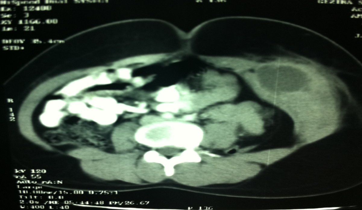
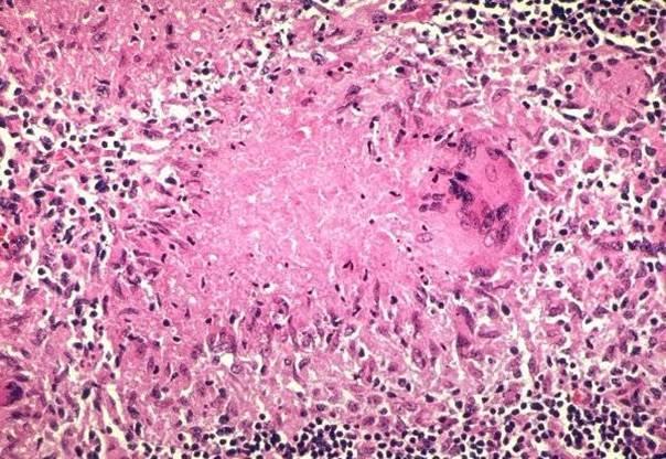
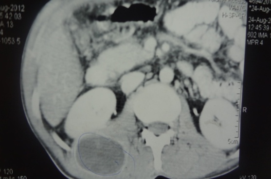
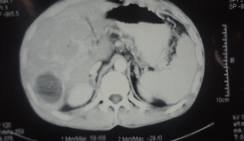
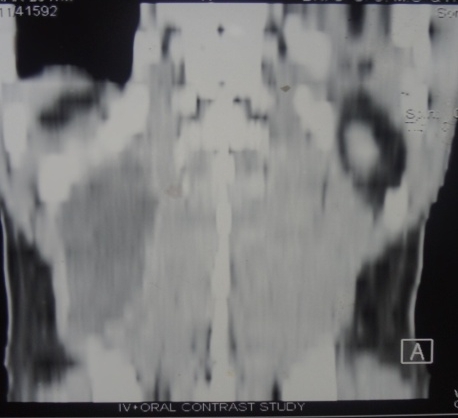
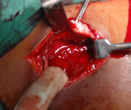
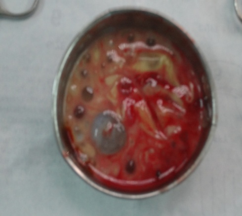
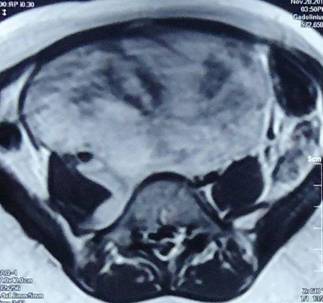
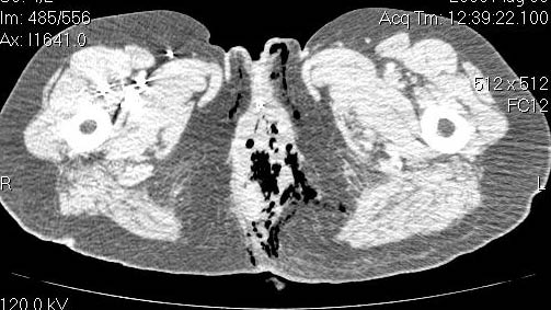
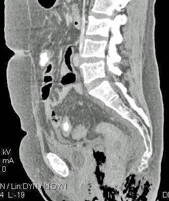
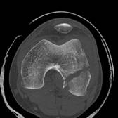
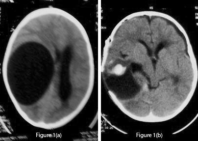
Fig 1 (a): Pre-operative unenhanced CT scan which shows a large CSF density cystic lesion on the right side causing mass effect and midline shift to the left. There is no peri-lesional oedema. Fig 1 (b): Post-operative CT scan of the lesion shows a large void which can lead to dangerous collapse. Mild haematoma is also seen. 
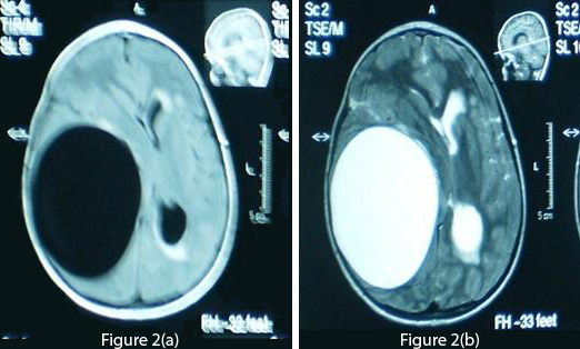
Fig 2 (a): T1-weighted axial MRI of the brain demonstrates a cyst density similar to CSF. Fig 2 (b): T2-weighted MRI shows no ring enhancement or oedema. The periventricular hyperintensity of the left side is probably due to obstructive hydrocephalus. 
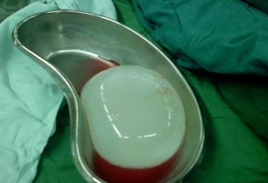
Fig 3: This shows the cyst removed in toto after operation. The cyst appears creamy and smooth. After summation of all the above data, the diagnosis of a hydatid cyst was made and a right frontotemporoparietal craniotomy was performed. A large cystic structure (14×14×12 cm) was delivered with utmost care to avoid rupture and spillage [fig 3]. A hydatid cyst was confirmed by pathology reports.  A post-operative CT scan showed a large space without any residual matter [fig 1(b)]. Post-operatively, albendazole 15 mg/kg was started and continued for four weeks. The patient showed marked improvement in his neurological deficit and was discharged after one week with close follow-up. Discussion/Review Of Literature Life CycleHydatidosis is caused by Echinococcus granulosus, which occurs mainly in dogs. Humans who act as intermediate hosts get infected incidentally by ingesting eggs from the faeces of the infected animal. The eggs hatch inside the intestines and penetrate the walls, entering blood vessels and eventually reach the liver where they may form cysts or move on towards the lungs. Even after pulmonary filter, a few still make it to the systemic circulation and can lodge in almost any part of the body, including the brain, heart and bones.2,3,8,14,16,26 Brain hydatid cysts are relatively rare and only account for up to 2% of total cases.4,5,7  The actual percentage may be higher than what we have in literature, due to under-reporting. Brain hydatid cysts can be primary (single) or secondary (multiple).2,3,4,5,7 The latter are thought to arise from the multiple scolices released from the left side of the heart following cyst rupture in the heart2,3,5,27 or due to spontaneous, traumatic or surgical rupture of a solitary cranial cyst.3,5 Cysts mostly involve the territory of the middle cerebral artery4,7 but other regions like intraventricular, posterior fossa and the orbit have also been reported.15,17,18,28 The wall of the cyst consists of an inner endocyst (germinal layer) and outer ectocyst (laminated layer). The host reacts to the cyst forming a pericyst (fibrous capsule), which provides nutrients to the parasite. In the brain, due to minimal reaction, the pericyst is very thin. The endocyst produce scolices which bud into the cyst cavity and may sediment within the hydatid cavity, commonly known as hydatid sand.3,14,29,30 Presentation and DiagnosisMost hydatid cysts are acquired in childhood and are manifested during early adulthood.8,29 Cysts develop insidiously, usually being asymptomatic initially, and present with protean clinical and imaging features.3,5,6 In previous studies the most common presenting symptoms were headache and vomiting.4,5,7,14,15,28 Also in the literature, patients reported ataxia, diplopia, hemiparesis, abducens nerve palsy and even coma.5,7,15,28 Surprisingly, in the present study the patient did not have a headache and presented with parasthesia and numbness of the toes. Later he developed left sided weakness, convulsions and finally ataxia, which correlate with previous studies. Diagnosis of a hydatid cyst can sometimes be confused with other space occupying lesions of the brain, especially abscesses, neoplasms and arachnoid cysts.14,31  In this study the patient had bilateral frank papilloedema which is also mentioned in earlier reports.4,28  The Casoni and Weinberg tests, indirect haemagglutination, eosinophilia and ELISA are used in diagnosing hydatid cysts, but as brain tissue evokes minimal response many results tend to be false negatives.2,5,8,25  In our case also, serology for hydatid cyst was negative. CT scan and MRI are used frequently in diagnosing the cystic lesions.3,8,14,23,32,33  However, MRI is considered superior in demonstrating the cyst rim.5,8,11,21,32,34  On CT scan, a solitary cyst appears as well-defined, spherical, smooth, thin-walled and homogeneous, with an inner density similar to CSF, and non-enhancing walls.11,29,32The wall may appear iso-dense to hyper-dense on CT scan3,8, and rarely, may become calcified.11,29,32 There is usually no surrounding brain parenchymal oedema, which if exists along with ring enhancement, indicates inflammation and infection. 7,11,32,33,34,35 Ring enhancement and peri-lesional oedema differentiates brain abscesses and cystic neoplasms from uncomplicated hydatid cysts.3,8 These findings can in fact sometimes cause dilemma and misdiagnosis and lead to catastrophic events.14 The cyst shows low signal intensity on T1-weighted, and high signal intensity on T2-weighted MRI.2 MRI may also show peri-lesional oedema not seen on regular CT scan imaging.7 MRI may prove superior in determining exact cyst location, presence of super-added infections and cystic contents, and also in surgical planning and ruling out other diagnostic possibilities.14,33 We strongly recommend MRI for better evaluation of cystic brain lesions. Spontaneous cystic rupture can lead to different appearances depending on which layers have been obliterated, and produce some specific signs.3 When only the endocyst ruptures, cyst contents are held by the outer pericyst giving a peculiar water lily sign, which is pathognomic.3,8 TreatmentThough still in infancy, medical therapy for small or inoperable brain hydatid cysts has been promising. Albendazole alone or in combination with other compounds, such as praziquantel, has been reported with favourable results as an adjunct and, in certain circumstances, as the primary mode of treatment.2,36,37,38 It is reported that albendazole results in the disappearance of up to 48% of cysts and a substantial reduction in size of the cysts in another 28%.2 The duration of the treatment is four weeks or more, and recently many authors have favoured a prolonged therapy. The change in levels of cyst markers such as alanine, succinate, acetate and lactate, measured before and during treatment on Proton Magnetic Resonance Spectroscopy (MRS), correlate well with shrinkage and resolution of cyst findings on conventional MRI and help in evaluating the efficacy of chemotherapy.39 Cysts may drain into ventricles or rupture completely, causing spillage of contents into the subarachnoid space, leading to fatal anaphylactic shock, meningitis or local recurrence.3,5,22,25 Surgery is the mainstay for treating intracranial hydatid cysts and the aim is to excise the cysts entirely without rupture, which can otherwise lead to catastrophic events as described earlier 2,3,14,25. The Dowling-Orlando technique remains the preferred method, in which the cyst can be delivered by lowering the head of the operating table and instilling warm saline between the cyst and the surrounding brain.40 Even minimal spillage can cause deleterious effects (1 ml of hydatid sand contains 400,000 scolices).14 The thin cyst wall, periventricular location and micro-adhesions to the parenchyma are the main problems encountered during the surgical procedure.1,22 The large cavity remaining after the cystic removal can lead to many serious complications, such as cortical collapse, hyperpyrexia, brain oedema and cardio-respiratory failure.5 Recurrence remains a major concern, which is managed by both antihelminthic chemotherapy and surgery. In a study conducted by Ciurea et al, 25% of the patients had recurrence, which highlights the need for long term follow up.23 In the present study, due to the huge size of the cyst and progressive neurological deficit, it was not wise to completely rely on medical therapy. Surgery was performed and post-operatively albendazole was started as an adjunct. We recommend that for treating brain hydatid cyst, the size of the cyst, multiplicity, location and neurological deficit must all be taken into consideration. 

Acknowledgements / Conflicts / Author Details
Competing Interests: 
None Declared
Details of Authors: 
ALI NEMATI MD; Chief Resident, Department Of Neurosurgery, Shiraz Medical School AHMAD KAMGARPOUR MD; Associate Professor, Department Of Neurosurgery, Shiraz Medical School MURTAZA RASHID MD; House Officer, Department Of Neurosurgery, Shiraz Medical School SAHAR SOHRABI NAZARI MD; House Officer, Department Of Neurosurgery, Shiraz Medical School
Corresponding Author Details: 
MURTAZA RASHID MD,Department of Neurosurgery, Shiraz University of Medical Sciences, Iran. P.O. Box: 71455-166 Tel: +98 917 910 5372
Corresponding Author Email: 
dr.murtazarashid@gmail.com
References
References: 

1. Budke CM. Global socioeconomic impact of Cystic echinococcosis. Emerg Infec Dis 2006;12(2):296-303.
2. Reddy DR. Managing cerebral and cranial hydatid disease. Neurol India 2009;57:116-118.
3. Kovoor JME, Thomas RD, Chandrashekhar HS, Jayakumar PN, Pillai S, Shankar SK. Neurohydatidosis. Australas Radiol 2007;51:406-411.
4. Ersahin Y, Mutluer S, Güzelbag E. Intracranial hydatid cysts in children. Neurosurgery 1993;33(2): 219-224.
5. Cavusoglu H, Tuncer C, Ozdilmaç A, Aydin Y. Multiple Intracranial Hydatid cysts in a boy. Turk Neurosurg 2009; 19(2):203-207.
6. Sierra J, Oviedo J, Berthier M, Leiguardo R. Growth rate of secondary hydatid cysts of the brain. Case report. J Neurosurg 1985;62:781-782.
7. Gana R, Skhissi M, Maaqili R, Bellakhdar F. Multiple infected cerebral hydatid cysts. J Clin Neurosci 2008;15(5):591-593.
8. Andronikou S, Welman C, Kader E. Classic and unusual appearances of hydatid disease in children. Pediatr Radiol 2002;32: 817-828
9. Afsar H, Yagci N, Aybasti N, et al. Hydatid disease of the kidney. Br J Urol 1994;73:17-22
10. Dahniya MH, Hanna RM, Ashebu S, et al. The imaging appearances of hydatid disease at some unusual sites. Br J Radiol 2001;74:283–289.
11. Pedrosa I, Saiz A, Arrazola J, et al. Hydatid disease: radiologic and pathologic features and complications. Radiographics 2001;20:795–817.
12. Rebhandl W, Turnbull J, Felberbauer F. Pulmonary echinococcosis (hydatidosis) in children: results of surgical treatment. Pediatr Pulmonol 1999;27:336–340.
13. Macedo AJ, Magalhaes MP, Jalles Tavares N, et al. Cardiac hydatid cyst in a child. Pediatr Cardiol 1997; 18:226–228.
14. Anvari M, Amirjamshidi A, Abbassioun K. Gradual and complete delivery of a hydatid cyst of the brain through a single burr hole, a wrong happening! Childs Nerv Syst 2009:25(12):1639-1642.
15. Kayaoglu CR. Giant hydatid cyst in the posterior fossa of a child: a case report. J Int Med Res 2008; 36(1)198-202.
16. Salehi M, Soleimani A. Cardiac echinococcosis with negative serologies: a report of two cases. Heart lung circ 2009;18(1):59-61.
17. Ergun R, Okten AI, Yuksel M, et al: Orbital hydatid cysts. report of four cases. Neurosurg Rev 1997;20:33–37
18. Karakaþ HM, Tokoðlu F, Kacar M, et al: Retrobulbar hydatid cyst:assessment of two cases. Australas Radiol 1997;41:179–180.
19. Mazyad MA, Mostafa MM, Morsy TA. Spinal cord hydatid cysts in Egypt. J Egypt Soc Parasitol 1998; 28:655–658.
20. Yurtçu M, Gündüz M, Toy H, Günel E. Spermatic cord hydatid cyst: an unusual localization. J Pediatr Surg 2007;42(12):e15-6.
21. Krajewski R, Stelmasiak Z. Cerebral hydatid cysts in children. Childs Nerv Syst 1991;7:154-155.
22. Izci Y, Tüzün Y, Seçer HI, Gönül E. Cerebral hydatid cysts: technique and pitfalls of surgical managemen.Neurosurg Focus 2008;24(6):E15.
23. Ciurea AV, Fountas KN, Coman TC, Machinis TG, Kapsalaki EZ, Fezoulidis NI, Robinson JS. Long-term Surgical outcome in patients with intracranial hydatid cyst. Acta Neurochir (wien) 2006:148(4):421-426.
24. Pearl M, Kosilimbos DG, Lehrer HZ, Rao AH, Fink H, Zaiman H. Cerebral echinococcosis, a pediatric disease. Report of 2 cases with one successful five year survival. Pediatrics 1987;61:915–920 .
25. Khaldi M, Mohamed S, Kallel J, Khouja N. Brain hydatidosis: report on 117 cases. Childs Nerv Syst 2000; 16:765-769.
26. Guillot J, Bouree P. Zoonotic worms from carnivorous pets: risk assessment and prevention. Bull Acad Natl Med 2007;191(1):67-78.
27. Turgut M, Benli K, Eryilmaz M. Secondary multiple intracranial hydatid cysts caused by intracerebral embolism of cardiac echinococcosis: an exceptional case of hydatidosis. J Neurosurg 1997;86:714–718.
28. Guzel A, Tatli M, Maciaczyk J, Altinors N; Primary Cerebral Intraventricular Hydatid Cyst. A Case Report and Review of the Literature. J Child Neurol 2008;23(5):585-588.
29. Haliloglu M, Saatcsi I, Akhan O, Ozmen MN, Besin A. Spectrum of imaging finding in pediatric hydatid disease. AJR 1997;169: 1627–1631.
30. Iyigun O, Uysal S, Sancak R, Hokelek M, Uyar Y, Bernay F, Ariturk E. Multiple organ involvement hydatid cysts in a 2-year-old boy . J Trop Pediatr 2004;50(6):374-376.
31. Bahloul K, Ouerchefani N, Kammoun B, Boudouara MZ . Unusual brain edema caused by an intracranial hydatid cyst: case report and literature review. Neurochirurgie 2009;55(1):53-56.
32. Tuzun M, Hekimoðlu B: Hydatid disease of the CNS. imaging features. AJR 1998;171:1497-1500.
33. El-Shamam O, Amer T, El-Atta MA. Magnetic resonance imaging of simple and infected hydatid cysts of the brain. Magn Reson Imaging 2004;22(9):1339-1340.
34. Altinörs N, Bavbek M, Caner HH, Erdogan B. Central nervous system hydatidosis in Turkey: A cooperative study and literature survey analysis of 458 cases. J Neurosurg 2000;93:1–8.
35. Behari S, Banerji D, Phadke RV, Shukla S, Krishnani N, Kumar D. Multiple infected extradural parasellar hydatid cysts. Surg Neurol 1997;48: 53–57.
36. Davis A, Dixon H, Pawloski ZS. Multicentre clinical trials of benzimidazole-carbamates in human cystic echinococcosis (phase 2), Bull World Health Organ 1989;67:503–508.
37. Singounas EG, Leventis AS, Sakas DE, Hadley DM, Lampadarios DA, Karvounis PC. Successful treatment of intracerebral hydatid cyst with albendazole: case report and review of the literature. Neurosurgery 1992;31:571–574.
38. Todorov T, Vutova K, Mechkov G, Tonchev Z, Georgiev P, Lazarova I. Experience in the chemotherapy of severe, inoperable echinococcosis in man. Infection 1992;20:19–24.
39. Seckin H, Yagmurlu B, Yigitkanli K, Kars HZ. Metabolic changes during successful medical therapy for brain hydatid cyst: case report. Surg Neurol 2008;70(2):186-189.
40. Carrea R, Dowling E Jr, Guevara JA. Surgical treatment of hydatid cysts of the central nervous system in the pediatric age (Dowling's technique). Childs Brain 1975;1(1):4-21.


A comparison of different methods of assessing cosmetic outcome following breast-conserving surgery and factors influencing cosmetic outcome

Authors
Charfare H, MacLatchie E, Cordier C , Bradley M, Eadie C, Byrtus A, Burnet K, Chapman D, Wishart GC and Purushotham AD
Article Citation and PDF Link
BJMP 2010;3(1):310
Abstract / Summary
Abstract: 

Methods to assess cosmesis following breast-conserving surgery are varied and assumed to yield similar results. The aim of this study was to compare three different methods of cosmetic assessment following breast-conserving surgery and to assess the impact of certain factors on cosmetic outcome.

One hundred and fifteen patients undergoing breast-conserving surgery had 3 view digital photographs taken for assessment of cosmesis at one year post-surgery. Subjective cosmetic assessment was performed by a 5 member panel and objective assessment by Breast Retraction Assessment (BRA) and Nipple Deviation (ND). Factors including tumour size, percentage breast volume excised, location of tumour and number of breast operations performed was correlated with final cosmetic outcome.

The majority of patients undergoing breast-conserving surgery demonstrated satisfactory cosmetic results. Inter-observer variation assessed using a kappa statistic for panel assessment gave a value of 0.42 with a 95% confidence interval (CI) of 0.37 to 0.47, indicating moderate agreement between observers. The kappa statistic for agreement between the three methods used for assessing cosmesis was -0.23 with 95% CI of -0.35, -0.11 indicating poor concordance between the three methods used. These methods however, may be complementary to each other and therefore these observations merit further investigation. Tumour location, tumour size and the number of operations performed did not influence cosmetic outcome. However, cosmetic outcome was related to percentage breast volume excised.

Keywords: 
breast-conserving surgery, cosmetic assessment
 
Introduction:
 
Cosmetic outcome following breast-conserving surgery depends on various factors including location of the tumour, weight of the specimen excised, number of surgical procedures, volume of breast, length of scar and postoperative adjuvant treatment1. The best method of cosmetic assessment following breast-conserving surgery is still unclear. However various objective and subjective methods in combination are known to give a good assessment of cosmesis2, 3, 4. It has been shown that photographic assessment is as effective as live assessment in the post-surgical setting5. Methods to assess cosmesis following breast-conserving surgery are varied and more recently computer software are being used to assess cosmesis following breast-conserving surgery.
 
The aim of this study was to compare three different methods of cosmetic assessment following breast-conserving surgery and to assess the influence of various factors on final cosmetic outcome.
 
Methods:
 
One hundred and fifteen patients underwent breast-conserving surgery for carcinoma of breast by wide local excision and level 2 axillary clearance. Following wide local excision, cavity shavings were taken to ensure adequate local excision. Breast drainage was not used but suction drains were used routinely following axillary clearance. All patients received adjuvant breast radiotherapy (46 Gy, 23 fractions with a cavity boost of 12 Gy in 4 fractions) administered over a period of 6 weeks.
 
bjmp-2010-3-1-310a.jpg
Figure-1: Measurement of Breast Retraction Assessment6 (reprinted with permission from Elsevier, ref 6 (page 670), copyright 1999)
 
Digital photographs were taken at one year in three views; frontal with arm by the side, frontal and oblique with arm abducted to 90 degrees. The photographs were used for subjective and objective assessment of cosmesis. The objective assessment of cosmesis was carried out using Breast Retraction Assessment (BRA) and Nipple Deviation (ND). BRA was calculated as indicated in figure 16. ND was calculated as a percentage difference from suprasternal notch to nipple on normal side compared with the operated side. BRA and ND were then categorised into three groups; BRA: (excellent to good <3.1 cm, fair 3.1-6.5, poor >6.5), ND: (difference of <5% - excellent to good, 5-10% fair and >10% poor). Subjective assessment was carried out using a panel consisting of a Consultant Breast Surgeon, Research Fellow, Secretary, Breast Care Nurse and Nurse Practitioner with each scoring independently. The method described by Harris et al7 with a score of 9-10 for excellent (no visible difference between two breasts), good (slight difference; score 7-8), fair (obvious difference but no major distortion; score 4-6) and poor (major distortion; score <4) was used to categorise patients.
 
Figure- 2: Measurement of breast volume (Sloane method) Formula for calculation of breast volume: 1/3 π r2h (reprinted with kind permission from Sloane project)
 
The volume of breast tissue excised was estimated with the length (L), width (W) and height (H) of the excised tissue specimen and the cavity shave measured by the pathologist and using the formulas for a prolate ellipse (V= 0.52* L* W* H); this was added on to the volume of cavity shave calculated using the formula 0.79* L* W* H. The total breast volume was estimated using the mammogram and applying the formula (1/3 πr2h) as shown in figure-2. Based on these measurements the percentage breast volume excised was calculated and compared with cosmetic outcome.
 
Statistics:
 
Multirater kappa statistics8 were used to assess inter-observer agreement between five different members of the panel and also to test agreement between the three different methods for assessing cosmesis. The average value given by the panel was used and categories good and excellent were combined in order to compare the three methods of cosmetic assessment. A kappa statistic of less than or equal to 0.20 was considered to demonstrate poor agreement, 0.21 to 0.40 fair agreement, 0.41 to 0.60 moderate agreement, 0.61 to 0.80 good agreement and 0.81-1.00 very good agreement9.
 
The effect of the percentage volume of the breast tissue excised and the tumour size on the three methods of cosmetic assessment was examined using where appropriate a Jonckhneere-Terpstra test for trend, a Kruskal Wallis test or a Mann-Whitney U test. The effect of the number of breast operations performed and the location of the tumour were assessed using Chi-square test or Fisher’s exact test when appropriate.
 
Results:
 
Of the 115 patients assessed using panel assessment 64 (56%) scored good to excellent, 39 (34%) scored fair and 12 (10%) scored poor. ND scored 50(43%) as good to excellent, 32 (28%) as fair and 33 (29%) as poor. Using BRA, the scores were 76 (66%), 38 (33%) and 1(1%) respectively. These results are shown graphically in figure-3.
 
Figure- 3: Number of patients classified into each of the three categories poor, fair and good/excellent for the three methods bra, nipple deviation and panel assessment. BRA= breast retraction assessment; Panel= assessment by different panel members; ND= nipple deviation
 
Taking the mean scores for these three methods of assessment and dichotomising the results into two categories of good to excellent and poor to fair, 52% of patients in this study had good to excellent cosmetic result and 48% were categorised as fair to poor cosmetic result. The Kappa statistic was calculated on 115 patients for the three methods of assessment and it was found to have a value of –0.23 (95% CI (–0.35, – 0.11) which falls within the poor agreement category.
 
 
Figure- 4: Comparison of panel assessment by different panel members. Pa, Ph, Pg, Pk and Pd= Codes for the different panel members
 
Examining the panel assessment using the kappa statistics for the 115 patients assessed there was moderate agreement between the panel members (Kappa statistic of 0.42; 95% confidence interval of (0.37, 0.47). This suggests there is moderate chance that the panel members will categorise each patient the same way. If one plots the panel assessment graphically one can see that excellent is used least by all and fair most frequently (figure 4).
 
Factors affecting cosmesis:
 
1) Percentage breast volume excised
 
 
Figure -5: Effect of percentage breast volume excised on cosmetic outcome using Panel assessment, BRA and ND
 
For panel assessment it appears that removal of a larger percentage volume gives a poor cosmetic result and a smaller percentage volume an excellent/good result (figure 5) as would be expected clinically. This is supported by a Jonckhneere-Terpstra test for trend (=0.01). Using ND median percentage volumes across the groups did not appear to differ (χ2=1.05 p=0.59, Kruskal Wallis test). However, for BRA, only one patient was classified as poor and no difference was seen between those with fair and good/excellent results (U=477, p=0.34). The median volume excised for different cosmetic outcome using the three methods is shown in table 1. 
 
Table-1: Medians volumes for the three measurements.
 
Panel assessment
BRA
Nipple deviation
Poor
 157.56
(only 1 poor value)
 100.61
Fair
 88.58
 93.11
 55.96
Good/Excellent
 68.33
 76.55
 81.33
BRA= breast retraction assessment
 
The percentage breast volume excised was then compared with cosmetic outcome using the three methods of assessment. As shown in table 2, 45-65% of patients with <10% estimated breast volume excised had good to excellent cosmetic result compared with 35-50% good to excellent result if >10% breast volume was excised.
 
2) Tumour location:
 
Tumour location was divided into inner or outer quadrants of the breast. The distribution of tumours in the breast and the cosmetic outcome with each of the three methods of assessment is shown in table 3. The location of tumour within the breast was not significantly associated with cosmetic outcome (χ2 =1.86, p=0.39 for panel assessment), (p=0.23, Fisher’s exact test for BRA) and (χ2 =0.21, p=0.90 for ND).
 
Table-2: Estimated percentage breast volume excised and cosmetic outcome
 
< 10% breast volume excised
> 10% breast volume excised
Panel Assessment
Good to excellent (%)
32 (65)
7 (35)
Fair (%)
15 (31)
6 (30)
Poor (%)
2 (4)
7 (35)
Breast Retraction Assessment
Good to excellent (%)
32 (65)
10 (50)
Fair (%)
16 (33)
10 (50)
Poor (%)
1 (2)
0
Nipple Deviation
Good to excellent (%)
22 (45)
8 (40)
Fair (%)
15 (31)
4 (20)
Poor (%)
12 (24)
8 (40)
 
3) Number of breast operations:
 
The influence of number of operations (1 vs 2) was examined for each of the three methods of assessment. Using BRA and Panel assessment there was no significant difference in the cosmetic outcome for patients who underwent one or two operations ( p=0.70 for panel assessment), (p=0.99, Fisher’s exact test for BRA). For ND there does appear to be a larger proportion in the poor group for those with two operations (p =0.30 Fisher’s exact test for ND). This is illustrated in Table 3.
 
Table-3: Factors affecting cosmesis
 
Panel
BRA
ND
Percentage volume excised
Poor (median (IQR))
Fair (median (IQR))
Good/Excellent (median (IQR))
 
13.8 (11.0,16.5)
8.4 (4.4,10.4)
5.8 (3.9,8.0)
 
-
8.0 (4.6,11.6)
6.9 (4.3,10.1)
 
8.5 (5.1,11.4)
5.8 (3.9,9.4)
7.2 (4.4,11.0)
Location
Poor (outer (n), inner (n))
Fair (outer (n), inner (n))
Good/Excellent (outer (n), inner (n))
 
8, 2
22,8
47,8
 
0,1
26,5
51,12
 
9,1
23,6
33,8
No. of Operations
Poor (One (n), Two (n))
Fair (One (n), Two (n))
Good/Excellent (One (n), Two (n))
 
9,1
24,6
48,8
 
1,0
26,5
54,10
 
20,5
27,2
34,8
Tumour size (mm)
Poor (median (IQR)
Fair (median (IQR)
Good/ Excellent (median (IQR)
 
12 (9, 15)
11 (9, 19)
12 (7, 15)
 
-
11 (8,15)
12 (7, 15)
 
12 (10, 15)
12 (8, 16)
9 (6,14)
Panel= panel assessment; BRA= breast retraction assessment; ND= nipple deviation; IQR= inter quartile range
 
4) Tumour size:
 
Table 3 shows the median tumour size and interquartile range for the three categories, good/ excellent, fair and poor and one can see that there is no significant difference in tumour size for these categories using panel assessment (Jonckheere-Terpstra p=0.31) or BRA (U =873, p=0.55). However, using ND there was evidence to suggest that large tumour size resulted in poor outcome (Jonckheere-Terpstra, p=0.04).
 
Thus, tumour size had a significant influence on the cosmetic outcome when ND was used as the method of assessment.
 
Discussion:
 
Cosmetic outcome following breast-conserving surgery is assessed using a combination of subjective and objective methods. The subjective method uses a panel of members from different backgrounds to assess overall cosmesis. However, Pezner et al10 showed relatively low level of agreement between observers when a four-point scale was used for assessment of overall cosmesis. The objective methods, which mainly compare the position of the nipple, are easy to reproduce but do not take into account skin changes and give poor assessment of cosmesis for lower quadrant tumours.
 
In this study the cosmetic outcome was assessed in 115 patients one year post-operatively. The mean cosmetic result using the three different methods of assessment was good to excellent in 55% of the patients, which compares favourably with other studies reported in the literature2, 4. Looking at inter-observer variation for the panel assessment, moderate agreement was found between different panel members. This compares favourably with an earlier study that looked at cosmetic outcome in the EORTC trial 22881/108826. However, when the three methods of cosmetic assessment were compared with each using kappa statistic there was poor concordance. Although some agreement was noted, this was likely to be due to chance as the kappa statistic was low. It is difficult to explain this finding as other authors1, 6 have reported moderate to good agreement between subjective and objective methods. One explanation for this lack of agreement is that each method assesses a different aspect of cosmesis.
 
The two objective methods of cosmetic assessment (BRA and ND) that are used to assess upward retraction of nipple have been found to be a very good determinant of cosmetic outcome and are easy to reproduce according to Fujishiro et al11. Furthermore, evaluation of nipple position has also been shown to be moderately representative of overall cosmetic result6. BRA is a two dimensional measurement of nipple position and some cosmetic factors such as volume, shape or skin changes cannot be accurately assessed11. This is probably the reason why BRA shows a better cosmetic outcome when compared with subjective assessment by panel members. In this study only one (1%) patient was deemed to have a poor cosmetic outcome using BRA compared with 12 (10%) using panel assessment.
 
A criticism of the current study is that patients’ perceptions of their own cosmetic outcome were not assessed. Previous studies have shown a significant correlation between patient satisfaction after breast-conserving surgery and their self-assessment of cosmesis12, 13. This study shows that there is need to find a reproducible method of cosmetic assessment which takes into account all the limitations of the methods currently used. More recently computer software like BCCT.core and Breast Analysing Tool have been developed and early results using these software are promising14, 15. There are various factors that are known to affect cosmesis following breast-conserving surgery. As expected larger percentage volume of excised breast tissue was associated with poorer cosmetic result. This was particularly evident from panel assessment. Such a relationship was less clear with BRA and ND. The effect of percentage volume of breast tissue excised and the outcome is consistent with a recent report that showed higher patient satisfaction if estimated percentage breast volume excised was < 10%16. Cosmetic outcome based on tumour location varies depending on the method of assessment used. BRA is adversely affected by tumour in the upper and outer quadrants of the breast, suggesting that surgery causes larger nipple deviation in this quadrant, while panel assessment gives poor scores for tumours located in inferior quadrant2, 11. In this study only 19% of patients had tumours located in the inner quadrant and the small number may explain why, no significant difference in cosmetic outcome was found. Tumour location or the number of operations performed did not appear to affect the cosmetic outcome in this study. The volume of breast tissue excised depends on tumour size. Since the majority of tumours in this study were small, the size of the tumour did not affect cosmetic outcome except when nipple deviation was used. This once again indicates that these three methods of assessment may be looking at different aspects of cosmesis.
 
In conclusion, cosmetic outcome following breast-conserving surgery is an important, measurable end point. However, the best method of assessment of cosmesis has not been devised17. Although, the objective methods are easier to apply and reproduce, they do not give a good assessment of global cosmetic results. Panel Assessment however, does appear to provide concordant results between different observers and may be a useful, simple measure of cosmetic assessment following breast-conserving surgery.
 
Acknowledgements / Conflicts / Author Details
Competing Interests: 
None declared
Details of Authors: 
CHARFARE H, Bedford Hospital NHS Trust, UK MACLATCHIE E, CORDIER C, EADIE C, Western Infirmary, University of Glasgow, UK BRADLEY M, Department of Public Health and Primary Care, University of Cambridge, UK BYRTUS A, PURUSHOTHAM AD, Department of Academic Oncology, King’s College London, UK BURNET K, CHAPMAN D, WISHART GC, Addenbrooke’s Hospital, Cambridge, UK
Corresponding Author Details: 
H CHARFARE, Bedford Hospital NHS Trust, South Wing, Kempston Road, Bedford Hospital NHS Trust, MK42 9DJ
Corresponding Author Email: 
habib.charfare@bedfordhospital.nhs.uk
References
References: 
1)Al-Ghazal SK, Blamey RW, Stewart Jet al. The cosmetic outcome in early breast cancer treated with breast conservation. Eur J Surg Oncol 1999; 26 (6): 566- 570
2)Van Limbergen E, Rijnders A, van der Scheuren et al. Cosmetic evaluation of breast conserving treatment for mammary cancer. 2. A quantitative analysis of the influence of radiation dose, fractionation schedules and surgical treatment techniques on cosmetic results. Radiother and Oncol 1989; 16: 253-267
3)Christie DRH, O’Brien MY, Christie JA et al. A comparison of methods of cosmetic assessment in breast conservation treatment. The Breast 1996; 5: 358- 367
4)Ash D V, Benson E A, Sainsbury J R et al. Seven year follow-up on 334 patients treated by breast-conserving surgery and short course of radical postoperative radiotherapy: a report of the Yorkshire breast cancer group. Clin Onco (R Coll Radiol) 1995; 7 (2): 93-96
5)Eadie C, Herd A, Stallard S: An investigation into digital imaging in assessing cosmetic outcome after breast surgery. J Audiovisual Media in Medicine 2000; 23 (1): 12- 16
6)Vrieling C, Collette L, Bartelink E et al. Validation of the methods of cosmetic assessment after breast-conserving therapy in the EORTC ‘ boost versus no boost’ trial. Int J Radiat Oncol Biol Phys 1999; 45 (3): 667- 676
7)Harris JR, Levene MB, Svensson G et al: Analysis of cosmetic results following primary radiation therapy for stage I and II carcinoma of the breast.
Int J Radiat Oncol Biol Phys 1979; 5: 257- 261
8)Siegel S, Castellan Jr NJ. Nonparametric statistics for the behavioural sciences 2nd Edition, McGraw-Hill, 1988, 284- 291.
9)Altman DG. Practical statistics for medical research, Chapman & Hall, 1991, 404.
10) Pezner RD, Lipsett JA, Vora NL et al. Limited usefulness of observer-based cosmesis scales employed to evaluate patients treated conservatively for breast cancer. Int J Radiat Oncol Biol Phys 1985; 11: 1117- 1119
11) Fujishiro S, Mitsumori M, Kokubo M et al. Cosmetic results and complications after breast-conserving therapy for early breast cancer. Breast Cancer 2000; 7 (1): 57- 63
12) Al-Ghazal SK, Fallowfield L, Blamey RW. Patient evaluation of cosmetic outcome after conserving surgery for treatment of primary breast cancer. Eur J Surg Oncol 1999; 25 (4): 344- 346
13) Kaija H, Rauni S, Jorma I et al. Consistency of patient-and doctor assessed cosmetic outcome after conservative treatment of breast cancer. Breast cancer Res Treat 1997; 45 (3): 225- 228
14) Fitzal F, Krois W, Trischler H et al. The use of breast symmetry index for objective evaluation of breast cosmesis.  The Breast 2007; 16: 429-435
15) Cardoso MJ, Cardoso J, Amaral N et al. Turning objective into subjective: The BCCT.core software for evaluation of cosmetic results in breast cancer conservative management. The Breast 2007; 16: 456-461
16) Cochrane RA, Valasiadou P, Wilson ARM et al. Cosmesis and satisfaction after breast-conserving surgery correlates with percentage breast volume excised. Br J Surg 2003; 90: 1505- 1509
17) A Munshi, S Kakkar, R bhutani et al. Factors influencing cosmetic outcome in breast conservation. Clin Oncol 2009; 21: 285-293

Current Management of Barrett’s Oesophagus

Authors
Sharif A Anwar, Senthooran Kathirgama Kanthan and Amjid Ali Riaz
Article Citation and PDF Link
BJMP 2009:2(4) 8-14
Abstract / Summary
Abbreviations: 
BO – Barrett’s Oesophagus, GORD - Gastro-Oesophageal Reflux Disease, LSBO - Long Segment Barrett’s Oesophagus, HGD - High Grade Dysplasia, EMR – Endoscopic Mucosal Resection, 5-ALA - 5-AminoLevulinic Acid, PDT - PhotoDynamic Therapy
Introduction
 
Barrett’s Oesophagus (BO) describes a histological abnormality of the lower oesophagus widely accepted to be associated with gastro-oesophageal reflux disease (GORD). The nature of this disease has been a subject of debate since its description by Tileston in 1906 as peptic ulceration of the oesophagus. Barrett himself initially theorised that the abnormal oesophagus was in fact stomach that had been pulled into the chest by a congenitally short oesophagus (1). This idea was ultimately challenged as the area in question lacked a peritoneal covering, contained submucosal glands and muscularis propria characteristic of the oesophagus (2). In 1976, Paull et al described a distinctive type of intestinal metaplasia the investigators called "specialised columnar epithelium”. Specialised intestinal metaplasia is now widely accepted to be the hallmark of BO with its presence predisposing to dysplasia and cancer regardless of its location within the oesophagus (3). 
 
Barrett’s Oesophagus, its identification and treatment continues to be an area of debate and interest. Although not sinister in itself, it is a known precursor to malignant disease and strongly associated with GORD. Barrett’s oesophagus is the most frequent predisposing risk factor for the progression to adenocarcinoma in the oesophagus. Sufferers have a 40 fold increased risk when compared to the general population (4).
The progression of GORD to BO appears to be related to exposure of oesophageal tissue to the acidic contents of the stomach. It is therefore seen in hiatus hernia, lower oesophageal dysfunction, delayed oesophageal acid clearance and duodenogastric reflux. Furthermore, it is the duration and not frequency of exposure to acidity that dictates erosive damage to the oesophagus. Levels of acidity also contribute. The damage to cells incurred leads to inflammatory infiltration and cell necrosis with replacement of oesophageal epithelium by metaplastic columnar cells.
 
Assessing severity of BO relies partly on endoscopic visualisation techniques and length of oesophagus involved. Long segment Barrett’s oesophagus (LSBO) indicated a >3cm segment of involvement with short segment disease involving <3cm. LSBO carries a higher risk of progression to adenocarcinoma. Its development is associated with long term symptoms, severe combined patterns of reflux (both erect and supine) on 24 hour pH monitoring and reduced lower oesophageal sphincter pressures. Patients are less sensitive to direct acid exposure than those with short segment disease. The latter group also tend to have shorter duration of symptoms, normal sphincter pressures and only upright reflux on 24 hour pH monitoring (3). 
 
The Prague C and M criteria is a recently developed classification system utilising the circumferential and maximal extent of oesophageal columnar tissue to assess disease severity endoscopically (5). Its accuracy is yet to be assessed clinically, however, it is believed to largely improve the overall assessment of Barrett’s (6). The further classification of disease severity is based on the degree of dysplasia, with high grade dysplasia carrying a higher risk of progression to malignancy. 
 
Barrett’s oesophagus is predominantly seen in the age group 55-65, with males being affected twice as frequently as females. The disease is more prevalent in the white population. Obesity, smoking and alcohol intake being further risk factors. H.pylori may be protective against Barrett’s oesophagus with two mechanisms postulated. Namely, the induction of atrophic gastritis, which results in decreased acid production and the production of neutralising ammonia independent of gastric atrophy (7). The duration of symptoms of GORD but not necessarily symptom severity is also associated with increased risk of progression to BO. The exact pathogenesis is not clearly understood and is believed to be a culmination of both hereditary and environmental factors. For example, some studies report a greater incidence of BO amongst first degree relatives in comparison to their unrelated counterparts (8). Other reports associate environmental factors such as a high body mass index, with an increasing risk of GORD and progression to BO (9). Underlying mechanisms include the proposition that central obesity predisposes to hiatus hernia formation (10) and subsequent gastric acid reflux. However further research is required to unlock the key processes that lead to the formation of BO; as these pathways may hold novel therapeutic targets.      
 
Prevalence of BO is difficult to ascertain due to the lack of population based studies. Studies from the United States involving patients aged over 40 years undergoing gastroscopy reported a prevalence of 6.8% in all patients (11). A Swedish study involving 1000 volunteers is the only available true population based study and found a prevalence of 1.6% (12). 
 
Endoscopic surveillance
 
The most appropriate method for both diagnosis and surveillance of Barrett’s Oesophagus is endoscopy. Its sensitivity is higher than other comparative techniques, such as barium based studies or CT/MRI. Endoscopic screening programmes can be beneficial in both highlighting patients with BO from those with chronic GORD, as well as monitoring patients with established disease who are at risk of progressing to adenocarcinoma of the oesophagus. The American College of Gastroenterologists identify older patients with chronic GORD symptoms as the most likely to benefit from endoscopic surveillance techniques. Studies have also shown that five year survival rates are generally greater for patients who have had their adenocarcinoma identified by surveillance in comparison to those who have not (13). Importance also lies in the method of surveillance, for example shorter endoscopic interval analysis for surveillance in low grade dysplasia, are associated with higher rates of detection of adeonacarcinoma (14).   
 
Although screening for Barrett’s oesophagus relies largely on established endoscopic techniques, it remains an area of contention for several reasons. These include low prevalence and the invasiveness of endoscopy, as well as a lack of an easily identifiable demographic group. Alternative methods include the use of capsule endoscopy which offers increased acceptability of screening, is less invasive and carries an increased uptake rate in comparison (15). However a study involving 96 patients demonstrated only 67% sensitivity and 84% specificity for identifying the condition using this technique (16). A recent meta-analysis of nine studies comprising 618 patients offers the most up to date evaluation of this technique. The pooled sensitivity and specificity for diagnosing BO using this method was found to be 77% and 86% respectively. Studies using OGD as reference demonstrated sensitivity 78% and specificity 90%. With intestinal metaplasia as the reference standard, sensitivity 78% and specificity 73% was discovered although the latter figure was particularly affected by one study with very low values for this (17). Capsule endoscopy offers benefits in patient tolerance and morbidity as well as cost as the capsule can be swallowed in an office, potentially under nursing supervision. Despite this latter point, cost-benefit analysis of this technique have proved equivocal. There are also several drawbacks. Views achieved are no longer under operator control and anatomical landmarks are more difficult and potentially impossible to identify. Oesophageal transition time has been demonstrated to be as short as 1 second and biopsy is not possible regardless of this. This greatly limits the use of capsule endoscopy in BO surveillance which relies on biopsy. Ultimately, the use of capsule endoscopy in diagnosis or screening of BO is unsupported at this current time and is an area for future research.    
 
Other methods include small calibre trans-nasal endoscopy, which involves inserting a small-calibre endoscope through the nose and oesophageal sphincter to visualise the oespophagus, stomach and duodenum. It has the advantage of not requiring any sedation only topical anesthesia, having a lower complication rate, requiring less nursing staff and being more cost effective in comparison to its more frequently performed counterpart. Capsule endoscopy, as described earlier, also has the advantage of lacking sedation, being less invasive and yielding lower complication rates. Other alternatives include narrow band imaging, which involves scanning large areas of mucosa for possible neoplasia and autoflourescence imaging in which dysplastic lesions are visualised by differences in colour. The usefulness of visualisation techniques including high-resolution magnification endoscopy and tissue staining with agents such as methylene blue or indigo carmine are still an area of debate. These techniques have been evaluated when used in combination and alone. Pit patterns identified using acetic acid chemoendoscopy were described in 2001 by Guelrud et al (18) and Sharma et al described differing mucosal patterns in BO (19). Numerous other agents and classification systems have been described. Currently the use of these techniques for diagnostic purposes has not been shown to offer superior results than the current gold standard of four quadrant biopsies. Comparison of biopsies taken with methylene blue directed biopsy versus conventional biopsy showed no significant benefit (20). The ability to identify areas of BO (particularly high-grade dysplasia) are not in question. However, low grade dysplasia may be missed and operator experience and skill must be greatly superior to utilise the benefits of these techniques. Staining techniques offer the additional complications and additional expense of carrying out the procedure. Methylene blue has been shown to induce cellular DNA damage in vitro via the generation of singlet oxygen when photoexcited by light (21) thereby potentially being carcinogenic in itself. Evidence to support non-biopsy detection of BO is currently not sufficient to replace the current gold standard but is another area of current and future research.   
 
The low prevalence of BO in the general population makes screening, with upper GI endoscopy, less viable on both a financial and logistic level. The general consensus is those individuals who suffer from chronic GORD are most susceptible to BO and would therefore benefit the most from upper GI endoscopy (22). However the factors involved in the progression of BO to dysplasia and subsequent adenocarcinoma remain unclear, and hence the value of endoscopic surveillance remains a point of discussion.  
 
Treatment options
 
The treatment options for BO must also be taken into consideration when addressing surveillance and burden of the disease. The treatment options can broadly be divided into three groups, which include conservative management with surveillance endoscopy, endoscopic therapy and surgical oesophagectomy. The pathways of treatment are governed by patient-specific factors as well as the degree of oesophageal dysplasia. Surveillance endoscopy forms an integral part of the management of BO, and this is largely due to studies which have demonstrated a greater five year survival and an earlier stage of detection of oesophageal carcinomas detected by surveillance endoscopy (13, 23). Current recommendations target individuals at high risk of BO, for example those with chronic GORD symptoms. If no dysplasia is found on biopsies from two endoscopies, surveillance intervals of 3 years are recommended. However, patients with low-grade dysplasia on biopsy should have an immediate repeat endocopy to confirm the diagnosis, and then yearly surveillance endoscopies until no dysplasia is observed. The management of patients with high-grade dysplasia is contentious and varies between centres. Recommendations include a repeat endoscopy to evaluate for cancerous progression, with some centres instituting regular three month surveillance with biopsies every 1-2cm of effected mucosa (6). Other centres, depending on the multi-focal extent of dysplasia recommend surgical intervention with oesophagectomy or endoscopic therapy; which includes mucosal resection, photodynamic therapy, argon plasma coagulation and endoscopic ablative techniques.     
 
Surgery
 
Oesophagectomy is normally reserved for the management of high grade dysplasia with the potential for malignant transformation. The percentage of high grade dysplasia which progress to adenocarcinoma vary throughout the literature from 5% to 59% up to seven years from initial diagnosis (7). Although oesophagectomy provides potential for complete resolution, it also carries increased number of adverse effects which include strictures, infections and anastomotic leaks. Mortality rates may also exceed 18% in centres which perform smaller amounts of the procedure on average every year (24) in comparison to high volume centres where the mortality rates can be lower than 5% (25); making the procedure very operator-dependent.    As a result less invasive therapeutic modalities are preferred in the management of lower grade oesophageal dysplasia.          
 
Endoscopic Therapy
 
Endoscopic treatment of Barrett’s oesophagus is currently an area of great interest. Endoscopic resection alone, or in combination with other treatments, have been investigated thoroughly in the past; however studies including large populations based and long term standardised protocol are lacking. The interpretation of these results is therefore very difficult. 
 
Endoscopic mucosal resection (EMR) for high grade dysplasia in BO was first reported in 2000 (26). The procedure involved initial identification of macroscopically visible or chemoendoscopically identifiable Barrett’s lesions. If the lesion showed no evidence of penetration into deeper tissue or metastasis, confirmed by ultrasound guidance, it would be open to resection (27). Ideal lesions include those easily identifiable by macroscopic techniques, limited in size and restricted to the mucosa. However, almost all reports realised the risk of incomplete treatment with recurrence of disease. Some authors advocated the use of circumferential endoscopic resection in order to minimise this risk (28). Endoscopic ultrasound has also been used, prior to treatment, to optimise therapy and has a degree of use in staging of oesophageal cancers (29). Post EMR data showed a low rate of complications with high rate of complete eradication of Barrett’s tissue in the short term. Larghi et al (30) investigated the long-term follow-up of patients undergoing EMR and complete Barrett’s eradication (CBE-EMR). This study involved 24 patients over a 3 year period. Histological eradication of Barrett’s oesophagus was achieved in 87.5% of patients. 3 patients suffered strictures which were endoscopically resolved. Other studies have shown similarly successful eradication with similar complications of bleeding and stricture formation (31, 32). Comparison with previous studies also demonstrated the need for long follow-up to identify potential disease recurrence. In order to minimise stricture formation, a maximum cirmcumference of 50% could be resected during each therapy. A median of 2 sessions was required for complete eradication. 13 patients also received argon plasma photocoagulation in order to ablate isolated islands of a few centimetres of BO. These studies highlight the use of mucosal resection either alone or in conjunction with other treatment modalities, such as argon plasma coagulation, in the treatment of BO. Other options are discussed below.
 
Argon Plasma Coagulation
 
This procedure involves the use of a high voltage current to ionise a jet of argon gas and treat the effected tissue. It is also used to treat bleeding lesions endoscopically hence the term coagulation. This procedure has been suggested to be of use in the treatment of BO (33, 34) and several studies have evaluated its efficacy (35-37). Conclusions have been mixed with some studies showing high rates of Barrett’s recurrence and others also suggesting poor rates of initial lesion ablation (37). Generally, rates of complete reversal of BO range in the region of 61-70% (38-42). Other studies have shown more successful results with complete ablation in 87-100% of patients (43-46). A later study evaluated these results as well as performing a further long-term follow-up of 66 patients with high-grade dysplasia undergoing APC with anti-reflux treatment. Histologically confirmed Barrett’s oesophagus was found in 12.1% of patients during further endoscopic surveillance. Patients were treated with anti-reflux therapy (both medical and surgical) and one repeat session of APC. No intraepithelial neoplasia or oesophageal adenocarcinoma was detected during the entire follow-up period of 51 months median (range 9-85) (35). 
 
The available evidence in relation to APC still remains slightly difficult to interpret. Even the larger trials do not involve extensive samples of patients. Furthermore there is a variance between studies with regard to patient selection and exclusion criteria, anti-reflux strategies and the procedure itself. Other pitfalls include the difficulty in assessing the precise depth of the lesion and whether the penetration during treatment was successful enough to ablate the entire lesion. There is also no histological confirmation to help correct insufficient ablation and for this reason some studies have reported an increased risk of progression to cancer and metastasis if invasion past the muscularis occurs (47). Low rates of recurrence seem to be related to the use of higher power settings for ablation, up to 90W as demonstrated by Madisch et al (35). This group also demonstrated the potential role for high dose proton pump inhibitor therapy using a total of 120mg daily in three divided doses to suppress acid for the duration of treatment. Surgical anti-reflux procedures were also found to be associated with reduced recurrence rates. As mentioned above, this procedure may in itself provide a form of treatment for BO and further progression. 
 
Although rare, complications of APC can be severe. Oesophageal perforation has been reported with 2 patient deaths as a consequence (40, 42). Mild oesophageal strictures amenable to endoscopic dilatation have been widely reported. Pleural effusions and bleeding ulcers have also been reported. Despite this, APC can be useful as an adjunct and also effective in the treatment of distinct groups of Barrett’s sufferers with amenable lesions.      
 
Photodynamic therapy
 
Photodynamic therapy (PDT) involves the systemic administration of a photosensitising drug, followed by irradiation with a controlled light source via an endoscope. The light, in the presence of oxygen, activates the photosensitiser causing photochemically induced tissue destruction (48). Although technically this sounds a difficult procedure, in practice it is actually one of the simplest to perform. However, as with surgical oesophagectomy, it is operator dependent with complication rates increasing within the community in comparison to specialist centres. 
 
The component parts of the photosensitisation process have also evolved with time.   
Several photosensitisers have undergone trial with varying results. Trials with Hematoporphyrin derivative as the photosensitising agent showed high rates of stenosis as well as prolonged sensitivity of the skin to light (49). More recently, 5-aminolevulinic acid (5-ALA) has shown promise with good therapeutic results and reduced side-effects in the short term (50-51). 5-ALA also has a reduced period of cutaneous photosensitivity of around one week, in comparison to previous photosensitisers such as sodium porfimer, in which patients would need to take precautions for thirty to ninety days (27). Several studies have demonstrated the effectiveness of photodynamic therapy in BO. The first randomised clinical trial looked at 485 patients with BO and high grade dysplasia (HGD) (52). 208 patients were accepted into the to-treat population, and received photodynamic therapy and omeprazole (PDT+OM); whilst 202 patients formed the control group and received omeprazole alone (OM). The study demonstrated a significant difference with 77% of the PDT+OM group, compared with 39% of the OM group, receiving complete ablation of HGD. The progression from HGD to adenocarcinoma was also significantly lower in the PDT+OM group (13% vs 28%). This study highlighted the effectiveness of photodynamic therapy in conjunction with medical antacid therapy, in ablating high grade dysplasia and reducing the incidence of oesophageal adenocarinoma.      
 
Pitfalls of PDT include the suggestion that lesions greater than 2mm in depth cannot be effectively removed (53), although the photosensitising agent used can influence this. For example, some studies have shown sodium porfimer to have an increased treatment depth of 3-4mm in comparison to other agents (54-55). However limited depth of penetration overall can compromise the ability of PDT to effectively treat high-grade dysplasia. Other common complications post PDT include stricture formation with some studies reporting rates as high as 30% overall and 50% in patients undergoing more than one procedure (56). Although high, long term complications related to this are not reported and most cases are relieved with endoscopic dilatation. Similarly other endoscopic techniques, photodynamic therapy may be inadequate at eliminating dysplastic tissue that is not visible on endoscopy. The issue of buried glands is an area of great interest due to the implication that a treated patient with macroscopically normal tissue may have dysplastic or even malignant tissue beneath. This highlights the importance of regular follow-up endoscopies with a thorough biopsy protocol. An additional complication with photodynamic therapy is the lack of histological samples post therapy, which might be used to assess the completeness of resection as in EMR.
 
Despite its limitations, photodynamic therapy has been proven an effective treatment for BO in numerous trials and case reports. Future directions include steps to improve photosensitiser agents, dosimetry, and light parameters which should help minimise the associated complication rate.    
 
Radiofrequency Ablation
 
Radiofrequency ablation is one of the newer endoscopic treatment modalities to show promise in preventing the progression of Barrett’s oesophagus and eliminating the lesion completely. The technique utilises a balloon, 3cm long and consisting of a 60 electrode rings spaced narrowly together every 500micrometres in a bipolar fashion (HALO360 system, Barrx Co, Sunnyvale, CA, USA). A sizing balloon is used to ascertain the circumference of the area to be treated before the ablation balloon is introduced. The system then delivers radiofrequency energy to the tissue circumferentially for 300milliseconds. A dose of 12J/cm2 has been shown to be effective in achieving depth penetration accurately above the muscularis mucosae thus limiting the complications involved with damaging deeper tissues (57). The close spacing of electrodes allows uniform penetration of the entire treated circumference and thus this technique can be used circumferentially with reports of stricture formation being minimal (58). This ability to control the depth of ablative penetration means that many other adverse side effects seen with alternative endoscopic techniques are greatly reduced. These include lower rates of chest pain, odynophagia, perforation and pneumothorax in comparison with laser and thermal ablation techniques. 
 
One recent paper reviewed the progress of 142 patients with endoscopically identifiable Barrett’s oesophagus and high-grade dysplasia managed at 16 separate academic and community centres. These patients underwent a total of 229 radiofrequency ablations and were followed up with repeat endoscopy and systematic biopsy for a median length of 12 months. The only adverse event of note was a stricture noticed on endoscopy in an asymptomatic patient. At follow-up, biopsy specimens were negative for high-grade dysplasia in 90% of patients. 80% of patients had no dysplasia on biopsy and 54% of patients were negative for intestinal metaplasia (59). These results are very encouraging, particularly as high-grade dysplasia carries the greatest risk of malignant progression.
 
Other benefits include minimal post-procedure discomfort with patients able to go home within hours of the procedure. Regarding the issue of buried glands, a study following 102 patients post circumferential ablation showed no evidence of buried glands in 4306 biopsy samples taken over a year follow-up (60). This once again highlights the advantages of RFA in comparison to other endoscopic techniques.   
 
Conclusion
 

The surveillance and treatment of Barrett’s Oesphagus remains an area of interest and controversy. This is heightened by the inability to discriminate those patients with BO which are most likely to progress to high grade dysplasia and then to adenocarcinoma of the oesophagus. This places greater emphasis on the endoscopic surveillance programme to identify this potentially pre-malignant state at an early stage. Future advances, particularly in endoscopic techniques, will help to increase efficacy of treatment and minimise complication rates. Further developments include progress in identification of genetic biomarkers which may help elucidate those patients at greatest risk. The management of Barrrett’s Oesophagus is becoming increasingly more important, particularly with the rise in incidence of oesophageal carcinomas in the Western world. The issues to address therefore include the identification and screening of at-risk groups and the further management from diagnosis of BO. Patients with chronic GORD symptoms are most in need of screening. Currently this should include the gold-standard four quadrant biopsy technique. This may include techniques to enhance visualisation as described above. In the authors opinion, non-biopsy screening does not carry enough evidence to support its use in replacement of biopsy as of yet. Medical treatment with PPI (if necessary in high-dose) as well as surgical treatment of GORD are essential considerations in the prevention and treatment of BO. Their use in combination with endoscopic therapy has proven benefits as outlined. Of the endoscopic therapies, the lack of complications combined with excellent post-procedure rates of disease elimination seen with RFA are most encouraging. Oesophagectomy should be reserved for those patients with disease not amenable to conservative or endoscopic therapy.  Continual research is required to help us gain more understanding into the pathogenesis of this condition, enabling us to effectively target and manage BO appropriately.   

Acknowledgements / Conflicts / Author Details
Competing Interests: 
None declared
Details of Authors: 
SHARIF A ANWAR, Watford General Hospital, Watford, UK SENTHOORAN KATHIRGAMA KANTHAN, Watford General Hospital, Watford, UK AMJID ALI RIAZ, Watford General Hospital, Watford, UK
Corresponding Author Details: 
AMJID ALI RIAZ, BSc(Hons) MBBS FRCS(Eng) FRCS(I) FRCS(Gen) PhD. Hunterian Professor RCS(Eng) & Consultant Upper GI, Laparoscopic and General Surgeon, Watford General Hospital, Watford, Hertfordshire, UK<br> Tel: 01923-244366 ext 7692<br> Fax: 01923-217962
Corresponding Author Email: 
mrariaz@hotmail.com
References
References: 
1. Barrett NR. Chronic peptic ulcer of the oesophagus and 'oesophagitis'. Br J Surg. Oct 1950;38(150):175-82.
2. Allison PR, Johnstone AS. The oesophagus lined with gastric mucous membrane. Thorax. Jun 1953;8(2):87-101.
3. Johnston MH, Eaststone JA, Barrett esophagus and barrett ulcer. http://emedicine.medscape.com/article/171002-overview.
4. Gamliel Z. Incidence, epidemiology and etiology of esophageal cancer. Chest Surg Clin North Am 2000; 10:441-450.
5. Sharma P, Dent J, Armstrong D, Bergmann JJGHM, Gossner L, Hoshihara Y, et al. The development and validation of an endoscopic grading system for barrett’s oesophagus: The Prague C & M criteria. Gastroenterology 2006; 131: 1392-1399.
6. Odze. Update on the diagnosis and treatment of Barrett esophagus and related neoplastic precursor lesions. Arch pathol lab med Vol 132.
7. Wood RK, Yang Y. Barrett’s Esophagus in 2008: an update; Keio J Med 2008; 57(3): 132-138.
8. Chak A, Lee T, Kinnard MF et al. Familial aggregation of Barrett’s oesophagus, oesophageal adenocarcinoma, and oesophagogastric junctional adenocarcinoma in Caucasian adults. Gut 2002; 51:323-328. 
9. Hampel H, Abraham NS, El-Serag HB. Meta-analysis:obesity and the risk for gastroesophageal reflux disease and its complications. Ann Intern Med 2005; 143:199-211.
10. Wilson LJ, Ma W, Hirschowitz BI. Association of obesity with hiatal hernia and oesophagitis. Am J Gastroenterol 1999;94: 2840-44.
11. Rex DK, Cummings OW, Shaw M, Cumings MD, Wong RKH, Vasudeva RS et al. Screening for barrett’s oesophagus in colonoscopy patients with and without heartburn. Gastroenterology 2003;125:1670-1677.
12. Ronkainen J, Aro P, Storkrubb T, Johansson S, Lind T, Bolling-Sternevald E, et al. Prevalence of barrett’s oesophagus in the general population: An endoscopic study. Gastroenterology 2005; 129: 1825-1831.
13. Corley DA, Levin TR, Habel LA, Weiss NS, Buffer PA. Surveillance and survival in Barrett’s adenocarcinoma: a population-based study. Gastroenterology 2002; 122:633-640.
14. Ramus JR, Gatenby PA, Caygill CP, Winslet MC, Watson A. Surveillance of Barrett’s columnar-lined oesophagus in the UK: endoscopic intervals and frequency of detection of dysplasia. Eur J Gastroenterol Hepatol. 2009; 21(6):636-41.
15. Swain P. The future of wirelss capsule endoscopy. World J Gastroenterology 2008: 14(26): 4142-4145.
16. Lin OS, Schrembre DB, Mergener K, Spaulding W, Lomah N, Ayub K, et al. Blinded comparison of esophageal capsule endoscopy for a diagnosis of barrett’s esophagus in patients with chronic gastroesophageal reflux. Gastrointestinal Endoscopy 2007; 65: 577-583.
17.A Meta-Analysis of the Diagnostic Accuracy of Esophageal Capsule Endoscopy for Barrett’s Esophagus in Patients with Gastroesophageal relux Disease; Am J Gastroent Apr 2009; 104:1533-1539
18. Guelrud M, Herrera I, Essenfeld H, Castro J.Enhanced magnification endoscopy: a new technique to identify specialized intestinal metaplasia in Barrett's esophagus; Gastrointest Endosc 2001 May;53(6):559-65
19. Sharma P, Weston AP, Topalovski M, et al. Magnification chromoendoscopy for the detection of intestinal metaplasia and dysplasia in Barrett's oesophagus;Gut. 2003 Jan;52(1):24-7
20. Wo JM, Ray MB, Mayfield-Stokes S, et al. Comparison of methylene blue-directed biopsies and conventional biopsises in the detection of intestinal metaplasia and dysplasia in Barrett’s esophagus: a preliminary study. Gastrointest Endosc 2001; 54:294-301
21. Boiteux S, Gajewski E, Laval J, et al. Substrate specificity of the Escherichia Coli FBG protein (formamidopyrimidine-DNA glycosylase): excison of purine lesions in DNA produced by ionising radiation of photosensitisation. Biochemistry 1992; 31:106-10
22. Sampliner RE. Practice Parameters Committee of the American College of gastroenterology: Updated guidelines for the diagnosis, surveillance, and therapy of barrett’s esophagus. Am J Gastroenterol 2002; 97:1888-1895.
23. Peters JH, Clark GWB, Ireland AP, Chandrasoma P, Smyrk TC, DeMeester TR. Outcome of adenocarcinoma arising in Barrett’s oesophagus in endoscopically surveyed and nonsurveyed patients. J Thorac Cardiovasc Surg. 1994; 108:813-822.
24. Birkmeyer JD, Stukel TA, Siewer AE, et al. Surgeon volume and operative mortality in the United States. N Engl J Med 2003:349;2117-2117.
25. Williams VA, Watson TJ, Herbella FA et al. Esophagectomy for high grade dysplasia is safe, curative, and results in good alimentary outcome. J Gastrointest Surg 2007; 11:1589-97.
26. Ell C, May A, Gossner L, Pech O et al. Endoscopic mucosal resection of early cancer and high grade dysplasia in Barrett’s esophagus. Gastroenterology 2000; 118: 670-667.
27. Wang KK. Current strategies in the Management of Barrett’s Esophagus. Curr Gastroenterol Rep. 2005 June; 7(3):196-201.
28. Seewald S, Akaraviputh T, Seitz U et al. Circumferential EMR and complete removal of Barrett’s epithelium; a new approach to management of Barrett’s esophagus containing high-grade intraepithelial neoplasia and intramucosal carcinoma. Gastrointest endosc 2003; 57: 854-859.
29. Familiari P, Marchese M, Larghi A, Spada C, Costamagna G. Staging of esophageal carcinoma: endoscopic ultrasonography rays. 2005 Oct-Dec; 30(4) 357-62.
30. Larghi A, Lightdale CJ, Ross AS et al. Long-term follow-up of complete Barrett’s eradication endoscopic mucosal resection (CBE-EMR) for the treatment of high grade dysplasia and intramucosal carcinoma. Endoscopy 2007; 39(12):1086-91.
31. Seewald A, Giovannini M, Bories E, Pesenti C et al. Circumferential endoscopic mucosal resection in Barrett’s esophagus with high-grade intraepithelial neoplasia or mucosal cancer. Preliminary results in 21 patients. Endoscopy 2004; 36; 782-787.   
32. Peters FP, Kara MA, Rosmolen WD et al. Stepwise radical endoscopic resection is effective for complete removal of Barrett’s esophagus with early neoplasia; A prospective study. Am J Gastroenterol 2006; 101:1449-1457.
33.   Dumoulin FL, Terjung B, Neubrand M, Schuerlan C, Fischer HP, Sauerbruch T. Treatment of Barrett’s esophagus by endoscopic argon plasma coagulation. Endoscopy 1997; 29:751-753. 
34. Deviere J. Argon plasma coagulation therapy of Barrett’s oesophagus. Gut 2002; 51: 763-764.
35. Madisch A, Miehlke S, Bayerdoerffer E et al. Long-term follow-up after complete ablation of Barrett’s esophagus with argon plasma coagulation. World J Gastroenterol 2005; 11(8): 1182-1186. 
36. Attwood S, Lewis C, Caplin S et al. Argon beam plasma coagulation as therapy for high-grade dysplasia in Barrett’s esophagus.  Clin Gastroenterol and Hepatology 2003; 1:258-263.
37.   Mork H, Al-Taie O, Berlin F et al. High recurrence rate of Barrett’s epithelium during long-term follow-up after argon plasma coagulation. Scand J of Gastroenterol 2007; 42: 23-27.
38. Kahaleh M, Van Laethem JL, Nagy N, Cremer M, Deviere J. Long-term follow-up and factors predictive of recurrence in Barrett’s esophagus treated by argon plasma coagulation and acid suppression. Endoscopy 2002; 34: 950-955.
39. Basu KK, Pick B, Bale R, West KP, de Caestecker JS. Efficacy and one year follow up of argon plasma coagulation therapy for ablation of Barrett’s esophagus: factors determining persistence and recurrence of Barrett’s epithelium. Gut 2002; 51: 776-780.
40.   Morris CD, Byrne JP, Armstrong GR, Attwood SE. Prevention of the neoplastic progression of Barrett’s esophagus by endoscopic argon beamer plasma ablation. Br J Surgery 2001; 88: 1357-1362.
41. Van Laethem JL, Cremer M, peny MO, Delhaye M, Deviere J. Eradication of Barrett’s mucosa with argon plasma coagulation and acid suppression: Immediate and mid term results. Gut 1998; 43: 747-751.
42. Byrne JP, Armstrong GR, Attwood SE. Restoration of the normal squamous lining in Barrett’s esophagus by argon beamer plasma coagulation. Am J gastroenterol 1998; 93: 1810-1815.
43. Pereira-Lima JC, Busnello JV, Saul C, Toneloto EB, Lopes CV, Rynkowski CB, Blaya C. High power setting argon plasma coagulation for the eradication of Barrett’s esophagus. Am J Gastroenterol 2000; 95: 1661-1668.
44. Schulz H, Miehlke S, Antos D, Schentke KU, Vieth M, Stolte M, Bayerdorffer E. Ablation of Barrett’s epithelium by endoscopic argon plasma coagulation in combination with high dose omeprazole. Gastrointest Endosc 2000; 51: 659-663.
45. Mork H, Barth T, Kreipe HH, Kraus M, Al-Taie O, Jakob F, Scheurlen M. reconstitution of squamous epithelium in barrett’s esophagus with endoscopic argon plasma coagulation; A prospective study. Scand J Gastroenterol 1998; 33: 1130-1134. 
46. Tigges H, Fuchs KH, Maroske J, fein M, freys SM, Muller J, Thiede. A combination of endoscopic argon plasma coagulation and antireflux surgery for treatment of Barrett’s esophagus. J Gastrointest Surg 2001; 5: 251-259.
47. May A, Gossner L, Pech O, et al. Local endoscopic therapy for intraepithelial high-grade neoplasia and early adeno-carcinoma in Barrett’s oesophagus:acute-phase and intermediate results of a new treatment approach. Eur J Gastroenterol Hepatol 2002;14:1085-1091.
48. Wang KK, Kim JY. Photodynamic therapy in Barrett’s esophagus. Gastrointest Endosc Clin N Am 2003;58:483-9.
49. Pech O, Gossner L, May A, Rabenstein T et al. Long-term results of photodynamic therapy with 5-aminolevulinic acid for superficial Barrett’s cancer and high grade intraepithelial neoplasia. Gastrointest Endosc 2005. 62;1 24-30.
50. Gondrie JJ, Pouw RE, Sondermeijer CM et al. Stepwise circumferential and focal ablation of Barrett's esophagus with high-grade dysplasia: results of the first prospective series of 11 patients. Endoscopy 2008; 40(5):359-69.
51.   Sharma VK, Jae Kim H, Das A, Wells CD, Nguyen CC, Fleischer DE. Circumferential and focal ablation of Barrett's esophagus containing dysplasia. Gastroenterol. 2009;104(2):310-7.              
52. Overholt BF, Lightdale CJ, Wang KK et al. Photodynamic therapy with porfimer sodium for ablation of high-grade dysplasia in Barrett’s esophagus: internationally, partially blinded, randomized phase III trial. Gastrointest Endosc 2005;62:488-498.
53. Gossner L, Stolte M, Sroka R, Rick K, May A, Hahn EG et al. Photodynamic ablation of high-grade dysplasia and early cancer in Barrett’s esophagus by means of 5-aminlevulinic acid. Gastroenterology 1998; 114:448-455.
54. Tang S-J, Marcon NE. Photodynamic therapy in the esophagus. Photdiagn Photodyn Ther 2004; 1:65-74. 
55. Kelty CJ, Marcus SL, Ackroyd R. Photodynamic therapy for Barrett’s esophagus: a review. Dis Esophagus 2002; 15:137-44.
56. Overholt BF, Panjehpour M, Haydek JM. Photodynamic therapy for Barrett’s esophagus; Follow-up in 100 patients. Gastrointestinal Endosc 1999; 49:1-7
57. Dunkin BJ, Martinez J, Bejarano PA et al. Thin layer ablation of human esophageal epithelium using bipolar radiofrequency balloon device. Surg Endosc 2006; 20:125-30.
58. Eldaif SM, Lin E, Singh KA, Force SD, Miller DL. Radiofrequency ablation of Barrett’s esophagus: Short-term results. Ann Thorac Surg 2009; 87:405-11.
59. Ganz RA, Overholt BF, Sharma VK et al. Circumferential ablation of Barrett’s esophagus that cntains high-grade dysplasia: a U.S. multicenter registry. Gastrointest Endosc 2008; 68:35-40. 
60. Sharma VK, Wang KK, Overholt BF et al. Balloon based circumferential, endoscopic radiofrequency ablation of Barrett’s esophagus: 1-year follow-up of 100 patients. Gastrointest Endosc 2007; 65: 185-202.

Open Mesh Repair Of Different Hernia. Is The Technique Free Of Complications?

Authors
Arshad M Malik, Asad Khan, K Altaf Hussain Talpur and Abdul Aziz Laghari
Article Citation and PDF Link
BJMP 2009: 2(3) 38-41
Abstract / Summary
Abstract: 

OBJECTIVES: To study mesh related complications in various hernias and their management.
METHODS: It’s a descriptive observational study of 638 hernia repairs of different types with mesh fixation in a teaching hospital as well as in private hospitals from June 2003 to June 2008. All the patients were operated by the open technique regardless of the type of hernia. Prolene mesh of varying sizes is used according to the size of defect and fixed in place by applying few stitches with prolene 3/o. Suction drain used selectively. The patients were followed up for three years and the complications associated with the use of mesh were noted. The data is statistically analyzed on SPSS version 12.
RESULTS: The mean age of the patients is 41 years with a range of 73 years and a Std of 19.089. There were 531 males (83%) and 107(16 %) females. Commonest variety is inguinal hernia (n=518, 81%) followed by Para-umbilical hernia (n=83, 13%). Wound infection was the commonest complication observed during the same admission. Mesh infection can lead to total failure and is difficult to manage. Recurrence occurred in 11(2 %) cases and 15(2%) patients complained of continued pain and discomfort over a long time.
CONCLUSION:
Mesh repair of hernias is not free from complications. Intensive broad spectrum antibiotics can help control mesh infection and failure of repair. Suction drainage plays a vital role in Para-umbilical and Incisional hernias

Keywords: 
Mesh repair, complications, suction drains, and conservative management

INTRODUCTION:

Implantation of prosthetic mesh in repair of different hernias has gained a global acceptance and popularity. It is thought to be a potent safeguard against recurrence of hernias 1, 2.The use of prosthetic mesh in different types of hernias has brought a phenomenal change in the out look of hernia patients all over the world 3. Today, an ever increasing number of hernias are repaired by implanting prosthetic meshes either by conventional open method or by laparoscopic approach 4. Beside its well known advantages and global acceptance in every day hernia surgery, a number of complications are thought to be associated with the use of mesh in different types of hernias 5-15. We present our experience of mesh related complications in different open hernia repairs and their management in this study. MARERIALS AND METHODS It’s a descriptive observational study over 5 years during which a total number of 1008 patients with different types of hernias were repaired electively    in a teaching hospital as well as in private hospitals by the same surgical team. Of these, 638 (63.29 %) hernias were repaired by implanting prolene mesh of different sizes depending on the size of the defect. A drain was left in place in selected cases .The patients were followed up to a period of three years after discharge from the hospital. The follow up visits were scheduled at 1 month, 6 months, and then yearly. Of the total patients , 432(67.71%) were followed up to three years while 159 (26%) patients were lost in the follow up from day of discharge from the hospital. Another 47 patients attended follow up up to 2 years only and then disappeared. Sixteen of these patients continued to complain about chronic pain and foreign body sensation up to two years and then they did not return while 4 of these patients developed recurrent hernia in one year time but they were lost in follow up. The data of every patient is recorded on a proforma and their follow up records are maintained. The variables studied included immediate, early and late post-operative complications in addition to the demographic details. The statistical analysis of the data is done on SPSS version 12. RESULTS The mean age of the patients is 41 years with a range of 73 years and a STD of 19.089. There is a male dominance (N=531, 83%) over females (N=107, 17 %). The commonest type of hernia in this series is inguinal hernia (n=518, 81%) followed by Para-umbilical hernia (n=83, 13 %). Distribution of different varieties of hernias among male and female study population is shown inTable-I. Table-1. Frequency of hernias and distribution among males and females 

  Type of hernia Total
  Inguinal Para- umbilical Incisional Epigastric  
Male 518 4 3 6 531
Female 0 79 26 2 107
Total 518 83 29 8 638

  Of the total number, 34 (5 %) hernias were recurrent hernias including 21 inguinal hernias, 11 Para-umbilical hernias and 2 Incisional hernias. The patients were operated after preliminary investigations and medical fitness. All the patients were operated by open conventional technique regardless of the type of hernia. Total duration of operation is shown in Figure -I with maximum patients (467, 73%) operated with in 60 minutes.  
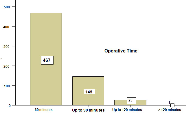
Figure-I.   Total duration of operation: Various early complications during the same admission occurred in 9 % (n=55) of the patients. Their distribution according to the type of hernia is displayed inTable- II. Table-II   Type of hernia * Complications during same hospitalization Cross tabulation 

Type of hernia Complications during same hospitalization Total
Wound infection Reaction Mechanical failure Seroma Pain Mesh infection Hematoma    
Inguinal 14 3 4 7 1 1 3 518
paraumbilical 5 1 2 3 0 3 3 83
Incisional 0 0 0 1 0 2 1 29
Epigastric 0 0 0 0 0 1 0 8
Total 19 4 6 11 1 7 7 638

 Wound infection has an alarmingly high incidence in our series (n=19, 3 %). Most of these infections are superficial and effectively controlled by intensive antibiotic therapy so that the failure of repair is saved and mesh rejection prevented effectively. Four of recurrences occurred during 2 years follow up but patient discontinued follow up afterwards. Remaining 07 recurrences occurred between 24 to 36 weeks. The complications observed during the follow up period of up to three years in different hernias are shown in Table-III.  Table-III    Type of hernia * Late Complications after discharge Cross tabulation 

  Complications after discharge Total
Type of hernia Pain Recurrence of hernia Thickening of spermatic cord Testicular atrophy  
Inguinal 10 4 7 2 518
paraumbilical 2 7 2 0 83
Incisional 2 0 0 0 29
Epigastric 1 0 0 0 8
Total 15 11 9 2 638

 A remarkably low incidence of wound infection in para-umbilical and incisional hernia is attributable to the placement of suction drain which prevents hematoma formation and secondary infection. Seven (2%) recurrences occurred in para-umbilical hernias where suction drain did not work. Mesh infection with massive reaction , although, treated conservatively in 5 patients with vigorous antibiotic therapy led to the formation of a weaker scar which later on gave way and recurrence of hernia occurred. Follow up was not 100% and about 26% patients were lost in the follow up. Majority of the patients (n=457, 90 %) were discharged within 4 days while patients who were complicated had a longer stay up to 15 days (P <0.001). Late complications were mostly chronic pain and foreign body sensations with 2 cases of unilateral testicular atrophy. DISCUSSION A number of non-infectious and infectious complications are attributed to the use of mesh in the repair of most of the hernias5, 13, 16. The type of mesh as well as their composition seems to affect complications following their use and specific materials are related to specific complications. We present 638 patients of different types of hernias repaired by implanting polypropylene mesh of varying sizes depending upon the size of defect. Prolene meshes are monofilaments, non-absorbable, inert, sterile and porous synthetic materials commonly used for this purpose. The use of mesh in hernia repair has brought a phenomenal change in the overall outcome of hernia repair especially in terms of recurrence of hernia 17. We experienced wound infection as the commonest complication in our series (n=19, 3%) and this was more so in the inguinal hernias. Most of the wound infections are superficial and very easy to control on conservative measures. On the contrary deep seated mesh infections are always dreaded complications and if not treated vigorously can lead to total failure and rejection of prosthetic mesh. A number of similar studies have reported an increased infection rate in mesh hernia repair testifying our results 18-28. Grant AM 29, however claims no difference in the incidence of infection between mesh and non-mesh repair of groin hernias. A severe local reaction occurred in 4 (0.62%) patients which resolved in few days without any consequences. Seroma and hematoma occurred in 11(2 %) and 7(1.09%) patients respectively. Only 3 seromas and one hematoma needed evacuation. We report a recurrence of 11(2 %) hernias of which 4 are inguinal and 7 are para-umbilical hernias. All of the recurrent para-umbilical hernias had severe reaction and wound infection in the post-operative period which was effectively controlled by vigorous antibiotic therapy and local wound care. There is probably a weaker scar after resolution of the infective process which resulted in recurrence. A significantly low recurrence rate in inguinal hernias in our study (0.77%) is consistent with the results of other similar reports 30-32. Chronic pain and a foreign body sensation poses a real problem and though it occurred in only two cases but a similar study by Bay-Nielsen M 33 claims 22.9% study population having chronic pain and more so in the adult males. CONCLUSION Although mesh repair is an efficient method of hernia repair, a number of complications may be associated and can lead to failure. An aggressive antibiotic therapy and wound care may control even massive infections.


Acknowledgements / Conflicts / Author Details
Details of Authors: 
<p>ARSHA M MALIK Assistant Professor Surgery, Liaquat University Of Medical and Health Sciences Jamshoro, Pakistan.<br /> ASAD KHAN Post-graduate student (Surgery), Liaquat University Of Medical and Health Sciences, Jamshoro.<br /> K ALTAF HUSSAIN TALPUR Associate Professor Surgery, ,Liaquat University Of Medical and Health Sciences Jamshoro, Pakistan.<br /> ABDUL AZIZ LAGHARI Associate Professor Surgery,Liaquat University Of Medical and Health Sciences, Jamshoro Pakistan.</p>
Corresponding Author Details: 
ARSHAD M MALIK, Assistant Professor, Department of Surgery, Liaquat University of Medical and Health Sciences, Jamshoro, Pakistan
Corresponding Author Email: 
arshadhamzapk@yahoo.com
References
References: 

 1.          Sanjay P, Reid TD, Davies EL, Arumugam PJ, Woodward A.Retrospective comparison of mesh and suture repair for adult umbilical hernias. Hernia 2005; 9(3):248-51.

2.          Cobb WS, Kerchief KW, Heniford BT. The argument for light weight polypropylene mesh in hernia repair.Surg innov,2005;12(1):63-9.3.          DeBord JR. The historical development of prosthetics in hernia surgery. Surg Clin North Am 1998; 78:973-1006.4.          Liem MSL, Van Der Graaf Y,Van Steensel CJ et al.Comparision of conventional anterior surgery and laparoscopic surgery for inguinal hernia repair.N Eng J Med 1997;336:1541-7.5.          Bliziotis IA, Kasiakou SK, Kapaskelis AM, Falagas ME. Mesh-related infection after hernia repair: Case report of an emerging type of foreign – body related infection. Infection 2006;34(1):16.          Chowbey PK, Bagchi N, Goel A, Sharma A, Khullar R, Sonni V, Baijal M.Mesh migration into the bladder after TEP repair: a rare case report. Surg Lapros Endosc Percutan Tech 2006; 16(5):377-8.7.          Delikoukos S, Tzovaras G, Liakou P, Mantzos F, Hatzetheofilou C. Late-onset deep mesh infection after inguinal hernia repair. Hernia 2007; 11(1):15-17.8.          Hoefkens MF, Vles WJ. A giant pseudocyst following repair of an incisional hernia by a propylene mesh. Ned Tijdschr Geneeskd 2008; 26; 152(4):216-20.9.          Celdran A, Esteban j, Manas j, Grazino JJ. Wound infections due to Mycobacterium Fortuitum after polypropylene mesh inguinal hernia repair. J Hosp Infect 2007; 66(4):374-77.10.       Singh-Ranger D, Taneja T, Sroden P, Peters J. A rare complication following laparoscopic TEP repair: Case report and discussion of literature. Hernia 2007; 11(5):453-56.11.       Bendavid R. complications of groin hernia surgery.Surg Clin North Am 1998; 78:1089-103.12.       Robinson TN, Clarke JH, Schoen J, Walsh MD. Major mesh related complications following hernia repair: events reported to the Food and Drug Administration. Surg endosc 2005; 19(12):1556-60.13.       White TJ, Santos MC, Thompson JS. Factors affecting wound complications repair of ventral hernias. Am Surg 1998; 64(3):276-80.14.       Sohail MR, Smilac JD. Hernia repair mesh –associatedMycobacterium goodie infection. J Clin Microbiol 2004; 42(6):2858-60.15.        Delikoukos S, Fafoulakis F, Christodoulidis G, Theodorpoulos T, Hatzitheofilou C. Re-operation due to severe late-onset persisting groin pain following anterior inguinal hernia repair with mesh. Hernia 2008 ;{ Epub ahead of print}.16.       Leber GE, Grab JL, Alexander AI, Reed WP. Long –term complications associated with prosthetic repair of Incisional hernias. Arch Surg.1998; 133(4):378-82.17.       Ove H, Kumar A. Complications of Incisional hernia repair: Delaminated mesh with incarcerated hernia.ANZJ Surg 2004; 74:705-6.18.       Furtschegger A, Sandbichler P, Judmaier W, Gstir H, Steiner E, Egender G. Sonography in the post-operative evaluation of laparoscopic inguinal hernia repair.J Ultrasound Med 1995;14:679-84.19.       Stoppa RE. The treatment of complicated groin and Incisional hernias .World J Surg 1989;13(5):54520.       Lujendijk RW, Hop WC, van den Tol MP, de Lange DC, Braaksma MM, Ijzemans JN et al.A comparison of suture repair with mesh repair for Incisional hernia.N Engl J Med 2000;343(6):392-398.21.         Cappelletti M, Attolini G, Cangioni G, Mascherini G, Taddeucci S, Gervino L. [The use of mesh in abdominal wall defects] Minerva Chir1999; 52(10):1169-76.22.        Van’t Riet M, de Vos van steenwijk PJ, Bonger HJ, Steyerberg EW, Jeekel J.Mesh repair for post-operative wound dehiscence in the presence of infection: is absorbable mesh safer than non-absorbable mesh? Hernia 2007; 11(5):409-13.23.       Falagas ME, Kasialkou SK.Mesh-related infections after hernia repair surgery. Clinic Microbio and infection 2004; 11(1):3-8.24.        Yerdel ,Mehmet A,Akin, Emin B, Dolalan ,Surku,Turkcapar, Ahmet G,et al.Effect of single dose prophylactic Ampicillin and Sulbactam on wound infection after tension free inguinal repair with polypropylene mesh: The randomized double-blind ,prospective trials. Annals of surgery 2001; 233(1):26-33.25.       Geoffrey E, Leber, Jane L, Garb, Alexender, William P, Reed.Long term complications associated with prosthetic repair of Incisional hernias. Arch surg 1998; 133:378-382.26.       Farrow B, Awad S, Berger DH, Albo D, Lee L, Subramanian A, Bellows CF. More than 150 consecutive open umbilical hernia repairs in a major veteran administration medical centre.Am J Surg 2008;196(5):647-51.27.       Cingi A; Manukyan MN; Gulluoglu BM; Barlas A; Yegen C; Yalin R; Yilmaz N; Aktan AO. Use of resterilized polypropylene mesh in inguinal hernia repair: a prospective, randomized study. J Am Coll Surg. 2005; 201(6):834-40.28.       Terzi C; Kilic D; Unek T; Hosgorler F; Fuzun M; Ergor G. Single-dose oral ciprofloxacin compared with single-dose intravenous cefazolin for prophylaxis in inguinal hernia repair: a controlled randomized clinical study. J Hosp Infect. 2005; 60(4):340-7.29.       Grant AM. Open mesh versus non-mesh repair of groin hernia: meta-analysis of randomized trials based on individual patient data [corrected]. Hernia. 2002; 6(3):130-6.30.       Kurzer M, Belsham PA, Kark AE.The Lichtenstein repair. Surg Clin North Am 1998; 78:1025-46.31.       Amid PK, Shulman AG, Lichtenstein IL, Simultaneous repair of bilateral inguinal hernias under local anesthesia. Ann Surg 1996; 223:249-52.32.       Scott NW, Mc Cormacmok K, Graham P, Go PM, Ross SJ, Grant AM. Open mesh versus non-mesh for repair of femoral and inguinal hernia. Cochrane database syst Rev 2002 ;( 4):CD002197.33.       BAY-NIELSEN M. (1) ; NILSSON E. (2) ; NORDIN P. (2) ; KEHLET H. Chronic pain after open mesh and sutured repair of indirect inguinal hernia in young males. Br j surg 2004. 91: 1372-1376.


A case report of a Giant Schwannoma of the Mesorectum

Authors
Gopall Jayprakash, Cheng Yong and Wang Yanqing
Article Citation and PDF Link
BJMP 2009: 2(3) 51-53
Abstract / Summary
Abstract: 

Introduction: Schwannoma is a peripheral nerve sheath tumour and commonly occurs on the head, neck and trunk. Giant schwannoma is rarely located in the retroperitoneum, the pelvis and rarer in the mesorectum.
Case presentation: A 36 year old Asian male patient presented with a lower abdominal mass since 2 months, urgency of defecation since 6 months, erectile dysfunction for more than 3 years.
Conclusion: We report this patient with a mesorectal schwannoma because of its giant size, rare location, vague symptoms and diagnostic dilemma.

Abbreviations: 
HIFU - high intensity focused ultrasound, CT – Computer Tomography, MRI – Magnetic Resonance Imaging, FNAC – Fine Needle Aspirate Cytology

Introduction

Schwannoma (neurilemmoma) is a peripheral nerve sheath tumour and commonly occurs on the head, neck and trunk. Giant schwannoma is rarely located in the retroperitoneum and the pelvis but rarer in the mesorectum. The majority of symptoms caused by the tumour are due to its mass effect. Surgical resection is enough to treat the tumour. Schwannoma is usually a benign tumour. Its malignant transformation is usually very rare. We report a 36 year old male patient with a giant mesorectal schwannoma who presented with vague symptoms.
 
Case Report
 
A 36 year old male patient presented with a lower abdominal mass since 2 months, urgency of defecation since 6 months and erectile dysfunction for more than 3 years. There was no history of urinary disturbances. General physical examination did not reveal any abnormality. Abdominal examination showed a non tender, firm, fixed mass with well defined borders over the suprapubic region which was extending into the pelvis. No organomegaly was detected. On rectal examination a hard, immobile mass was felt 5 cm away from the anal verge and lying anterior to the rectum.
His urine analysis, routine hematological investigations and renal function test were within normal limits. Total bilirubin was raised to 24.9 µmol/l (3.42-20.52 µmol/l), direct bilirubin was also elevated 8.2 µmol/l (0-6.8 µmol/l) while all other results were within normal range. The patient was positively screened for Hepatitis B and Syphilis. CT scan (figure 1) of abdomen revealed a large soft tissue mass measuring 10x11 cm in the pelvis. It was a well defined mass without any infiltration to the following structures: sigmoid, rectum, prostate gland, the bladder and the sacral bone.
 
         
Figure 1 shows a large soft tissue mass measuring 10x11 cm which compresses the rectum.
 
FNAC of the mass which was taken 3 cm from the anal verge showed isolated fibrotic tumor.
The patient was advised for HIFU therapy (high intensity focused ultrasound) in view of eventual surgery. In HIFU therapy, the ultrasound beams are focused on the diseased tissue and due to the significant energy deposition at the focus, temperature within the tissue rises to 65° to 85°C, destroying the diseased tissue by coagulation necrosis and hence facilitating the dissection of the mass during surgery. After HIFU therapy, the patient no longer complained of urgency of defecation. But there was no significant change in the size of the mass after a rectal examination.
Hence, explorative laparotomy was carried out. The abdomen was opened through the lower midline incision. A huge mesorectal mass of 15x11x8 cm (figure 2), was identified on the right side of the rectum and behind the prostate gland extending upwards, without any posterior infiltration into the sacral bone. Complete excision of the mass was achieved by blunt dissection and the mass was sent for histopathological examination. The operation was uneventful.
 
Figure 2 shows the complete excision of a well encapsulated mass of 15x11x8 cm. The external surface of the mass was gray-white and smooth.
 
Figure 3: Histology showing typical pallisading arrangement of spindle cells (Antoni-A/Verocay Body) and less cellular myxoid areas (Antoni-B)

Discussion
 
Schwannoma (Neurilemmoma) is a benign neoplasm arising from the myelinated nerve sheaths. Malignant schwannoma despite their name, never arise from malignant degeneration of schwannomas. Instead they occur from transformation of a plexiform neurofibromatosis. This fact provides the basis for their association with neurofibromatosis1.
 
Schwannomas are frequently present in patients aged 20 to 50 years with a male predominance. They comprise 5% of all benign soft tissue tumors and have a predilection for the head and neck2, 3, 4 and flexor surfaces of the upper and lower extremities5. Pelvic schwannoma is rare and accounts for less than 1% of all benign schwannomas6. The rarity of this tumor and the lack of clinical manifestations make a pre-surgical diagnosis very difficult.
 
These tumors slowly increase in size before becoming symptomatic. Clinical signs or symptoms manifest themselves only by the compression or infiltration of adjacent organs. Therefore, detection is often accidental.
Schwannoma typically appears as a solitary, well encapsulated, firm and smooth-surfaced round mass. In large tumors (>8-10 cm), a degenerative pattern (cystic areas, calcifications, interstitial fibrosis, and hyalinization) is commonly found that identifies the “ancient” variant7. Histologically, typical schwannomas are composed of inter mixed Antoni A components (cellular and arranged in short bundles or interlacing fascicles) and Antoni B areas (less cellular and organized with more myxoid components).Schwannomas show uniform and intense staining for S 100 protein8.
 
Malignant transformation is very rare and controversial9. Malignant schwannomas are large in size and highly aggressive tumours. They are painful, and may cause many different symptoms depending on the location and size. They infiltrate into the adjacent structures and metastasize to distant sites. Histologically, the nuclear palisading may be a striking feature. There will be perineural and intraneural spread of tumour, lesional proliferation, or herniation into the lumina of the vessels10.
 
A combination of clinical, pathological and immuno-histochemial studies help to make the diagnosis.
However preoperative diagnosis of schwannomas is not easy owing to the difficulty in finding imaging features (with Ultrasonography, CT, or MRI) that enable the discrimination of schwannomas from other soft tissue tumors (fibrosarcoma or liposarcoma) and benign forms from malignant ones11. Schwannomas can frequently be misdiagnosed as psoas abscesses, pancreatic cysts, neoplasms, adnexal masses, adrenal, kidney or hepatic tumors, as reported in the literature12, 13, 14. Yet, radiological imaging is helpful in therapeutic planning, as it gives information about the tumor’s size, location, and possible invasion of other structures. Fine-needle aspiration biopsy may theoretically be useful if Schwann cells are found in the sample, but the tissue specimen collected for diagnosis is often inadequate and may be misleading because of cellular pleomorphism in degenerated areas which might be interpreted as consistent with malignancy15.
 
Hughes MJ et al11, in their largest radiological series of abdominal or pelvic schwannomas, showed that a smooth well defined border, ovoid or spherical shape and location in the pre sacral region or lower retro peritoneum at the pelvic brim, are frequent findings in primary abdominal or pelvic schwannomas ; 11 of 13 cases showed all these features. Evidence of degeneration was common, with 8 schwannomas showing cystic change and 3 showing areas of calcification. The cystic schwannomas tended to be larger, with a mean diameter of 10.5cm.
 
It is difficult to make an accurate pre operative diagnosis. The treatment depends solely on surgery. Malignant schwannomas are insensitive to chemotherapy and radiation, resulting in poor prognosis16.
 
Conclusion
 
Schwannoma is a benign encapsulating neoplasm with an overall low incidence, occurring mostly in the head, neck and trunk regions. Giant schwannoma is rarely located in the retroperitoneum, the pelvis and rarer in the mesorectum.
 
Schwannomas pose a difficult diagnostic challenge. Radiological findings are often non-specific. Ultrasonography can differentiate between solid and cystic tumours. CT can be helpful in determining the size, location, local involvement and distant spread. MRI provides similarly useful information as CT, but yields better visualization of the tumour. FNAC is not often helpful because the tissue architectural information required is not obtainable from cytological specimen. The only gold standard diagnostic investigation is histology of either biopsy or excised specimen.
Surgical excision has remained the mainstay of treatment. Although benign, large and incompletely excised lesions are capable of recurrence, malignant change is exceedingly rare.


Acknowledgements / Conflicts / Author Details
Competing Interests: 
None declared
Details of Authors: 
<p>GOPALL JAYPRAKASH-General Surgery department, The First Affiliated Hospital, Chongqing Medical University, Chongqing 400016, China.<br /> CHENG YONG-Associate Professor, Department of General Surgery, The First Affiliated Hospital of Chongqing Medical University, Chongqing 400016, China.<br /> WANG YANQING-Professor, Department of General Surgery, The First Affiliated Hospital of Chongqing Medical University, Chongqing 400016, China.</p>
Corresponding Author Details: 
CHENG YONG, Associate Prof, Department of General Surgery, The First Affiliated hospital, Chongqing University of medical science, Chongqing 400016, China.
Corresponding Author Email: 
vishall_76@yahoo.com
References
References: 

1.Rai BR1, Chaudhary D1, Thapa P. Ancient cystic pelvic schwannoma presenting as a right iliac fossa mass. Kathmandu University Medical Journal (2005) Vol. 3, No. 3, Issue 11, 285-288.2.Jayaraj SM, Levine T, Frosh AC. Ancient schwannoma masquerading as parotid pleomorphic adenoma. J Laryngol Otol 1997; 111:1088–1090. 3.Ogren FP, Wisecarver JL, Lydiatt DD. Ancient neurilemmoma of the infratemporal fossa: a case report. Head Neck 1991; 13:243–246. 4.Dayan D, Buchner A, Hirschberg A. Ancient neurilemmoma (schwannoma) of the oral cavity. J Craniomaxillofac Surg 1989; 17: 280–282. 5.  Graviet S, Sinclair G, Kajani N. Ancient Schwannoma of the foot. J Foot Ankle Surg 1995; 34:46–50. 6.Ng KJ, Sherif A, McClinton S. Giant ancient schwannoma of the urinary bladder presenting as a pelvic mass. Br J Urol 1993; 72:513–514. 7.Peter T Chan, Sankalp Tripathi , Su E Low. Ancient schwannoma of the scrotum. BMC Urology 2007, 7:1doi:10.1186/1471-2490-7-1 8.S.W. Weiss and J.R. Goldblum. Benign tumours of peripheral nerves. In: S.W. Weiss and J.R. Goldblum. Editors, Enzinger & Weiss’s soft tissue tumours. Mosby, Messouri, USA (2001): 1146-68. 9.Rasbridge SA, Browse NL, Tighe JR. Malignant nerve sheath tumor arising in benign ancient schwannoma. Histopathology 1989; 14:525–528. 10. J. Nakashima, M. Ueno, K. Nakamura. Differential diagnosis of primary benign and malignant retroperitoneal tumours. Int J Urol 1997; 4: 441-46. 11. Hughes MJ, Thomas JM, Fisher C. Imaging features of retroperitoneal and pelvic schwannomas. Clin Radiol. 2005:60:886-893. 12. Kishi Y, Kajiwara S, Seta S. Retroperitoneal schwannoma misdiagnosed as a psoas abscess: report of a case. Surg Today.2002; 32:849-852. 13. Ibraheim M, Ikomi A, Khan F. A pelvic retroperitoneal schwannoma mimicking an ovarian dermoid cyst in pregnancy. J Obstet Gynaecol. 2005; 25:620-621. 14. Duran B, Guvenal T, Yildiz E. An unusual cause of adnexal mass: fallopian tube schwannoma. Gynecol Oncol. 2004; 92:343-346. 15. Daneshmand S, Youssefzadeh D, Chamie K, et al. Benign retroperitoneal schwannoma: a case series and review of the literature. Urology. 2003; 62:993-997. 16. Qiang Li, Chuntao Gao, Jonathan T. Analysis of 82 cases of retroperitoneal schwannoma. ANZ Journal of surgery 2007; 77 (4); 237-40. 

Blocked percutaneous endoscopic gastrostomy tube - an unusual cause

Authors
Vijay Joshi and Ashis Banerjee
Article Citation and PDF Link
BJMP 2009:2(2) 46-47


Case report

An 82 year old lady, who had suffered multiple strokes in the past and was currently on long term percutaneous endoscopic gastrostomy( PEG) feeding, was admitted as an emergency from a nursing home with a two week history of productive cough and fever. She had been on PEG feeding since her first stroke six years previously. The first PEG tube (placed in 2001) subsequently fell out of position, and a second tube (15 French Frecka PEG tube) was inserted in 2003.

On admission, she was pyrexial, dehydrated, and hypoxic on room air. Chest examination revealed bilateral crackles and neurological examination revealed expressive dysphasia, and spastic weakness in both lower limbs. Abdominal examination revealed an inflamed PEG site with purulent discharge. Blood tests revealed raised inflammatory markers with neutrophilia (WBC 20 x 109 /L with a neutrophil count of 12 x 109/L) and a raised C-reactive protein at 193 mg/L.

She was managed with intravenous fluids and antimicrobial therapy (tazocin and metronidazole) for possible aspiration pneumonia. Vancomycin was subsequently commenced as methicillin resistant staphylococcus aureus (MRSA) was isolated from the PEG site. As she remained stable, PEG feeding was recommenced.

A week following her admission she became unwell with an episode of vomiting and choking following PEG feeding. This was associated with difficulty in infusing feeds and medications through the PEG tube. Multiple flushes through the tube were unsuccessful. The tube was found to be persistently blocked and lacked free mobility within the tract.

Urgent upper gastro intestinal endoscopy revealed a buried bumper as the cause of blockage of the PEG tube. This necessitated insertion of a new PEG tube (9 French Frecka) for enteral feeding. The old PEG tube was removed surgically under local anaesthesia in due course. As the removal of the buried bumper was found to be very difficult endoscopically, and surgical intervention was deemed to be inadvisable in view of co morbidities, the bumper was left in situ. Feeding was recommenced through a new tube. In view of persistent discharge through the PEG site, abdominal ultrasound examination was performed, revealing a possible gastro-cutaneous fistula. No local collection was seen around the PEG wound.

As the patient remained clinically stable, she was discharged home with necessary instructions to her carers for regular flushing of the PEG tube with water, before and after each feed, to prevent further blockages.

Fig 1: Buried bumper (stomach-lower body)


Fig 2: New peg and buried bumper

Discussion

PEG is primarily used for long term (longer than 6 weeks) enteral alimentation for patients with impaired swallowing (e.g. from stroke, degenerative neurological disease, head injury, and oropharyngeal malignancy). However numerous complications have been reported since its introduction in 1980.

Buried bumper syndrome (BBS) is an uncommon but well documented complication of PEG insertion, first described in 1988 1. It is usually a late complication occurring up to 3 years post PEG insertion and reported to occur in 0.3-2.4 % of patients2.

The internal bumper of the PEG tube should normally sit snugly against the anterior gastric wall, and this is confirmed endoscopically at the time of initial placement. BBS develops when there is migration of the internal bumper/flange through or into the anterior abdominal wall. This probably occurs as a result of excessive tension between the internal and external bumpers, from over-tightening of the external flange, leading to gastric wall erosion. During migration it becomes lodged along the gastrostomy tube tract between the gastric and abdominal walls. Once epithelialisation occurs the bumper gets covered with gastric mucosa3.

The diagnosis of BBS should be suspected if localised abdominal pain, peri-tubal leakage or inability to infuse feed occurs. Initial measures to deal with a blocked tube include flushing with warmed water, and occasionally passage of a flexible wire through the lumen, in order to unblock any obstruction. Tube obstruction is usually related to the administration of protein-enriched formulae or medications, especially if the tube size is 9 French. Fungal colonisation may also lead to tube blockage, requiring specific solutions for flushing the tube4. Tube exchange should only be considered if the gastrocutaneous tract is mature (6 weeks or longer after placement of the tube).

Endoscopy is confirmatory in cases of BBS. The internal bumper is not seen, and the site of the PEG is indicated by an elevated area of submucosa with a central depression. Failure to recognise BBS can result in gastric perforation and gastrointestinal haemorrhage or intra abdominal sepsis, peritonitis and even death5.

Ideally, the buried bumper should be removed even if the patient is asymptomatic, to avoid potential complications from continued tube migration until it is completely impacted in the abdominal wall. The literature describes various methods of dealing with this complication. Endoscopic ultrasound of the gastric wall with a catheter US probe can facilitate the localisation of the bumper and also provides information regarding feasibility of surgical or endoscopic removal of PEG tube6.

Regular and optimal PEG care has been vital in identifying and prevention of this complication. During daily cleaning of the external PEG site, the PEG should be pushed in approximately 1 cm and rotated prior to repositioning of the external bumper. The length of the tube outside the abdominal wall should be examined at regular intervals so that migration can be recognised5.

This report reinforces the fact that physicians should be aware of this recognised risk of PEG feeding and prompt referral for endoscopy is necessary to avoid serious consequences including gastro-intestinal bleeding, peritonitis and death. Similarly specific instructions should be given to carers for prevention of BBS.


COMPETING INTERESTS
None Declared
AUTHOR DETAILS
VIJAY JOSHI, Trust registrar in integrated medicine, Chase Farm Hospital, Enfield, UK
ASHIS BANERJEE, Consultant in emergency medicine, Chase Farm Hospital, Enfield, UK
CORRESPONDENCE: MR ASHIS BANERJEE, Consultant/honorary senior lecturer in emergency medicine, Chase Farm Hospital, The Ridgeway, Enfield EN2 8JL United Kingdom
E-mail: libra19542003@yahoo.co.uk


References

  1. Shallman RW, Norfleet RG, Hardache JM. Percutaneous endoscopic gastrostomy feeding tube migration and impaction in the abdominal wall. Gastrointest Endosc 1988; 34: 367–68.
  2. Venu RP, Brown RD. Pastika BJ Erickson LW. The buried bumper syndrome: a simple management approach in two patients. Gastrointest Endosc 2002; 56: 582–84.
  3. Anagnostopoulos GK, Kostopoulos P, Arvanitidis DM. Buried Bumper Syndrome with a fatal outcome, presenting early as gastrointestinal bleeding after percutaneous endoscopic gastrostomy placement. J Postgrad Med 2003; 49:325–27
  4. Iber, FL, Lusak, A, Patel, M Importance of fungus colonization in failure of silicone rubber percutaneous gastrostomy tubes (PEGs) Dig Dis Sci, 1996, 41: 226-231
  5. Braden B, Brandstaetter M, Caspary WF, Seifert H. Buried bumper syndrome: treatment guided by catheter probe US. Gastrointest Endosc 2003;57:747-51
  6. Ma MM, Semalacher EA, Fedorak RN, Llor EA, Duerksen DR et al The buried gastrostomy bumper syndrome: prevention and endoscopic approaches to removal. Gastrointest Endosc 1995 ; 41:505-8

Laporoscopic Fundoplication: Not a simple wrap

Authors
Riaz AA, Kosmoliaptsis V and Meyrick-Thomas J
Article Citation and PDF Link
BJMP 2009:2(2) 25-29
Abstract / Summary
Abstract: 

Introduction
Laparoscopic fundoplication (LF) has been emerging as the procedure of choice for selected patients with symptomatic and problematic reflux disease. The aim of this study was to investigate post-operative complications associated with LF. Furthermore we wanted to look at the impact of a surgeons learning curve on post operative morbidity and investigate patient satisfaction after LF.
Methods
A single surgeon’s series of 75 patients who underwent laparoscopic fundoplication were included in the study. The data was collected prospectively but analysed retrospectively. A satisfaction survey was performed with one to eight years follow-up.
Results
Laparoscopic fundoplication was associated with a learning curve. One of the major aims of the study was to record post-operative complications. Interestingly, post-operative dysphagia occurred in up to 40/75 of our patients. Our initial policy was to perform an OGD and dilate prophylactically; however, this was abandoned halfway through the study as it was found that the dysphagia settled in all patients with conservative management.
Conclusion
Our study confirms a real learning curve for LF. Furthermore, it has also clearly highlighted that post operative dysphagia is common and affects a significant number of patients. In our study we found that this was best managed conservatively.


Laparoscopic fundoplication (LF) has been emerging as the procedure of choice for selected patients with symptomatic and problematic reflux disease since the first described case by Dallemagne in 1991 (1). This was followed by a rapid expansion into routine clinical practice shortly afterwards. With increased acceptance and availability of laparoscopy as a safe surgical modality there has been a huge increase in the number of patients undergoing LF. This has probably been due to increased willingness of patients and referring doctors to consider the less invasive procedure, rather than the older ‘open’ surgical treatment, with its more rapid recovery, smaller incisions and earlier return to work and to normal daily activities.

Patient referral patterns have also changed over the last decade with the main indication for consideration of LF being patient choice, a general unwillingness to take long term medication as well as ineffective or intolerance of medications and relapse of symptoms (5-6). Several advantages of LF have been described by recent data including shorter hospital stay, less requirement for analgesia, and sooner return to work (7-9). These have to be offset against procedure specific complications including gastro-oesophageal perforation, pneumothorax, dysphagia, and bleeding (2-4). However, the other main downside to LF is the learning curve during which there are an increased number of complications. Previous reports have suggested the learning curve to be around 20 for an individual surgeon and 50 for an institution (10-12).

In a recent meeting of the Upper GI group at the Royal College of Surgeons there was discussion concerning the incidence and management of dysphagia following laparoscopic fundoplication (13 Bill Owen Day RCS (Eng) 2004). There is a paucity of data available in this regard with a general paucity of negative or unequivocal results in the literature equating to a selection bias towards only positive data and positive reporting of good results.

Therefore the aim of the present study was to look critically at the learning curve and, with respect to operative complications, with specific regard to the incidence and management of dysphagia in a personal series of patients who underwent a laparoscopic fundoplication in a District General Hospital in the United Kingdom.

Patients and Methods

From December 1997 to February 2004, 75 patients who underwent laparoscopic fundoplication under the care of a single consultant surgeon in a district general hospital were included in this study. It became routine practice for LF to be performed by one dedicated surgical team (JMT) who kept a complete prospective list of procedures. This series was complete and the hand written records of all the procedures were used to identify patients undergoing operations for LF with or without reduction and repair of Para-oesophageal hernia during the study period. These records were cross-checked with the theatre logbook, hospital computer system as well as with the surgeon’s own record. This ensured full inclusion in the analysis of patients.

The groups were not randomised and ‘all comers’ where included in the study. However, Group 1 consisted of the first 20 LF whereas Group 2 included LF (numbers 21-75). Informed consent was obtained in writing prior to surgery. At least a single dose of prophylactic antibiotics (of either a third generation cephalosporin or co-amoxiclav) was administered at induction; all patients received standard thromboprophylaxis (subcutaneous clexane, TEDS, intermittent pneumatic calf compression).

Patients underwent a laparoscopic fundoplication as briefly described below. The patient was placed in the lithotomy position with reversed Trendelberg tilt; a pneumoperitoneum was created and 4 ports inserted. The liver was elevated using a ‘Nathanson’ liver retractor placed through a 5 mm epigastric incision. Initially the right and left limbs of the right crus are dissected alongside the pancreaticogastric and phrenogastric ligaments. A window is created behind the distal oesophagus. A Penrose drain is passed through the gap. The short gastric vessels are divided using a harmonic scalpel (Ethicon, USA), if required. The crural limbs are approximated using 2, 2-0 ethibond sutures to leave a hiatus 1 cm wider than the oesophagus. A laparoscopic babcock is placed behind the oesophagus and the gastric fundus is brought left to right behind the oesophagus and bought round to meet with the remaining portion of the fundus anteriorly. Two (or rarely three) sutures of 2-0 ethibond were used for the fundoplication. The upper suture included a bite of the anterior hiatal margin to anchor the wrap. Of note, the important feature of the procedure is the creation of a ‘floppy’ tension free fundoplication. This is hugely aided by good mobilisation of the gastric fundus with its associated ligaments and if required, division of the short gastric vessels. Notably, one must carefully create a window behind the oesophagus and an overlap of no longer than 3 cms length and one must stay high up on the fundus in order to avoid the creation of a ‘2’ compartment stomach syndrome’.

Post-operatively all patients were treated in an identical manner. As soon as tolerated after the operations the patients were allowed the consumption of water; diet and analgesia were made available as soon after surgery as required by individual patients. Complications were noted as they occurred during the follow-up period. Both general and specific complications were documented for at least 6 months. Furthermore, patient demographics, details of operations, all complications and follow-up data were kept. Follow up for the purpose of this audit involved completion of a proforma at a minimum post operative period of one year and a maximum of eight years.

All patients where regularly reviewed daily on the ward whilst they were in-patients and in the outpatients at 4-6 week intervals or sooner should the need arise by the surgical team. Thereafter they were given a “see on request” appointment. Data on patient and procedure related morbidity and acceptability was also collected.

At the time of the study contact was made via the telephone and a questionnaire was completed. A telephone survey asking the patient four questions,

  1. Where they happy with the operation
  2. Would they reccomend the procedure to a friend
  3. Had there symptoms resolved
  4. If they had had post op dysphagia had it resolved

The data was reviewed and analysed in conjunction with our department of medical statistics. Analysis was performed using the Mann-U test. Multivariate analysis of the means was performed using the Kruskal-Wallis Test.

Results

Overall the 75 patients who underwent laparoscopic fundoplication consisted of 44 males (59%) and 31 females (41%). The mean age was 47.0 years (range 22-80 years). Group 1, which consisted of the first 20 LF cases included 11 male and 9 females. The mean age was 53.25 years (range 32-80). Group 2, which consisted of the LF cases numbering 21-75 included 33 male and 22 females. The mean age was 44.8 years (range 22-78). Both groups were well matched across the above parameters with no statistical differences (Table A). Only 4 patients were obese (5%), smoker (n=10, 14%), 7 patients suffering with Hypertension (10%) and one with diabetes mellitus (2%) and were equivalently represented in both groups (data not shown).

Presenting features are shown in Table B. Notably, the commonest presenting complaints included regurgitation of acid/ food in 79% (n=59), heartburn in 73% (n=55) and pain and discomfort 53% (n=35). Other complaints included dysphagia 21% (n=16), cough/wheeze 19% (n=14) and excess salivation 18% (n=13).


Table A: Patient Demographics

 Group 1Group 2P value
Numbers2055NS
Male: Female9: 1122: 33NS
Age range (mean) yrs32-80 (53)22-78 (45)NS
Operating time (median) mins120-240 (190)75-195 (144)<0.05
Sliding/No sliding1144NS
Type II/Type III hiatus hernia911NS


Table B: Presenting Features

 No. of cases
Regurgitation (acid/food)59 (79%)
Heartburn55 (73%)
Dysphagia16 (21%)
Cough/wheeze14 (19%)
Excess Salivation13 (17%)
Nausea10 (14%)
Epigastric pain6 (8%)
TOTAL173 (100%)


Patients underwent pre-operative evaluation with Upper GI endoscopy 88% (n=66), pH manometry 47% (n=35) and barium swallow 32% (n=24). Previous to this procedure all patients (100%) were on or had been during some part of their illness on therapeutic doses of proton pump inhibitors.

There was a significant difference between the operating times in the two groups. Thus in Group 1 the average operating time was 190 minutes (range120-240 minutes) whereas in Group 2 the average operating time was 144 minutes (range 75-195 minutes, P <0.05).


Table C: Post Op Mild and Moderate Complications<

 Cases (n=75)
Dysphagia40
Regurgitation7
Nausea4
Diarrhoea3
Heartburn5
Excess Salivation2
Portsite discomfort6
Gas Bloat4
Total71


One of the major aims of the study was to record post-operative complications. Table C shows all mild and moderate complications which resolved completely with conservative management. Of note Table F shows that dysphagia occurred in up to 40 of our patients. Table F shows the distribution of dysphagia in both groups. In group A (n=20) the are 15 patients who complained with dysphagia (75%) of which 5 settled spontaneously and 10 required further investigation with OGD +/- dilation. in group B (n=55), 25 patients suffered with dysphagia all of which settled with conservative management. Our initial policy was to perform an OGD and dilate prophylactically; however, this was abandoned halfway through the study. It was found that the dysphagia settled in all patients with conservative management. Furthermore, other mild/moderate complications, of note included regurgitation of stomach contents (7/75) and port-site discomfort (6/75), all of which also resolved spontaneously


Table D: Major Complications Groups 1 & 2

 Group 1Group 2P value
PE1 (5%)0NS
Pneumonia1 (5%)1 (2%)NS
Oesophageal Perforation1 (5%)1 (2%)NS
Stomach perforation01 (2%)NS
Pneumothorax01 (2%)NS
Major desaturation01 (2%)NS
TOTAL3 (15%)5 (9%)NS


Table E: Conversion to Open Surgery

 Group 1(n=20)Group 2(n=55)P Value
Desaturation01 (2%)NS
Perforation viscus1 (5%)1 (2%)NS
Pneumothorax01 (2%)NS
Technical (Obese/previous surgery)2 (10%)2 (4%)NS
TOTAL3 (15%)5 (9%)NS


Major complications are shown in Table D. Of note there are three conversions to ‘Open’ surgery in Group 1 and five in Group 2 (Table E). In detail, four cases (two in each group) were converted early because of poor or very difficult access. They included difficulty in reducing the stomach and omentum from the mediastinum into the abdomen, unable to reach the hiatus despite placing the ports as high as possible, dense adhesions between the liver and stomach and G-O junction thereby leaving no access to the hiatus and simply impossible access to the upper stomach. Further, oesophageal perforation which was caused by intra-operative insertion of a nasogastric bougie (Group 1), this was repaired laparoscopically with no sequelae. Other reasons for conversions included perforation of the greater curve of the stomach due to the fact that there was thickened fatty tissue around the greater curve of the stomach and spleen which produced a perforation while dissecting the stomach free. Also in Group 2 there was one pneumothorax and in another patient there was marked desaturation on creation of the pneumoperitoneum and in both cases it was deemed safer to open the patient.


Table F: Dysphagia Group

 Group 1(n=20)Group 2 (n=55)P value
Dysphagia1525NS
Resolved Spontaneously525<0.05
Investigated (swallow and OGD)100<0.05

Relaparotomy occurred in three patients; one developed severe pain and clinical shock at 24 hours and it was found on laparotomy to have a perforated oesophagus, the second patient we found disruption of the wrap (requiring re-operation and refashioning the wrap) and a final patient developed small bowel obstruction.

Finally a telephone survey at the conclusion of the study managed to contact 70/75 patients. It was found that overall 68/70 patients were satisfied with their procedure and would recommend the procedure to a relative or friend.

Discussion

Gastro-oesophageal reflux disease (GORD) is the commonest disorder of the Upper GI tract affecting approximately between 10-40% of most western populations and with rising incidence (11). In Australia it has shown to consume around 10% of the national expenditure on prescription drugs. Fortunately the majority of patients settle with simple measures including weight loss and reductions in smoking, caffeine and chocolate consumptions. Furthermore, better timing of meals as well as increasing the number of pillows and raising the head end of the bed can lead to improved symptoms. The advent of H2- receptor antagonist (H2RA) and later proton pump inhibitors (PPIs) has led to symptom control in the majority of patients. However, patients on maximum therapy who remain symptomatic or who develop complications (i.e. haemorrhage, oesophagitis, strictures) or those who refuse long term medication are deemed candidates for surgical intervention.

LF has emerged as the procedure of choice for GORD. The present study, which is a personal single surgeon series, shows that laparoscopic fundoplication is a safe and effective procedure with low rates of long-term complications. Importantly, post operatively these patients may develop dysphagia which settles with conservative measures in the vast majority of cases (13).

There is no doubt that for LF a ‘learning curve’ exists but there is debate about the actual numbers. Most studies suggest that it is around 20 for an individual and around 50 for a department (12), thus in the present study we compared our first 20 (classically thought to be within the learning curve) with the next 55 in order to assess major complications, conversions to open procedure. We found that in Group 1 there were 3 major complications (15%) whereas in the next 55 cases there were five (9%). Indeed only one major complication in Group 1 and two in Group 2 could be considered as technical, they were oesophageal and gastric perforations the rest being post operative pneumonia, PE and major desaturation. There were no deaths in either group. This was in keeping with previously published series (14-16).

Previously published data and our own observations revealed that there were significant post operative rates of dysphagia. In the present series dysphagia was the single commonest complication experienced by 53% of patients (n=40). Initially these were investigated with barium swallow and OGD and treated aggressively with early dilation (Group 1) however this strategy was abandoned after it was found that the vast majority of our patients resolved their dysphagia with conservative treatment. From thereon we adopted a very conservative approach reassuring the patient and keeping within close contact until the dysphagia resolved. We reserved dilatation for only highly resistant dysphagia or patients who where non-compliant with conservative treatment. We found that clear explanations pre-operatively, regular reassurance and assessment was generally all that was required. In Group 2 (21-75) none of the patients required dilation for dysphagia. It is almost universal that patients undergoing LF will have a degree of dysphagia. However what is now accepted and reflected from the experience from the present study is that dysphagia after LF should only cause concern if severe, presenting with severe pain, uncontrolled retching and vomiting requiring immediate surgical revision (17). Most commonly this is due to over-tightening of the hiatus or with poor mobilisation and a 360 degree wrap. This may be related to the learning curve, being more common in the earlier cases in a personal series. In our series 53% of patients (n=40) experienced dysphagia. This is comparable to previous reported data (2,16). Notably, Fontaumard et al reported a dysphagia rate of 78% (40/51). The reason for this post operative dysphagia has been thought to be related to the type of procedure. In our series all patients underwent a Nissen type of repair however evidence is now emerging that the incidence of dysphagia and gas related complications are reduced following anterior partial fundoplication (19,20). This is shown from the data of two recent randomised controlled studies. Baigrie et al (18) in a double-blind, randomized study compared laparoscopic Nissen total fundoplication and anterior partial fundoplication. There were no differences in mean heartburn scores between groups but dysphagia scores for both liquids and solids were lower after anterior fundoplication. Also Ludemann et al (19) compared total fundoplication for gastro-oesophageal reflux disease with an anterior 180 degrees partial fundoplication. Both achieved effective reflux control but the partial wrap was associated with fewer side-effects in the short term than total fundoplication. After 5 years, dysphagia, measured by a visual analogue score for solid food and a composite dysphagia score, was worse at 5 years after total fundoplication.

Our study confirms what has previously been shown with the learning curve for LF and its acceptability. It has also clearly highlighted that post operative dysphagia is common and affects a significant number of patients post operatively. However in our study we found that this was best managed conservatively and the almost all resolved spontaneously. There is no evidence to support early intervention unless the symptoms are very severe and occur very soon after surgery, when the patient should be taken back to theatre for another look. The specific causes of the dysphagia are not known but it is postulated that it is due to increased pressure as the upper part of the wrap augments the pressure of the lower oesophageal sphincter causing it to become over competent. Over time this mechanism relaxes leading to an improvement in dysphagia with simple conservative therapy. Furthermore, there was a hugely positive satisfaction score on a simple telephone survey suggesting symptom control from this procedure.

COMPETING INTERESTS
None Declared
AUTHOR DETAILS
A A RIAZ, FRCS(I) FRCS(Eng) FRCS(Gen) PhD, Hunterian Professor RCS(Eng) and Senior Visiting Clinical Fellow, University of Hertfordshire, UK
CORRESPONDENCE: A A RIAZ, Consultant Upper GI, Laparoscopic and General Surgeon, West Hertfordshire Hospitals NHS Trust, Waverley Road
Hertfordshire, AL3 5PN, UK
Email: mrariaz@hotmail.com


References

  1. Dallemagne B, Weerts JM, Jehacs C, Markiewid S, Lombard R. Laparoscopic Nissens Fundoplication: preliminary report. Surg Laparosc Endosc 1991:1;138-143.
  2. Watson DI, Johnson AG, Reed MWR, Stoddard CJ. Laparoscopic Fundoplication for gastroesophageal reflux. Ann R Coll Surg Engl 1994:76; 264-68.
  3. Schauer PR, Meyers WC, Eubanks S et al. Mechanisms of avoidance of esophageal perforation during laparoscopic Nissens fundopliaction. Ann Surg 1996; 223:43-52.
  4. Reid DB, Winning T, Bell G. Pneumothorax during laparoscopic dissection of the diaphragmatic hiatus. Br J Surg 1993;80:670
  5. Perdikis G, Hinder RA. Lund J. Laparoscopic Nissens Fundoplication: where do we stand? Surg Laparosc Endosc 1997; 7; 17-21.
  6. Laine S, Rantala A, Gullichsen R, Ovaska J. Laparoscopic vs. conventional Nissens Fundoplication. Surg Endosc 1997; 11:441-44.
  7. Zacharoulis D, O'Boyle CJ, Sedman PC, Brough WA, Royston CM. Laparoscopic fundoplication: a 10-year learning curve. Surg Endosc. 2006; 20(11):1662-70.
  8. Martin RC 2nd, Kehdy FJ, Allen JW. Formal training in advanced surgical technologies enhances the surgical residency. Am J Surg. 2005 Aug; 190(2):244-8.
  9. Hwang H, Turner LJ, Blair NP. Examining the learning curve of laparoscopic fundoplications at an urban community hospital. Am J Surg. 2005 ;189(5):522-6;
  10. 7th Bill Owen Oesophago-Gastric Symposium 'Meet-the-Experts' The Royal College of Surgeons of England, London March 2005.
  11. Speechler SJ. Barrett's esophagus. Semin Oncol. 1994 Aug; 21(4):431-7.
  12. Watson DI, Baigrie RJ, and Jamieson GG. A learning curve for laparoscopic fundoplication. Definable, avoidable, or a waste of time? Ann Surg. 1996 August; 224(2): 198–203.
  13. Champault GG, Barrat C, Rozon RC, Rizk N, Catheline JM. The effect of the learning curve on the outcome of laparoscopic treatment for gastroesophageal reflux. Surg Laparosc Endosc Percutan Tech. 1999 Dec; 9(6):375-81.
  14. Cuschieri A, Hunter J, Wolfe B, Swanstrom LL, Hutson W. Multicenter prospective evaluation of laparoscopic antireflux surgery. Preliminary report. Surg Endosc. 1993; 7(6):505-10.
  15. Jones NJ, Soper DB. Laparoscopic Nissens Fundoplication. Surg Rounds 1994:17;573-81
  16. Gotley DC, Smithers BM, Rhodes M, Menzies B, Branicki FJ, Nathanson L. Laparoscopic Nissen fundoplication--200 consecutive cases.
    Gut. 1996 Apr; 38(4):487-91.
  17. Watson DI, Jamieson GG, Mitchell PC, Devitt PG, Britten-Jones R.Stenosis of the esophageal hiatus following laparoscopic fundoplication. Arch Surg. 1995 Sep;130(9):1014-16.
  18. Baigrie RJ, Cullis SN, Ndhluni AJ, Cariem A. Randomized double-blind trial of laparoscopic Nissen fundoplication versus anterior partial fundoplication. Br J Surg. 2005 Jul; 92(7):819-23.
  19. Ludemann R, Watson DI, Jamieson GG, Game PA, Devitt PG. Five-year follow-up of a randomized clinical trial of laparoscopic total versus anterior 180 degrees fundoplication. Br J Surg. 2005 Feb; 92(2):240-3.
  20. Watson DI, Jamieson GG, Lally C, Archer S, Bessell JR, Booth M, Cade R, Cullingford G, Devitt PG, Fletcher DR, Hurley J, Kiroff G, Martin CJ, Martin IJ, Nathanson LK, Windsor JA; International Society for Diseases of the Esophagus--Australasian Section. Multicenter, prospective, double-blind, randomized trial of laparoscopic nissen vs anterior 90 degrees partial fundoplication. Arch Surg. 2004 Nov; 139(11):1160-7.

Photographic documentation of open fractures: A survey of current practice and proposed recommendations.

Authors
R Ahmad, SKM Annamalai, SMY Ahmed, SA Joseph and M Bould
Article Citation and PDF Link
BJMP 2009:2(1) 32-34


Abstract

The primary objective of this survey was to check the availability and use of cameras in documentation of open fractures in emergency departments in England, Wales and Scotland. We also checked the use of mobile phone cameras in emergency departments without cameras. A telephone questionnaire posed questions to the first on-call orthopaedic doctor aboutthe availability of a functional camera in the emergency department. Altogether, 102 doctors replied in 115 hospitals that were surveyed. Only 63 emergency departments had a camera available for photography of open fractures, in which 53 orthopaedic doctors would always take photographs of an open fracture. In 7 emergency departments a mobile phone camera was used for documentation of open fractures, as a camera was not available.

Subsequently, recommendations for wound documentation using a mobile phone camera have been proposed as it is easily accessible and portable with good picture quality that can be transmitted to offsite senior colleagues for advice. This would prevent wound infection and also act as an adjunct to a narrative description of the wound.


Open fractures are quite common and account for a considerable number of admissions to the emergency department with a frequency of approximately 23 per 100000 patients per year1. Infections of open musculoskeletal injuries, especially open fractures, continue to pose a challenge to the Orthopaedic and Trauma surgeons. To reduce the risk of nosocomial infections these wounds need to be instantly photographed before the application of a sterile dressing which has to be left undisturbed until the surgical intervention in the theatre2.

The British Orthopaedic Association and British Association of Plastic surgeons recommend that a Polaroid photograph of every open fracture should be taken, this recommendation is not followed due to non availability of the equipment in some institutions1. This leads to unnecessary disturbance of the wound dressing, distress to the patient and nosocomial infection.

The objective of this study was to look at the availability and use of cameras in compound fractures in Hospitals across England, Wales and Scotland by using a telephone questionnaire.

Methods

We randomly selected 115 NHS hospitals with A&E departments from all the regions of the England, Wales and Scotland using the www.specialistinfo.com website and selected every third hospital on the list. The survey was conduction by the means of a telephone questionnaire (Table 1); the questions were answered by the first on call Orthopaedic doctor. 89% (102/115) first on-call doctors answered the questionnaire.

TABLE 1 Telephonic questionnaire

1. Do you have access to a camera in the A&E?

2. If yes, is the camera available 24/7?

3. Have you found a functional camera available every time you have wanted to use it?

4. Do you always take a picture of an open fracture?

5. Do you think that a picture is a useful tool in the assessment of a wound?

6. If a camera is not available in the A&E do you use your mobile phone camera?

Y/N

Y/N

Y/N

Y/N

Y/N

Y/N


The questionnaire was meant to investigate if the doctor had access to a functional camera that was available round the clock in the Emergency department. It also questioned whether the doctor on call would always take a picture of an open fracture. They were also asked whether they thought that the picture was helpful in assessment of the wounds. Finally the respondents were asked whether they used a mobile phone camera in case of non availability of a camera in the emergency department.

Results

Of the 102 first on call Orthopaedic doctors questioned, 38% (39/102) stated that they were unable to photograph an open fracture due to non availability of a camera in the emergency department. A functional camera was not available in both trauma centers and small hospitals. Of the 61% (63/102) who had a camera available 22% (14/63) did not have 24 hour access to the camera. 33% (21/63) who have a Polaroid camera available have found occasions when they needed a camera but it was not functional. (Figure 2)

Figure 1

Figure 2 Availability of cameras in A&E.

84% (53/63) who had a camera available said that they always photographed an open fracture. (Figure 3) 28% (11/39) doctors who did not have a camera in the emergency department would use a mobile phone camera to photograph an open fracture (Fig 1). 4 doctors used it even though they had a Polaroid camera in the Accident and Emergency department (A&E) as they found a mobile phone camera easy to use and readily accessible.

All the doctors who responded to the questionnaire thought that a camera was a valuable tool in the documentation of an open fracture.

Discussion

The objective of this study was to look at the availability and use of cameras in compound fractures in hospitals across England, Wales and Scotland.

Figure 3 Percentage of doctors photographing open fractures in A&E.

We found that 38% of hospitals did not have access to a camera at all, in a further 15% there wasnt 24 hour access and another 21% had a camera that was not functional when needed at some time in the Emergency department. Thus 74% of hospitals did not have a functional camera available in the emergency department at all times. Therefore the photographic documentation of an open fracture as recommended by the British Orthopaedic association and the British association of Plastic Surgeons working party could not be implemented2. 13% (8/63) respondents who had a camera in the Emergency department did not routinely photograph an open fracture. A similar study done in London and south-east UK hospitals found that 10% hospitals did not have a camera to photograph an open fracture and another 19% did not have it available 24 hours1.

The potential reasons for failure of photographic wound documentation could be difficulty to obtain funding for purchase of a camera, lack of secure storage space and unfamiliarity of the junior doctors with the guidelines of photographic wound documentation

Open wounds of the musculoskeletal system are usually contaminated at presentation with pathogenic organisms3. In the A&E the wound should be assessed and dressed until the formal debridement in the theatre. The ideal practice is to photograph the wound before application of the sterile dressing. These photographs are a vital tool as they act as an adjunct to a narrative description allowing the wound to be accurately described to colleagues. This avoids unnecessary disturbance of the sterile dressing over the wound which has a risk of nosocomial infection2.

The problem of non availability of Polaroid cameras in the emergency department can be overcome by using mobile phone cameras. We faced the same problem of non availability of a camera in our A & E department and we started using our camera phones for photographing open fractures. The authors found that images on mobile phone cameras allowed a useful presentation in the morning trauma meetings. The incidence of re- examining of the wound by the senior surgeons also dropped significantly. Mobile phones can be used in A & E as the maximum distance at which any phone causes interference is only two meters4.

We found the mobile phone camera a valuable alternative as it is easy to use and has excellent portability with an image quality that is sufficient for interpretation. In our study it is already being used by many doctors in different hospitals. With the advancement in technology the mobile phones have become more user friendly with the power to process medical images5. The advantage over a Polaroid picture is that these images can be transferred to another mobile phone or sent as an e-mail attachment for assessment by the offsite registrars and consultants for immediate advice. This is even more useful in hospitals with hospital at night where second on call doctors are usually nonresident. It has been shown that it takes only 3-4 minutes from image taking to remote reception6. The Integrated Service Digital Network (IDSN) used for image transfer in some hospitals is expensive and mobile camera phones are a suitable inexpensive substitute.

We recommend supplying a mobile phone camera to the on call doctor. Unlike Polaroid cameras they dont need a constant supply of batteries and expensive film and the risk of theft is negligible as they are smaller, lighter with excellent portability. The picture can be magnified and is easier to store than traditional cameras. Images must be stored in a manner that prevents tampering and preserves confidentiality. This could be done by deleting the images after they have been assessed, hard copies produced and kept in the case notes as done with pictures taken with a Polaroid camera or storing them by using specific software packages that do not allow image manipulation7 .

There have been two studies assessing the accuracy of the use of mobile phone cameras in diagnosing intracranial pathologies and interpretation of emergency Ear Nose and Throat (ENT) radiological investigations. 5, 8 They have also been used to take pictures of injured digits and then transmitted to camera phones of plastic surgeons for assessment 6. Tsai et al. in their study on teleconsultation by using the mobile camera phone showed that the camera phone is valuable for remote management of the extremity wound9 .Lam TK et al. showed that low cost and the ease of use make the mobile camera phones easily incorporated into clinical practice10.

Conclusion

A functional camera was not available at all the times for photographic documentation of open fractures in the majority of the hospitals. We also feel that mobile phone cameras may be used as a powerful graphic adjunct to a verbal clinical presentation to other health professionals located on or off site.
 

COMPETING INTERESTS

None Declared

AUTHOR DETAILS

MR R AHMAD, MBBS, MRCS, MR S K M ANNAMALAI, MBBS, MRCS, Registrars in T & O, Weston General Hospital, UK

MR SMY AHMED, Registrar T & O, Great Western Hospital, Swindon, UK

MR SA JOSEPH MBBS MRCS, Registrar T & , Wolverhampton General Hospital, UK

MR M BOULD FRCS, Consultant Orthopaedic Surgeon, Weston General Hospital, UK

CORRESPONDENCE: MR. SKM ANNAMALAI, Registrar, T & O, House 18, Staff Residence, Weston General Hospital, Weston Super Mare, BS23 4TQ

Email: suresh.annamalai@nhs.net


References

1. Solan MC, Calder JD, Gibbons CE, Ricketts DM. Photographic wound documentation after open fracture. Injury 2001; 32: 33-35.

2. The management of Open Tibial Fractures. British Orthopaedic Association and British association of Plastic Working Party, London, 1997.

3. Akinyoola AL, Ako-Nai AK, Dosumu O, Aboderin AO, Kassim OO. Microbial isolates in early swabs of open musculoskeletal injuries. Niger Postgrad Med J 2006; 13: 176-81.

4. Tri JL, Hayes DL, Smith TT, Severson RP. Cellular phone interference with external cardiopulmonary monitoring devices. Mayo Clin Proc 2001; 76: 11-5.

5. Eze N, Lo S, Bray D, Toma AG. The use of camera mobile phone to assess emergency ENT radiological investigations. Clin Otolaryngol 2005; 30: 230-3.

6. Hsieh CH, Tsai HH, Yin JW, Chen CY, Yang JC, Jeng SF. Teleconsultation with the mobile camera-phone in digital soft-tissue injury: a feasibility study. Plast Reconstr Surg 2004; 114:1776-82.

7. Bhangoo P, Maconochie IK, Batrick N, Henry E. Clinicians taking pictures a survey of current practice in emergency departments and proposed recommendations of best practice. Emerg Med J 2005; 22: 761-5.

8. Yamada M, Watarai H, Andou T, Sakai N. Emergency image transfer system through a mobile telephone in Japan: technical note. Neurosurgery 2003; 52: 986-8.

9. Tsai HH, Pong YP, Liang CC, Lin PY, Hsieh CH. Teleconsultation by using the mobile camera phone for remote management of the extremity wound: a pilot study. Ann Plast Surg 2004; 53:584-7.

10. Lam TK, Preketes A, Gates R.Mobile phone photo messaging assisted communication in the assessment of hand trauma. Lam TK, Preketes A, Gates R. ANZ J Surg 2004; 74:598-60

Back Pain: How to Avoid Surgery?

Authors
Yili Zhou and Stephen Irwin
Article Citation and PDF Link
BJMP 2009:2(1) 4-5

Treatment of low back pain remains a dilemma. In the USA more than 300 thousand back surgeries are performed each year. For about 10% to 39% patients, pain may continue or even get worse after back surgeries1. This condition is called failed back surgery syndrome. In the USA, about 80,000 new cases of failed back surgery syndrome are accumulated each year2. Pathological changes such as recurrent disc herniation, arachnoiditis, scar tissue formation, poor surgical indication, misdiagnosis, and surgical technique failure can all contribute to the failure of surgery. Pain after back surgery is difficult to treat. Many patients have to live with pain for the rest of their lives with severe disability.

Over the last several decades, our understanding of the causes of low back pain has been challenged. With a sensitivity up to 95%3, MRI has been used a gold standard for the diagnosis of spine disease such as lumbar disc herniation. With the MRI evidence of a disc herniation and nerve root compression, patients are more easily convinced surgery is their best and only option. However, the reliability of MRI as the evidence for surgical decision has been questioned. An early study found that in a group of asymptomatic volunteers at age of 60 years or older, about 57% had abnormal MRI findings including disc herniation and spinal stenosis4. Follow up studies have yielded similar results. Now it is widely accepted that degenerative disc disease, such as disc herniation is a common finding in asymptomatic adults. Even though at the age of 60 years or older, 57% or more may have abnormal MRI findings in the lumbar spine, however, only less than 20% of this group of people have chronic low back pain. A recent study also suggested a lack of correlation between imaging findings of spine degenerative change and back pain5. Simply, degenerative change in the lumbar spine, such as a herniated disc, is not necessary painful.

The results of these studies have changed our belief in the relationship between lumbar disc herniation and back pain. It is believed that back and leg pain in the presence of acute disc herniation is not merely the result of a pinched nerve root, rather it is more related to the inflammation of the nerve roots and nerve endings around the herniated disc or it may be the combined results of chemical inflammation and mechanical compression6. A herniated disc is not a sole indication for back surgery and up to 70% to 95% of patients may be pain free after 12 months without major intervention7. The primary goal of treatment of lumbar radicular pain should be the suppression of inflammation, relieving the pain and restoring function rather than removal of the herniated disc. Before one chooses an open back surgery the following options should be considered:

Diagnosis: Low back pain can be related to a herniated disc, nerve root irritation, annular tear, facet joint arthritis, muscle spasm, injuries to the ligament, sacroiliac joint arthritis and referred pain from visceral organs. An MRI finding of a herniated disc, no matter how large, is not enough to justify surgery. A thorough history and physical examination is tantamount to judge whether the herniated disc is the real source for the ongoing pain.

Medications: Non-steroid anti-inflammatory medications should be offered as the first line medication to patients with mild back pain. Early administration of oral steroid medication in patients with acute sciatica may lead to slightly more rapid improvement in pain, mental well-being, and disability scores8. Anti-depressants, especially tricyclic antidepressants, are often used to treat patients with chronic back pain.

Physical therapy, massage therapy and chiropractic management have been widely used for treatment of back pain and lumbar radicular pain, even though the value of these treatment modalities have yet to be proven.

Spine injections: Multiple double blind, clinical controlled studies have confirmed the clinical efficacy of lumbar epidural steroid injection (LESI) in relieving the acute radicular pain due to herniated nucleus pulposus, speeding the rate of recovery and return to function9. The pain relieving effect of LESI may last up to three months. Inflammatory mediators, such as phospholipase A2, have been implicated in lumbar radiculopathy and disc herniation and have been the focus of recent research. Lumbar epidural steroid injections can decrease pain by suppressing the function of inflammatory mediators. As long as the patient is pain free and is without any neurological deficits, a herniated disc should not be a clinical concern. Even though LESI alone may not decrease the necessity of back surgery, it will be intriguing to investigate whether a combination of LESI and other treatment such as physical therapy and life style modification will decrease the need for surgery.

Minimally invasive surgery: Minimally invasive surgery offers another alternative in the treatment of back pain. These treatments include chymopapaine, percutaneous nucleotome, automated percutaneous lumbar discectomy, laser discectomy, neucleoplasty and disc deKompressor. The advantage of the minimally invasive techniques is that it leaves no or minimal scar after the surgery. Among the minimal invasive techniques, laser discectomy has a reported success rate of 80% to 90%10. Neucleoplasty and disc deKompressor have been recently introduced with early non-controlled studies showing success rates up to 78%11. These procedures are still not widely accepted and more studies are needed to confirm their clinical efficacy.

Life style modification: Low back pain can often be the result of improper lifestyle choices. Smoking can increase the risk of low back pain12. Obesity can worsen back pain and contribute to disk degeneration13. Heavy lifting, sport related injuries and motor vehicle accidents can cause back pain. Education to patients with low back pain is critical to help them recover from back pain and prevent future back pain. Smoking cessation and weight control should be strongly recommended to back pain patients. Proper exercise techniques should be taught. Patients, especially those with spinal stenosis often have difficulty walking due to neurological claudication. Treadmills and long distance walking exercise may exacerbate back pain. Some studies suggested therapeutic aquatic exercise is potentially beneficial to patients suffering from chronic low back pain14.

Conclusion: Lumbar spine surgery can potentially provide quick pain relief and functional recovery. There are many downsides to surgery however that would include post laminectomy surgery syndrome and a lack of proven long term benefit. Because of these risks one should be very careful in determining surgical candidacy. A preliminary study15 has provided the evidence that the rate of back surgery can potentially be decreased through appropriate education and application of evidence-based medicine for patients, general practitioners and spine surgeons. Conservative treatment with the combination of medications, physical therapy, spinal injections and life style modification should be tried before surgery is considered.


COMPETING INTERESTS

None Declared

AUTHOR DETAILS

YILI ZHOU, MD, PH.D, Comprehensive Pain Management of North Florida, USA

STEPHEN IRWIN, MD, Comprehensive Pain Management of North Florida, USA

CORRESPONDENCE: YILI ZHOU, MD, PH.D, Medical Director, Comprehensive Pain Management of North Florida, Gainesville, USA, FL32608

Email: Yilizhoumd@yahoo.com


References:

  1. Graz B, Wietlisbach V, Porchet F, et al. Prognosis or "curabo effect?": physician prediction and patient outcome of surgery for low back pain and sciatica. Spine 2005 June 15;30(12):1448-52.

  2. Ragab A, Deshazo RD. Management of back pain in patients with previous back surgery. Am J Med 2008 April;121(4):272-8.

  3. Mullin WJ, Heithoff KB, Gilbert TJ, Jr., et al. Magnetic resonance evaluation of recurrent disc herniation: is gadolinium necessary? Spine 2000 June 15;25(12):1493-9.

  4. Boden SD, Davis DO, Dina TS, et al. Abnormal magnetic-resonance scans of the lumbar spine in asymptomatic subjects. A prospective investigation. J Bone Joint Surg Am 1990 March;72(3):403-8.

  5. Kalichman L, Kim DH, Li L, Guermazi A, et al. Spondylolysis and spondylolisthesis: prevalence and association with low back pain in the adult community-based population. Spine 2009 January 15;34(2):199-205.

  6. Roberts S, Butler RC. Inflammatory mediators as potential therapeutic targets in the spine. Curr Drug Targets Inflamm Allergy 2005 April;4(2):257-66.

  7. Legrand E, Bouvard B, Audran M, et al. Sciatica from disk herniation: Medical treatment or surgery? Joint Bone Spine 2007 December;74(6):530-5.

  8. Holve RL, Barkan H. Oral steroids in initial treatment of acute sciatica. J Am Board Fam Med 2008 September;21(5):469-74.

  9. Sethee J, Rathmell JP. Epidural steroid injections are useful for the treatment of low back pain and radicular symptoms: pro. Curr Pain Headache Rep 2009 February;13(1):31-4.

  10. Goupille P, Mulleman D, Mammou S, et al. Percutaneous laser disc decompression for the treatment of lumbar disc herniation: a review. Semin Arthritis Rheum 2007 August;37(1):20-30.

  11. Al-Zain F, Lemcke J, Killeen T, et al. Minimally invasive spinal surgery using nucleoplasty: a 1-year follow-up study. Acta Neurochir (Wien ) 2008 December;150(12):1257-62.

  12. Mikkonen P, Leino-Arjas P, Remes J, et al. Is smoking a risk factor for low back pain in adolescents? A prospective cohort study. Spine 2008 March 1;33(5):527-32.

  13. Hangai M, Kaneoka K, Kuno S, et al. Factors associated with lumbar intervertebral disc degeneration in the elderly. Spine J 2008 September;8(5):732-40.

  14. Waller B, Lambeck J, Daly D. Therapeutic aquatic exercise in the treatment of low back pain: a systematic review. Clin Rehabil 2009 January;23(1):3-14.

  15. Goldberg HI, Deyo RA, Taylor VM, et al. Can evidence change the rate of back surgery? A randomized trial of community-based education. Eff Clin Pract 2001 May;4(3):95-104.

The Hoffa Fracture: a case report

Authors
SKM Annamalai, JR Berstock and MN Shannon
Article Citation and PDF Link
BJMP 2008:1(2) 36-37


ABSTRACT

We report a case of the rare coronal unicondylar fracture of the distal femur called Hoffa fracture. Hoffa fracture is a rare injury consisting of unicondylar tangential posterior fracture of the distal femur. These fractures are due to high energy trauma and sometime not easy to visualise on routine imaging, and therefore could represent a diagnostic challenge to the accident department and to the orthopaedics surgeon. Clinically however, our patient had obvious knee swelling, localised tenderness and was unable to weight bear. Plain radiograph and CT scan confirmed the fracture and was treated surgically with cancellous screws. 
 

Case Presentation

A thirty-four year old male injured his left knee whilst turning on a motocross bike. He put his left leg on to the ground with his knee in 20 degrees of flexion to stabilise himself whilst turning a left hand corner at about 30 mph. He did not crash, but was unable to weight bear afterwards.

The initial AP and lateral radiographs showed a coronal fracture of lateral condyle of the distal femur.


[FIG 1: AP radiograph right knee / Lateral radiograph right knee].

This fracture could have been easily missed on the plain radiograph. Further imaging with the CT scan confirmed the fracture and its pattern.


[FIG2: Axial CT of distal femur / Saggital CT of distal femur].

He had an open reduction and internal fixation via a lateral approach, vastus lateralis was reflected off the lateral intermuscular septum and the knee joint opened. Maintaining the knee flexed during the surgery relaxes the posterior capsule, gastrocnemius and protects the neurovascular structures. Soft tissue attachments of the fractured fragment constitute the sole source of blood supply and must be preserved. The joint was carefully inspected for associated injuries. After reduction the fragments were temporarily fixed with Kirschner wires. Partially threaded cancellous screws were used in the lag mode to secure compression across the fracture.


FIG3: Image Intensifier of ORIF with cancellous screw]

Discussion

This fracture pattern was initially described by Hoffa in 1904 1 hence the name for this fracture. The Hoffa fracture is an intra-articular fracture of the knee analogous to the capitellum fracture of the elbow 2. This injury is the result of violent force and generally occurs in young adults. There is usually a combination of forces: direct trauma, possibly with an element of abduction, the ground reaction is transmitted through the tibial plateau and the axial compression on a flexed knee concentrates the force in the posterior half of the femoral condyles1. In flexion the lateral condyle is the leading part of the knee to receive the impact1.  Although the Hoffa fracture may be of either condyle4 the preponderance of lateral condylar fractures suggests an anatomic-biomechanical vulnerability due to the physiological valgus.

Few cases have been reported in literature with associated femoral shaft fracture 6, ligament entrapment with irreducible knee dislocation5, open and bicondylar fractures7. Our case is unique as it is a closed injury, uniconylar fracture with no associated ligamentous or meniscal disruptions. Open reduction has been shown to be mandatory for good long-term function 2, 3. High index of suspicion, further imaging with CT scan / 3D reconstruction, open reduction and internal fixation is necessary for good outcome following these types of fractures.

Acknowledgements / Conflicts / Author Details
Competing Interests: 
None Declared
Details of Authors: 
SURESH KM ANNAMALAI, MBBS, MRCS. Registrar in Orthopaedics, Weston General Hospital, United Kingdom JR BERSTOCK, MBBCh, Senior House Officer in Orthopaedics, Weston General Hospital, United Kingdom MN SHANNON, FRCS(South Africa, Ortho), Consultant Orthopaedic Surgeon, Weston General Hospital, United Kingdom
Corresponding Author Details: 
SURESH KM ANNAMALAI, Registrar in Orthopaedics, Department of Orthopaedics, Weston Area Healthcare Trust, Weston General Hospital, Grange Road South, Uphill, Weston Super Mare, BS23 4TQ Tel: 01937 636363
Corresponding Author Email: 
sureshkumar.annamalai@gmail.com
References
References: 

1. Hoffa A: Lehrbuch der Frakturen und Luxationen.4th ed.Stuttgart: Ferdinand Enke-Verlag 1904, 453.

2. Lewis SL, Pozo JL, Muirhead-Allwood WFG: Coronal fractures of the lateral femoral condyle. Journal of Bone and Joint Surgery (Br) 1989, 71:118–120.

3. Ostermann PAW, Neumann K, Ekkernkamp A, Muhr G. Long-term results of unicondylar fractures of the femur. Journal of Orthopaedics and Trauma 1994, 8(2):142–146.

4. Heuschen UA, Göhring U, Meeder PJ Bilateral Hoffa fracture--a rarity. Aktuelle Traumatol 1994 May; 24(3):83-6.

5. Shetty GM, Wang JH, Kim SK, Park JH, Park JW, Kim JG, Ahn JH  Incarcerated Patellar tendon in Hoffa fracture: an unusual cause of irreducible knee dislocation Knee Surg Sports Traumatol Arthrosc. 2007 Oct 24 

6. Miyamoto R, Fornari E, Tejwani NC.  Hoffa fragment associated with a femora shaft fracture. A case report J Bone Joint Surg Am. 2006 Oct; 88(10):2270-4.

7. Calmet J, Mellado JM, García Forcada IL, Giné J.  Open bicondylar Hoffa fracture associated with extensor mechanism injury. J Orthop Trauma. 2004 May-Jun; 18(5):323-5.

Stapled haemorrhoidectomy: A day case procedure for symptomatic haemorrhoids

Authors
Riaz AA, Singh A, Patel A, Ali A and Livingstone JI
Article Citation and PDF Link
BJMP 2008:1(2) 23-27


Introduction: 
Since Longo first described it in 1998, Stapled Haemorrhoidectomy (SH) has been emerging as the procedure of choice for symptomatic haemorrhoids (1). Several studies have shown it to be a safe, effective and relative complication free procedure (2). The aim of this study was to determine the suitability of SH as a day case procedure at a District General Hospital.


Methods 
From June 2001 to May 2005, 66 patients who underwent stapled haemorrhoidectomy were included in this study. Parameters recorded included post-operative complications, analgesic requirements, cost effectiveness, duration of hospital stay and patient satisfaction. Follow-up was performed at 4 weeks with a further telephone follow-up up to 4 years after.


Results 
Of the 66 patients that underwent a stapled haemorrhoidectomy 43 (65%) were male and 23 (35%) were female. The mean age was 49.8 years (range 16-78 years). 11% (n=7) of patients were discharged the same day and 88% (n=58) had overnight stay. Nearly 50% had complete resolution of symptoms and returned to work within a week. The satisfaction data showed that 90% of patients were completely satisfied with the procedure at initial follow-up, which increased to 98% after 6 months-4 years follow-up.


Conclusion 
Our present study shows that stapled haemorrhoidectomy is a safe and very well tolerated procedure with low post-operative analgesic requirements, high patient satisfaction and early return to work. The majority of patients could avoid an overnight stay which would make this procedure suitable for day surgery.  
 

Introduction

Since Longo first described it in 1998 (1), Stapled Haemorrhoidectomy (SH) has been emerging as the procedure of choice for symptomatic haemorrhoids. Several studies have shown it to be a safe, effective and relative complication free procedure with fewer days off work, reduced requirement for analgesia and rapid discharge (2-4). Historically symptomatic haemorrhoids have been dealt with by simple dietary modification, injection sclerotherapy, cryotherapy, band ligation and surgery (5-7).

Unfortunately there is no single optimum therapeutic option. Surgery for symptomatic haemorrhoids was popularised by the open Milligan–Morgan technique in the late 1930’s or one of its variations. Unfortunately, this has been associated with postoperative pain, the risk of severe haemorrhage, and more concerning the risk of anal stenosis (especially if skin bridges are not maintained) and sphincter injury.

Controversy exists as regards to the overall safety and acceptability of SH. On the one hand recent reports of SH have been positive especially in regards reduced postoperative pain and recovery and adverse functional sequelae. A study by Pavlitidsi  et al (8) included 80 patients with second to fourth degree hemorrhoidal disease in which patients were randomly allocated to undergo either the stapled Longo procedure (group 1) or Milligan-Morgan hemorrhoidectomy (group 2) under epidural anesthesia. SH had better postoperative pain scores with lower mean epidural morphine requirement and mean hospital stay.  Conversely, a recent review from New Zealand (9) suggested that SH was more expensive, and the results should be looked upon with caution.

Several studies have suggested that SH may be safely performed as a Day case procedure. Patients following SH had reduced, post operative pain, hospital stay, analgesic requirements and earlier return to work (21-23). Day Surgery procedures have been at the forefront of recent changes within the NHS in the fight to reduce waiting times and better patient care. It is not only popular with patients who are able to recover and convalesce at home in a familiar environment but also reduce the chances of a hospital acquired infection and large cost saving implications for the NHS. However, not every surgical procedure is amenable for Day surgery, and thus procedures which require only moderate amounts of analgesia, reduced post operative stay and few complications and further in 2001 the Audit Commission included Haemorrhoidectomy as one of its 25 procedures suitable for  Day Surgery  (24).

Therefore the present study was to look critically at the learning curve, operative complications, duration of hospital stay, analgesic requirements, cost effectiveness and patient satisfaction in at the personal series of the first 66 patients who underwent SH at Watford General Hospital, a District General Hospital in Hertfordshire, United Kingdom. The aim of the present work was to determine the suitability of SH as a routine day surgery procedure which is not routine in the UK.

Patients and Methods

From June 2001 to May 2005, 66 patients who underwent stapled haemorrhoidectomy were included in this study. It was routine practice that stapled haemorrhoidectomy was performed by one dedicated surgical team (JIL).

Informed consent was obtained in writing prior to surgery. During induction at least a single dose of prophylactic antibiotics (of either a third generation cephalosporin or co-amoxiclav) was administered.

In brief, under general anaesthesia, the patient is put in lithotomy position and a rigid sigmoidoscopy done to exclude any rectal lesions. Stay-sutures with 2-0 silk are applied at the 3, 6, 9 & 12 o’clock. The anus is dilated using with a proctoscope.

An anal ring is applied and fixed to the anal verge by the previously taken stay-sutures. The inner end of the ring must be reaching beyond the dentate line.

Purse-strung sutures are taken all around the anal mucosa on top of the haemorrhoids (beyond the dentate line) followed by a per-rectal examination to make sure that the muscle layer is not taken within the sutures so as to avoid postoperative anal stenosis. Similarly, in female patients, per-vaginal examination is done to insure that vaginal wall is not taken within the sutures. A specialized circular stapler is introduced into the anal canal and the two ends of the purse-strung sutures are passed through special holes in the stapler and tied. Stapler is tightened (the indicator reads between 3 & 4 cm depth) and fired. The stapler is kept closed in place compressing them for 30 seconds in order to encourage haemostasis. The staples line then is checked for bleeding points, for which 2/0 vicryl under-running sutures may be use for further haemostasis. Finally, the anus is packed with ‘spongistan’ as well as flagyl and voltarol suppositories.            

Post-operatively both groups were discharged when comfortable. Complications where noted as they occurred during the follow-up period at 4 weeks. Furthermore, a further telephone follow-up during July 2005 was done results from the survey are shown in table II.

The data was reviewed and analysed in conjunction with our department of medical statistics. Analysis was performed using the Mann-U test. Multivariate analysis of the means was performed using the Krushal-Wallis Test.

Results

Of the 66 patients that underwent a stapled haemorrhoidectomy 43 (65%) were male and 23 (35%) were female. The mean age was 49.8 years (range 16-78 years). Only 7 patients suffered with hypertension and one with diabetes mellitus.

There presenting complaints included rectal bleeding (bright red) in 86% (n=57) and pain and discomfort 53% (n=35). Other complaints included a sensation of something coming down (n=3), change in bowel habit (n=2), constipation (n=1), and incontinence (n=1).

All patients underwent evaluation with proctoscopy and rigid sigmoidoscopy. Further evaluation with colonoscopy (14%, n=9),  flexible sigmoidoscopy (9%, n=6), barium enema (9%, n=6) and anorectal MRI (1%, n=1) to rule out other associated pathologies accounting for their symptoms.

Previous to SH, 57 patients (87%) had undergone previous therapeutic manoeuvres in the form of injection sclerotherapy (76%) and banding of haemorrhoidal tissue (11%).

The operating time ranged between 15-40 minutes with an average of 24 minutes. There were no major complications although the majority of patients warranted oversewing of bleeding points around the staple line after the stapling procedure. 

Post-operative hospital stay revealed that 88%, (n=58) went home after overnight stay (within 24 hours) and only 1 patient had a 48 hours stay with only moderate strength analgesics (Voltarol) (Table 1).

At routine follow-up at 1 month, we found that nine patients (14%) had had minor degrees of faecal urgency, frequency and soiling rectal bleeding, all of which subsequently resolved. Only one patient had developed a peri-anal heamatoma, which was evacuated under local anaesthesia in the Outpatient Department. Further, only two patients had significant problems. One patient complained of pain and discomfort at four-week follow-up, which we think resulted from too low application of the stapler which resolved completely over a six month period with simple analgesics and a single case of rectal stenosis resulting from too high a stapling, which was referred to a specialised centre for further management (Table 2).  

Indeed, only 3 patients had recurrence of symptoms, which were treated with further sclerotherapy injection in the Out-patients department and one requiring re-stapled haemorrhoidectomy.

Follow-up was in the form of Out-patient review 4-6 weeks after the procedure and a telephone questionnaire up to 4 years after the operation the results of which are shown in Table 3-5. We can see that a third of the patients did not require or use analgesia after discharge and further 44% (n=29) needed just Voltarol. As regards to symptoms we can see that approximately two thirds of patients where off analgesics, and half had complete resolution and returned to work within a week. The satisfaction data shows that the immediately 90% of patients were completely satisfied with the procedure which increased to 98% on our follow up (6 months-4 years). 

Discussion

The present study shows that stapled haemorrhoidectomy is a safe and very well tolerated procedure which is amenable for Day Case Surgery. We have shown that this approach is significantly quicker than the classical conventional haemorrhoidectomy and better tolerated with reduced post-operative pain and analgesic requirements, good patient satisfaction and early return to work. Further the vast majority of patients (65/66) were discharged after overnight stay with mild and moderate oral analgesics. We have now adopted this as our technique of choice for haemorrhoidal disease. 

The optimal mode for haemorrhoidal disease has been an ongoing debate for over 20 years and can be achieved by either open conventional Milligan Morgan and associated procedures and more recently stapled haemorrhoidectomy.  Open haemorrhoidectomy has been associated with pain, discomfort, anal stenosis and a poor satisfaction scores (13-14). Several randomised clinical trials have compared Open haemorrhoidectomy with SH and have suggested an advantage with SH (2,3,15,16) however due to the fact that this is a new procedure there is a paucity of long term data.  

In our study we looked at the complications and patient related factors associated with SH these are shown on Tables 3-5. Minor complications included rectal bleeding, fecal urgency and perianal hematoma all of which resolved conservatively. One must note that although every effort is made to ensure that at the end of the procedure the operating site is haemostatically secure some passage of blood per rectum is considered inevitable, our data is from patient self assessments who may interpret this differently.  We were unable to consistently quantify this and relied upon patient testimonies. Also Van De Stadt (12) noted a 55% rate of persistent or recurrent symptoms as well as a 20% requiring recurrent or redo surgery and Thaha et al (11) showed post-defecation syndrome rate of 4%.

Major complications associated with SH have been reported mainly in the form of case reports. In our series of 66 patients we had only two major complications. One patient had quite a lot of pain and discomfort post-operatively and upon assessment in the Out-patient department it was found that the staple line was too low (i.e. less than 3 cms) and this may be encroaching on the dentate line. Fortunately the patient was very tolerant and resolved over six months with moderate analgesics. Furthermore, our other major complication the patient developed rectal stenosis and was referred to a specialised unit where he underwent a resection. We feel that this patients staple line was too high (i.e. above 4 cms). Thus the message is that although SH is a simple procedure the critical step is the application of the purse-string so that the staple line is between 3-3.5 cms as complications can arise if you are too high or too low which was recently confirmed in a study of acute haemorrhoidal crisis (17).

Several papers have expressed concern with post-operative pain, faecal urgency after SH (18-19). This has been postulated to be due to incorporation of the muscle layers in the purse-string. However a recent paper by Kam et al (20) in a series of 33 patients found no association between amount of resected muscle and incontinence.  Indeed, it is important to keep the purse-string suture superficial and not encroaching on the other bowel wall layers. 

Our study shows good patient satisfaction scores and symptom control (Tables 3-5). We have shown that almost all 98% (65/66) of patients were discharged after a maximum overnight stay. This has huge cost saving consequences and although the equipment costs are high for SH this is compounded by a short duration of the operation, shorter hospital stay, reduced analgesic requirement and quicker return to work with the Health Service coming up on top overall. Of note, we can see that over 50% (n=34) of patients were back to work within a week of the operation and this is further supported by the fact that the majority of patients required simple analgesics for short durations.

This study highlights the excellent results after SH, with all the advantages such as reduced pain and hospital stay, and an earlier return to work. Importantly there does not seem to be a learning curve and once a surgeon has attended a training day or supervised there seems to be no increase in complications. The complication rates are low and as we have shown earlier only a single complication requiring intervention.

In conclusion, our study demonstrates that the SH is a safe, effective and well tolerated procedure which seems to have all the requirements for Day case surgery. We have also shown that it is quick procedure and post operatively less painful and requiring only simple analgesia for a short period.  Thus we have adopted this technique as our procedure of choice and will consider it as a Day Case prodedure for symptomatic haemorrhoidal disease.

Table 1 : Duration Of Stay

Same Day 7
Overnight 58
2 Days 1
Total 66

 Table 2 : Complications

Minor  
Rectal Bleeding 9
Faecal Urgency 10
Perinanal Hematoma 1
Periananl Lump 1
Total 21
Major  
Pain And Discomfort 1
Rectal Stenosis 1
Total 2

 Table 3 : Analgesia

Nil 22
Pracetamol 8
Voltarol 29
Coproxamol 4
Paracetamol & Voltarol 3
Total 66

 Table 4 : Analgesia

  Duration Of Analgesics Complete Resolution Return To Work
Nil 22 0 20
<1 Week 20 30 14
1-2 Weeks 16 22 24
>2 Weeks 6 14 8

 Table 5 : Patient Satisfaction

  Immediate 4-6 Weeks >6 Months
Satisfied 90% 95% 98%
Not Satisfied 10% 5% 2%


COMPETING INTERESTS

None Declared


AUTHOR DETAILS

PROFESSOR AA RIAZ BSc FRCS(I) FRCS (Eng) FRCS(Gen) PhD, Watford General Hospital, United kingdom

CORRESPONDENCE: Professor AA Riaz, Department of Surgery, Watford General Hospital, Vicarage Road

Watford Hertfordshire, WG1, UK.

Tel: 01923-244366 ext 7692

Fax: 01923-217962

Email: mrariaz@hotmail.com


 References

1. Longo A. Treatment of haemorrhoidal disease by reduction of mucosa and haemorrhoidal prolapse with a circular stapling device: a new procedure. 6th World Congress of Endoscopic Surgery. Mundozzi Editore: Naples, 1998;777-84.

 2. Boccasanta P, Capretti PG, Venturi M, Cioffi U, De Simone M, Salamina G, Contessini-Avesani E, Peracchia A. Randomised controlled trial between stapled circumferential mucosectomy and conventional circular hemorrhoidectomy in advanced hemorrhoids with external mucosal prolapse. Am J Surg. 2001;182(1):64-8.

 3. Shalaby R, Desoky A. Randomized clinical trial of stapled versus Milligan-Morgan haemorrhoidectomy. Br J Surg. 2001 Aug;88(8):1049-53.

 4. Ascanelli S, Gregorio C, Tonini G, Baccarini M, Azzena G. Long stapled haemorrhoidectomy versus Milligan-Morgan procedure: short- and long-term results of a randomised, controlled, prospective trial. Chir Ital. 2005 Jul-Aug;57(4):439-47.

5. Alonso-Coello P, Guyatt G, Heels-Ansdell D, Johanson J, Lopez-Yarto M, Mills E, Zhou Q, Alonso-Coello P. Laxatives for the treatment of hemorrhoids. Cochrane Database Syst Rev. 2005 Oct 19;(4):CD004649.

6. Ramzisham AR, Sagap I, Nadeson S, Ali IM, Hasni MJ. Prospective randomized clinical trial on suction elastic band ligator versus forceps ligator in the treatment of haemorrhoids. Asian J Surg. 2005 Oct;28(4):241-5.

7. Hardy A, Chan CL, Cohen CR. The surgical management of haemorrhoids--a review. Dig Surg. 2005;22(1-2):26-33.

8.Pavlidis T, Papaziogas B, Souparis A, Patsas A, Koutelidakis I, Papaziogas T. Modern stapled Longo procedure vs. conventional Milligan-Morgan hemorrhoidectomy: randomized controlled trial. Int J Colorectal Dis.2002;17(1):50-3.

9. Hill A. Stapled haemorrhoidectomy--no pain, no gain? N Z Med J. 2004 8;117(1203):U1104.

10. Peng BC, Jayne DG, Ho YH. Randomized trial of rubber band ligation vs. stapled hemorrhoidectomy for prolapsed piles. Dis Colon Rectum. 2003;46(3):291-7; discussion 296-7.

11. Shanmugam V, Thaha MA, Rabindranath KS, Campbell KL, Steele RJ, Loudon MA.Rubber band ligation versus excisional haemorrhoidectomy for haemorrhoids. Cochrane Database Syst Rev. 2005 Jul 20;(3):CD005034. Review.

12. Van de Stadt J, D'Hoore A, Duinslaeger M, Chasse E, Penninckx F; Belgian Section of Colorectal Surgery Royal Belgian Society for Surgery. Long-term results after excision haemorrhoidectomy versus stapled haemorrhoidopexy for prolapsing haemorrhoids; a Belgian prospective randomized trial. Acta Chir Belg. 2005 Feb;105(1):44-52.

13. Uba AF, Ihezue CH, Obekpa PO, Iya D, Legbo JN.Open haemorrhoidectomy revisited. Niger J Med. 2001;10(4):185-8

14. Arroyo A, Perez F, Miranda E, Serrano P, Candela F, Lacueva J, Hernandez H, Calpena R.Milligan-Morgan haemorrhoidectomy with ultrasonic scalpel. G Chir. 2003;24(11-12):422-7.

15. Khalil KH, O'Bichere A, Sellu D. Randomized clinical trial of sutured versus stapled closed haemorrhoidectomy. Br J Surg. 2000;87(10):1352-5

16. Hetzer FH, Demartines N, Handschin AE, Clavien PA. Stapled vs excision hemorrhoidectomy: long-term results of a prospective randomized trial. Arch Surg. 2002 ;137(3):337-40.

17. Kang JC, Chung MH, Chao PC, Lee CC, Hsiao CW, Jao SW.Emergency stapled haemorrhoidectomy for haemorrhoidal crisis. Br J Surg. 2005;92(8):1014-6.

18. Cheetham MJ, Mortensen NJ, Nystrom PO, Kamm MA, Phillips RK. Persistent pain and faecal urgency after stapled haemorrhoidectomy. Lancet. 2000 26;356:730-3.

19. Rowsell M, Bello M, Hemingway DM. Pain after stapled haemorrhoidectomy. Lancet. 2000 Dec 23-30;356(9248):2188; author reply 2190

20. Ravo B, Amato A, Bianco V, Boccasanta P, Bottini C, Carriero A, Milito G, Dodi G, Mascagni D, Orsini S, Pietroletti R, Ripetti V, Tagariello GB. Complications after stapled hemorrhoidectomy: can they be prevented? Tech Coloproctol. 2002;6(2):83-8.

21. Law WL, Tung HM, Chu KW, Lee FC. Ambulatory stapled haemorrhoidectomy: a safe and feasible surgical technique. Hong Kong Med J. 2003 Apr;9(2):103-7,

22. Ong CH, Chee Boon Foo E, Keng V. Ambulatory circular stapled haemorrhoidectomy under local anaesthesia versus circular stapled haemorrhoidectomy under regional anaesthesia. ANZ J Surg. 2005 Apr;75(4):184-6.

23.  Ascanelli S, Gregorio C, Tonini G, Baccarini M, Azzena G. Long stapled haemorrhoidectomy versus Milligan-Morgan procedure: short- and long-term results of a randomised, controlled, prospective trial. Chir Ital. 2005 Jul-Aug;57(4):439-47

24. Royal College of Surgeons of England (1992) Guidelines of day case surgery (revised edition), London: RCS.

Pathological Fractures as the Presenting Symptom of Parathyroid Adenoma: A Report of Three Cases

Authors
Rajesh Rachha
Article Citation and PDF Link
BJMP 2008:1(1) 26-29
Abstract / Summary
Abstract: 
Primary Hyperparathyroidism is usually diagnosed as an incidental finding of hypercalcemia in blood tests or due to symptoms secondary to the high calcium. Overt bone disease is an extremely rare presentation. Fractures in hyperparathyroidism are unusual and usually affect the vertebrae. Pathological fractures have been described in patients with parathyroid carcinoma. We report on three patients with benign parathyroid adenoma who primarily presented with pathological fractures of long bones. Excision of the parathyroid adenoma and immobilization of the fractures resulted in the return of the serum chemistry to normal, improvement of bone density and fracture union.

INTRODUCTION

Patients who have untreated primary hyperparathyroidism with Ostitis Fibrosa Cystica have become a rarity. Primary hyperparathyroidism is usually diagnosed as a result of chance finding of raised serum calcium or complications associated with hypercalcemia such as polyuria, polydipsia, muscle weakness, gastrointestinal upsets and renal stone formation. Bone disease is rarely overt. Radiographic manifestations are seen in less than 2% of patients and include subperiosteal erosions, diffuse osteoporosis, cystic lesions (brown tumours), pathological fractures, ‘salt and pepper’ mottling of skull and loss of lamina dura in the mandible1.

The reported incidence of fractures in hyperparathyroidism is quite low, about 10% in two large series2 and apart from vertebral compression fractures, no characteristic fracture pattern have been described.

Extensive bony involvement with pathological fractures as a presenting feature due to parathyroid carcinoma has been documented3, but multiple pathological fractures, as a presenting feature of primary hyperparathyroidism due to parathyroid adenoma is extremely rare. Here we describe three patients seen at our institution in the period from 2001 to 2004 who presented with pathological fractures due to parathyroid adenoma.

CASE 1

A 50-year-old housewife was admitted with painful right thigh and inability to weight bear following a twisting injury of her right leg. She also complained of generalized weakness, lethargy and muscle pain over the past few months. Radiographs revealed segmental fracture of right femur (figure 1a) and gross osteopenia with a cortical index of 0.2 (normal: > 0.45). She also sustained a fracture of left femur following a very trivial injury while transferring her from trolley to bed.

Investigations (table–1) revealed hypercalcemia (12Mg/dl), hypophosphatemia (2.6Mg/dl) and elevated parathyroid hormone level (70 Pmmol/l). 25-hydroxyvitamin D value was in the lower limit of normal (22ng/ml). Renal parameters were normal.

Figure 1a. Gross osteopenia with segmental pathological fractures of right femur.

High-resolution ultrasound (HRUS) of neck revealed a hypoechoic mass measuring 4.1 x 1.7 cm in the posterior aspect of right thyroid lobe, suggestive of parathyroid adenoma. Bone scan showed patchy tracer uptake in almost entire skeleton with generalised osteoporosis and microfractures, suggestive of metabolic bone disease.

Figure1b. 12 Months post Parathyroid adenectomy showing improved bone density and Mallunion of fractures.

Patient underwent parathyroid adenectomy under the general surgeons. Histopathology confirmed the diagnosis of parathyroid adenoma. Following surgery patient developed hypocalcemic tetany and seizures, treated with intravenous calcium gluconate and followed by oral calcium supplements. Fractures were treated conservatively by splinting in groin to toe casts, as the bone quality was very poor. The biochemical tests came back to normal three months after surgery and radiographs revealed fracture healing and improvement in bone density. Casts were removed and mobilization commenced. Over next six months fractures healed but were mall-united (figure 1b). At eighteen months the patient was fully weight bearing with minimal functional disability.

CASE 2

A 32-year-old female patient presented with pain in the right thigh and inability to weight bear after a trivial fall at home. Radiographs revealed fracture middle one-third of right femur, osteopenia and with subperiosteal resorption (figure 2a)

Figure 2a. severe osteopenia with pathological fracture of right femur.

Biochemical tests and parathyroid hormone assay (see table-1) was suggestive of primary hyperparathyroidism. 25-hydroxyvitamin D was within normal limits (35ng/ml). High-resolution ultrasound of neck revealed hypoechoic lobulated lesion measuring 8.4x1.1x1.3 cm, on the inferior and posterolateral aspect of the left lobe of thyroid suggestive of parathyroid adenoma. Bones scan showed tracer uptake at the site of fracture and patchy sclerosis of femur, and increased uptake in left sacroiliac joint.

The patient underwent parathyroid adenectomy under the care of general surgeons. Histopathology confirmed the diagnosis of parathyroid adenoma. Patient received parentaral calcium supplements in the immediate post operative period and later by oral route. Femur fracture was initially treated in groin to toe cast and the calcium levels were controlled, after 2 months bone quality improved, despite callus formation there was mobility at the fracture site, and hence the fracture was managed with intramedullary nail and bone grafting (figure 2b).

Figure 2b. Fracture union with intramedullary nail insitu.

It took ten months for the fracture to unite.

CASE 3

A 20-year-old female patient presented with diffuse pain in the left elbow of two months duration following a trivial injury to her elbow. Plain radiograph of the elbow showed a well-defined lytic lesion with sub cortical erosions and with break in the cortex suggestive of Brown tumour with pathological fracture (figure 3a).

Figure 3a. Brown tumour lower end of humerus with pathological fracture

Biochemical analysis revealed hypercalcemia and hypophosphatemia and parathyroid hormone assay was suggestive of primary hyperparathyroidism (table-1). 25-hydroxyvitamin D levels were within normal limit (40ng/ml). High-resolution ultrasound neck showed 2.6 X 1.1 X 1.6 cm hypo echoic mass lesion inferior to lower pole of thyroid suggestive of left parathyroid adenoma.

Bone scan showed patchy increased tracer concentration in entire skeleton with increased tracer concentration in the distal end of humerus.

She underwent parathyroid adenectomy and histopathological examination confirmed the diagnosis of parathyroid adenoma. The fracture was managed in a cast.

Figure 3b. One year post parathyroid adenectomy shows healed fracture with increased bone density and sclerosis.

Patient was followed up with regular check on her calcium levels and serial radiographs of elbow. By 12 months the fracture was completely healed and brown tumour resolved with increased bone density and sclerosis (figure 3b).


Ca++

Mg/dl

Phosphorus

Mg/dl

Alk. Phos.

IU/L

Intact PTH

Pmmol/l

25-hydroxy Vit D

ng/ml

Normal

8 -10

3 - 4.5

34-135

1.1 – 6.5

20 - 56

Case 1

12

2.6

3000

70

22

Case 2

11.5

2.0

745

56

35

Case 3

10.5

2.5

1184

48

40

TABLE –1 Serum biochemical parameters in the patients at presentation

DISCUSSION:

Primary hyperparathyroidism is a well-recognised entity identified almost more than a century ago by Von Recklinghausen. He and his co-workers coined the term Osteitis fibrosa cystica1.

This condition is more common in females. Peak age incidence is between 30 to 50 years and incidence increases with age, though patient aged as young as 14 years was documented. In U.S.A annual incidence is around 0.2% in patients > 60 years1. All the patients in our series are females with florid changes observed in-patient aged 52 years other 2 patients are aged 20 years and 32 years.

Disease results from excessive secretion of parathyroid hormone either due to solitary (50-85%) or multiple (10%) adenomas, hyperplasia (10-40%), or rarely due to a carcinoma of a single parathyroid gland. Extensive bony involvement with pathological fractures as a presenting feature due to parathyroid carcinoma has been documented3. In our series of 3 patients all of them were diagnosed to have solitary parathyroid adenoma. Our first patient aged 52 years had coexisting vitamin D deficiency, which explains severe osteomalacia and multiple fractures. Coexistence of vitamin D deficiency in patients with Primary Hyperparathyroidism may put the patient at a significant higher risk of loosing bone mineral density and development of osteoporosis.

Two distinct types of bone lesions are described in primary hyperparathyroidism4. The slowly progressive type- leads to cortical thinning and osteoporosis & the rapidly progressive type. Pathological fractures may occur through a cyst or in a weakened long bone. A principal test at present is the ‘Immunoassay’ for PTH 1-84 as it distinguishes the hypercalcaemia of malignancy from that of hyperparathyroidism5.

Once the diagnosis of primary hyperparathyroidism has been made by biochemical analysis, the site or sites of adenomatous or hyperplastic parathyroid tissue must be identified6. Some authors advocated ‘Exploratory neck operation’ as most adenomas are localized in the neck7. CT scan and Thallium subtraction scans are useful for detecting parathyroid pathology in normal as well as ectopic locations.

USG of neck can be helpful in picking abnormal parathyroid tissue but CT scan and MRI are more sensitive to assess ectopic sites8. In our series of 3 cases, High Resolution Ultrasound Scan (HRUS) of neck helped in localizing the parathyroid adenoma (90% of adenomas are in the neck). Excised parathyroid gland has to be subjected to histopathological examination to confirm and differentiate adenoma, hyperplasia and malignancy. Histopathological examination in all our 3 cases confirmed the diagnosis of parathyroid adenoma.

Our experience with fractures in primary hyperparathyroidism revealed that these take longer to heal and are prone to malunion unless splinted internally or externally. Average time taken for fracture union in our series was 12 months. Non-union of fractures is rare and healing proceeds uneventfully after excision of an adenoma.

Bone histology returns to normal within 5-6 weeks6. Brown tumours usually resolve with increase in bone density and sclerosis after parathyroid adenectomy.

The extensive skeletal involvement due to hyperparathyroidism has rarely been reported. The substantial improvement in bone density, in promotion of fracture healing and in preventing pathological fractures after successful parathyroid adenectomy has been demonstrated in our series of 3 cases.

CONCLUSION:

In conclusion, a high index of suspicion is necessary to diagnose this unusual presentation of primary hyperparathyroidism. A pathological fracture in young lady with marked osteopenia is highly suggestive. A combination of biochemical tests, including serum levels of calcium, phosphorus, alkaline phosphatase and parathormone assay will help in diagnosing primary hyperparathyroidism in 90% of the cases. All patients with Primary Hyperparathyroidism should have Vitamin D level assessment in order to exclude the coexistance of Vitamin D deficiency with Primary Hyperparathyroidism. High Resolution Ultrasound Scan of neck provides valuable preoperative information in selected cases especially in those undergoing minimally invasive parathyroid surgery. Surgical excision and calcium supplementation along with external or internal splinting of fractures allowed the fractures to heal.

CONFLICT OF INTERESTS:
None declared

ACKNOWLEDGEMENTS:
Department of Orthopaedics, Nizam’s Institute of Medical Sciences (NIMS), HYDERABAD, INDIA.

AUTHOR DETAILS:
RAJESH RACHHA, Diploma (Orthopaedics), MRCS. Senior Clinical Fellow, Bedford hospital NHS trust, United Kingdom
CORESSPONDENCE: Dr R Rachha, Bedford Hospital, Kempston Road, Bedford, MK42 9DJ.
Email: drrajeshracha@yahoo.com


REFERENCES:

    • Henry,J.Mankin.: An instruction course lecture- Metabolic bone disease. The American Academy of orthopaedic surgeons. Journal of Bone & Joint Surgery; 1994;Vol- 76A, No.5; 760-788.

    • Chalmers J, Irvine GB: Fractures of the femoral neck in elderly patients with hyperparathyroidism. Clin Orthop Related Res. 1988 Apr;(229): 125-130

    • R.G.Deshmukh., S.A.L.Alsagoff., S.Krishnan et al. Primary hyperparathyroidism presenting with pathological fracture. The Royal College of Surgeons of Edinburgh, December 1998; Vol-43, 424-427.

    • Lancourt JE, Hochberg F. Delayed fracture healing in primary hyperparathyroidism Clin OrthoP 1977; 124: 214-218

    • Nussbaum,S.R., & Polt,J.T., Jr.: Immunoassays for parathyroid hormone 1-84 in the diagnosis of hyperparathyroidism. J Bone and Min. Res., 6 (supplement 2); s43- s50, 1991.

    • George,D.C., Incaro,S.J., Devlin,J.T et al .: Histology of bone after parathyroid adenectomy – A case report. J Bone & Joint Surgery .1990;Vol 72A, No: 10 ; pp 1558- 1561.

    • Satava,R.M., Jr., Beahrs, O.H., and Scholz, D.A.: Success rate of cervical exploration for hyper parathyroidism. Arch.Surg., 1975;Vol-110. 625-628.

    • Winzelberg,G.G.: Parathyroid imaging. Ann.Intern. Med.,1987;Vol-107: 64-70.

share
Syndicate content