


ISSN 1757-8515
Perineal Necrotising Fasciitis
Stephen O’Neill, Syed Imran Hussain Andrabi and Michael Whiteside
Cite this article as: BJMP 2011;4(2):a413
|
We present a case of a 48-year-old lady with a history of bony metastatic breast carcinoma who presented with abdominal pain, diarrhoea and bleeding per rectum. She had recently finished a course of chemotherapy 2 weeks ago.
On examination, she was febrile with a temperature of 38.4°C. Her blood pressure was 84/54mmHg and pulse rate was 130/min. She had lower abdominal tenderness with bowel sounds present and a small perineal haematoma. Per rectal examination revealed a small amount of fresh blood, but no surrounding crepitus or induration. Rectoscopic examination was not performed.
Initial haematological investigations revealed a haemoglobin of 11g/dl, white cell count 0.3x10⁹/litre, neutrophil count 0.05x10⁹/litre and a C-reactive protein of 171mg/L. A provisional diagnosis of neutropenic sepsis was made. She was managed with analgesia, intravenous fluids and broad spectrum intravenous antibiotics (piperacillin and tazobactam 4.5g 3-times per day). An urgent CT of abdomen and pelvis was arranged for that morning. It showed rectal wall thickening with air in the pelvis but no tumour or diverticulae (see figure 1).
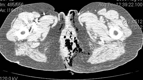
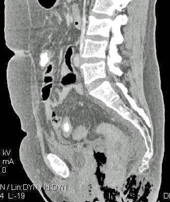
Fig 1: CT scan of abdomen and pelvis showing free air around rectum
Explanation:
Stercoral perforation of the colon is caused by progressive ischemic necrosis of the bowel wall by a faecal mass. It is the least likely diagnosis here as it usually occurs on the antimesenteric border of the sigmoid colon and is usually associated with a history of chronic constipation and megacolon.
Typhylitis is a potentially life threatening inflammatory bowel process that is a recognised complication of systemic chemotherapy. It can progress to bowel necrosis and perforation but is usually characterised by involvement of the caecum or ascending colon and the rectum is rarely involved.
Clostridial gas gangrene infection occurs with tissue inoculation in a low oxygen tension environment. Approximately 80% of patients without trauma have a malignancy of which 40% are hematologic, however the vast majority of cases are preceded by trauma of which there was no history of in this case 1.
Perineal Necrotizing fasciitis is a rare condition with an estimated 500 cases each year in the UK2. It can affect healthy individuals of any age but carcinoma and immunosupression are known to increase susceptibility3. The initial lack of obvious skin findings make this condition difficult to diagnosis but exquisite pain, especially pain that is disproportionate to what would be expected from the clinical findings is seen 2, 4. Where concurrent signs of sepsis exist, a high index of suspicion is required.
As the disease progresses, the skin may begin to appear smooth, shiny and swollen. Blistering and serous bullae may develop, and a haemorrhage into bullae may occur and giving the appearance of a haematoma as in our case. Crepitus, induration and foul smelling watery discharge secondary to liquefactive necrosis can also become apparent2. On CT scans, fascial thickening, fat stranding and gas tracking may be seen in nearly 80%2, 5 of cases, and was seen in this case as well.
Discussion
Necrotizing fasciitis is a lethal soft tissue infection characterised by rapidly progressive inflammation and necrosis of the subcutaneous fascial tissues. The adjacent skin and muscle are relatively spared until late in the course of the disease. Treatment with surgical debridement must be instigated without delay or the patient inevitably succumbs to sepsis and multi-organ failure2.
A 24 hour delay in treatment has been shown to increase mortality by 18% and further surgery is usually indicated with an average of 3.8 debridements needed overall5, 6. Surgical treatment should be instigated in conjunction with broad spectrum intravenous antibiotics and intensive care. The antibiotics selected should be effective against gram-positive, gram negative and anaerobic organisms. Adjuvant therapies like hyperbaric oxygen, intravenous immunoglobulin and activated protein C are of uncertain value.
Following surgery the patient is invariably left with a large tissue defect. Perineal wounds are particularly complex and present multiple challenges including the risk of infection from faecal contamination. Thus diverting colostomies are advised and a Vacuum Assisted Closure (VAC) system may facilitate wound healing 5.
|
Acknowledgements The authors would like to acknowledge the patient on whom the case report was based. Written informed consent was obtained. Competing Interests None declared Author Details S O’NEILL, Surgical Registrar, MB BCh BAO MRCS S I ANDRABI, Surgical Registrar, MB FRCS M C WHITESIDE, Consultant Surgeon, MD FRCS CORRESPONDENCE: STEPHAN O'NEILL, MB BCh BAO MRCS 6 Dean Park Mews, Edinburgh EH4 1EF Email: stephenoneill@doctors.org.uk |
References
1. San Ildefonso A, Maruri I, Facal C, Casal E. Clostridium septicum infection associated with perforation of colon diverticulum. Rev Esp Enferm Dig 2002;94(6):361-6.
2. Hasham S, Matteucci P, Stanley PR, Hart NB. Necrotising fasciitis. BMJ 2005;330(7495):830-3.
3. Kumar S, O'Donnell ME, Khan K, Dunne G, Carey PD, Lee J. Successful treatment of perineal necrotising fasciitis and associated pubic bone osteomyelitis with the vacuum assisted closure system. World J Surg Oncol 2008;6:67.
4. Young MH, Aronoff DM, Engleberg NC. Necrotizing fasciitis: pathogenesis and treatment. Expert Rev Anti Infect Ther 2005;3(2):279-94.
5. Elliott DC, Kufera JA, Myers RA. Necrotizing soft tissue infections. Risk factors for mortality and strategies for management. Ann Surg 1996;224(5):672-83.
6. Wong CH, Chang HC, Pasupathy S, Khin LW, Tan JL, Low CO. Necrotizing fasciitis: clinical presentation, microbiology, and determinants of mortality. J Bone Joint Surg Am 2003;85-A(8):1454-60.

The above article is licensed under a Creative Commons Attribution-NonCommercial-NoDerivatives 4.0 International License.