Anaesthesia

Training for anaesthetists in a new healthcare system: pathway to learning & development

Authors
Purnima Narasimhan & Krishnan Melarkode
Article Citation and PDF Link
BJMP 2021;14(1):a001
Abstract / Summary
Abstract: 

Every year, a number of overseas anaesthetists move to the UK under MTI, which is a training programme to assist doctors with proven capability in anaesthesia/Intensive Care/Pain Medicine from low and middle income countries to undertake further training in anaesthesia in the UK. This initiative benefits both the trainees and the NHS. The whole process involves a lot of work and is stressful too, as the trainee has to adapt to a new environment. The awareness of these is essential for both the trainees and the supervisors, as these form barriers to learning and need to be addressed before any learning begins. The MTI trainees are adult learners with a self-image and pre-existing knowledge. Therefore, any learning strategy for them should begin with assessment of the educational needs followed by constructivism, feedback and reflection without criticism. They should be encouraged to use the same learning platform as the UK trainees for an all-round development. The supervisors can provide advance structures upon which the MTIs can continue to build opportunities and gain confidence to rehearse and apply their new knowledge. An organised induction programme, a period of familiarisation and good mentorship with patience helps to remove the barriers to learning for the MTI trainees. A dynamic trainee-supervisor relationship to accommodate the changing educational goals and an appropriate mix of strategies can help the MTI trainees attain medical competence.


Abbreviations: 
MTI- Medical Training Initiative, NHS- National Health Service, UK- United Kingdom
Keywords: 
Anaesthesia, Adult learning, Medical Training Initiative

What is the MTI scheme?

The Medical Training Initiative (MTI) is a training programme to assist doctors with proven capability in anaesthesia/Intensive Care/Pain Medicine from low and middle income countries to undertake further anaesthesia training in the UK, for a maximum of 24 months1.

Why MTI?

It offers an opportunity not only to fine-tune their clinical acumen, but also to assimilate non-clinical skills (medical education, leadership and management, quality improvement projects) 2. The exposure most of the MTIs receive overseas is heterogeneous - in terms of level of supervision/independence, access to modern equipment and medications, lines of management, level of expectations and communication or interaction with patients. Funding received by training hospitals overseas can be variable thereby impacting on the resources available to provide standardised training. Under the MTI scheme, anaesthetic trainees can also take the FRCA examination.

From home to UK

A general awareness of the scheme helps the department to provide the MTIs with an appropriate support system. Details of the MTI scheme are available on the Royal College of Anaesthetists (RCoA) website.

It takes about 3-6 months after verification of the educational qualification by Educational Commission for Foreign Medical Graduates (ECFMG) via Electronic Portfolio of International Credentials (EPIC). The planning involves resignation from the current job, applying for a Tier 5 Visa to be in time for the GMC identity check (3 month deadline) and collecting the Biometric Residence Permit to be able to start work in the UK.

Medical staffing has more paperwork, one of which is the Disclosure and Barring Service (DBS). Prior intimation to the MTIs on the need for police verification from their home country would be of great benefit to make the process smoother. Hospital accommodation should be offered and organised in advance.

Acquaintance with the system

The MTI trainees often join at a time that doesn’t coincide with the UK training programme. Hence, a one-to-one induction customised towards overseas doctors will be beneficial. In addition to a named Educational supervisor (a mandatory requirement stipulated by the RCoA), the MTIs will benefit from having a nominated mentor within the department. The trainees can also get familiarised to the new healthcare system via the RCoA approved training courses – ‘Simulation for MTIs’ and ‘New to NHS’.

Allocating MTIs to theatre lists with only a select number of consultants in the initial stages helps them to settle in a new healthcare environment before they commence on-call (out of hours) duties. The MTIs should be encouraged to attend resuscitation courses like Advanced Life Support (ALS) as most of them follow the Advanced Cardiac Life Support (ACLS). They should be encouraged to document their progress like any other UK trainee via the RCoA Lifelong Learning Platform (LLP).

Anaesthetic training in the UK is very structured. The three stages of training (core, intermediate and higher/advanced) are well defined. The curriculum is well laid out and assists trainees to not only develop clinical but also gain non-technical skills. A six-monthly ARCP (Annual Review of Competence Progression) like assessment with annual anonymised multi-source feedback helps to create professional development plans, monitor progress and put supportive plans in place (if needed ) for a struggling trainee. The curriculum provides an opportunity for all-round development to every overseas trainee.

Gaining experience in non-technical skills (leadership and management, medical education and QI/audit projects) can be lacking in some home countries as the curriculum back home could be heavily biased towards the development of clinical acumen only.

What to expect from an MTI?

The MTIs have at least 3-5 years (may be more) of anaesthetic experience. The NHS benefits from their skills and experience. Their experience helps the department to allocate them to provide out of hours work (on-call) sooner than a UK trainee after an appropriate period of induction. The journey of patient experience also improves with the presence of experienced staff on the shop floor.

Departments gain from increased service provision too. As an example, after obtaining the initial assessment of competency (IAC), the MTI anaesthetists can be allocated to do solo theatre lists with a named supervising consultant anaesthetist present within the theatre suite. The reliance on locum staff is reduced thereby reducing unnecessary cancellations of theatre lists for lack of permanent staff. It reduces the financial burden on the NHS as staffing the department with locums can add to increased costs.

Patient safety is of paramount importance in any healthcare setting. Since the MTIs have a two year working contract, they are familiar with the department policies and guidelines unlike a locum doctor who does the odd shift in a hospital.

Equally, new skills gained by the MTIs are ultrasound guided regional anaesthesia, using a fibre-optic scope and different airway gadgets, ICU training, experience in geriatric and bariatric anaesthesia, total intravenous anaesthesia (TIVA) / target controlled infusion (TCI) alongside access to new medications like remifentanil, sugammadex which may not be available in low to middle income countries. The NHS provides excellent opportunities in simulation training and teaching courses.

However, a system of protocols can be unnerving to the MTIs. One may find them taking a step back when it comes to ‘decision making’ as they are not sure if it would be approved or criticised. At times, some of the MTIs may come across as unyielding despite adequate teaching. It is essential to remember that the process of unlearning to re-learning takes time, and therefore, patience is the key. This is where the concept of teaching experienced medical practitioners with knowledge comes in handy.

Training learners with knowledge

Medical education comprises three inter-linked domains - knowledge, skills and attitude.3 Though trainees may differ in terms of their motivation for learning, it can manifest only after the basic needs are satisfied - the external barriers to motivation such as life events and transitions, opportunities, and barriers to learning or obtaining information are addressed and they feel respected in the educational environment. The MTIs are essentially adult learners with pre-existing knowledge, who bring a great deal of first-hand experience to any work-place. Learning should, therefore, be integrative, which forms the basis of constructivism theory of learning4. New knowledge and skills should be integrated into the existing bank of knowledge. They also have pre-set strong tastes and habits which can be a real asset or a hindrance to effective learning. The educational supervisor should be able to encourage or curb them accordingly5.

Being adults, MTIs enter training situations with a self-image as independent, mature beings as they have already passed the qualifying exams in their home country. They can direct their own learning, including decision making and plans for taking examinations. The supervisors should engage with the trainees in activities that create a sense of self-responsibility to facilitate better learning opportunities.

Many adult learners suffer from a fear of failure and living up to expectations6 and thus, educational supervisors should be cautious to avoid unnecessary criticism. Instead the focus should be to offer constructive positive feedback. Any educational plan for them should start with an awareness of their prior acquired knowledge, an assessment of their educational needs along with room for motivation and reflection. This helps the trainees retain the original “frame of reference” while continuing to constantly challenge and transform practice via reflection on-action and reflection in-action. The educational supervisors need to provide a supporting educational environment, a structured guide for reflection and constructive feedback to develop the trainees’ reflective practice7.

A simple multi-step approach involving active participation from both the trainee and the educational supervisor can be summarised into a model as below (Figure 1) 8. This model begins with the trainee’s prior knowledge.


Figure 1: Multi-step approach to training

The RCoA LLP, work-place based assessments and multi-source feedback along with the six-monthly meeting with educational supervisor are useful tools to deliver a holistic learning experience. It helps to refine the existing knowledge, reflect and provide constructive feedback. The supervisor can provide advance structures upon which the MTI can continue to build opportunities and gain confidence to rehearse and apply their new knowledge.

Summary

An organised induction programme, a period of familiarisation and good mentorship with patience helps to remove the barriers to learning for the MTI trainees. A dynamic trainee-supervisor relationship to accommodate the changing educational goals and an appropriate mix of strategies can help the MTI trainees attain medical competence, which is defined as “the habitual and judicious use of communication, knowledge, technical skills, clinical reasoning, emotions, values and reflection in daily practice for the benefit of the individual and the community being served3.”

 
Acknowledgements / Conflicts / Author Details
Competing Interests: 
None declared
Details of Authors: 
PURNIMA NARASIMHAN, MBBS, MD, DNB Anaesthesiology, Pinderfields General Hospital, Wakfield, UK. KRISHNAN MELARKODE, MBBS, MD, DNB, FRCA, PGCert (medical education), Pinderfields General Hospital, Wakfield, UK.
Corresponding Author Details: 
Dr PURNIMA NARASIMHAN, Department of Anaesthesia, Pinderfields General Hospital, Wakefield, UK WF1 4DG
Corresponding Author Email: 
purni89@yahoo.com
References
References: 
  1. RCoA. https://www.rcoa.ac.uk/about-college/global-partnerships/overseas-doctors-training-uk/medical-training-initiative
  2. World Federation of Societies of Anaesthesiologists. Medical Training Initiative. https://www.wfsahq.org/medical-training-initiative
  3. Abela J. Adult Learning Theories and Medical Education: a Review. Malta Medical Journal.2009; 21:11-18
  4. Badyal DK, Singh T. Learning Theories: The Basics to Learn in Medical Education. Int J Appl Basic Med Res. 2017 Dec; 7(Suppl 1): S1–S3.doi: 10.4103/ijabmr.IJABMR_385_17
  5. Thoms KJ. They’re not just big kids: motivating adult learners. http://www.mtsu.edu/∼itconf/proceed01/22.pdf
  6. Kennedy RC. Applying Principles of Adult Learning: The Key to More Effective Training Programs. http://www.ncjrs.gov/App/publications/abstract.aspx?ID=199990
  7. Mukhalalati BA, Taylor A. Adult Learning Theories in Context: A Quick Guide for Healthcare Professional Educators. Journal of Medical Education and Curricular Development. January 2019. doi: 10.1177/2382120519840332
  8. David C. M. Taylor & Hossam Hamdy (2013) Adult learning theories: Implications for learning and teaching in medical education: AMEE Guide No. 83, Medical Teacher, 35:11, e1561-e1572, doi: 10.3109/0142159X.2013.828153


Ketamine and Propofol Combination Used in Deep Sedation Reduces Opioid Use and Adverse Outcomes in Pediatric Esophagogastroduodenoscopy in Ages 2-18 Years

Authors
David C Mari & Abhik K Biswas
Article Citation and PDF Link
BJMP 2019;12(3):a020
Abstract / Summary
Abstract: 

Background: EGD procedures can elicit significant pain and cause complications in children. Therefore it is essential to use sedation agent(s) that reduce pain and are safe.
Aims: Primary aim is to determine the differences in adverse cardiopulmonary events, vital sign parameters, objective pain, and adjunct fentanyl use during EGD procedures between propofol and ketofol (ketamine-propofol mixture) sedation agents. Secondary aim is to determine the differences in site performance metrics between the two agents.
Methods: Retrospective analysis of 90 patients undergoing deep sedation for same-day EGD procedures at a military academic medical centre. Patients were analysed separately via age groups of 2-11 years and 12-18 years. Forty-one patients underwent sedation via propofol, and 49 patients underwent sedation via 1:5 ratio of ketamine to propofol as main agents. Main agents were administered via intravenous loading dose based on 1 mg/kg of propofol followed by a 200-250 mcg/kg/minute infusion. One mcg/kg fentanyl bolus was given when a sedationist perceived patient pain.
Results: Ketofol reduced fentanyl use by ~1 mcg/kg compared to propofol in all age groups (p<0.008). Ketofol compared to propofol had less apnoea requiring intervention (3.7% versus 53.8%, p<0.001, ages 2-11 years; and 0% versus 33.3%, p=0.005, ages 12-18 years), and less hypotensive events (3.7% versus 15.4%, p=0.192, ages 2-11 years; and 0% versus 13.3%, p=0.144, ages 12-18 years). There was no difference in LOS between main sedation agents (p>0.008).
Conclusions: Ketofol is effective at reducing opioid demand and adverse cardiopulmonary events during EGD procedures compared to propofol in ages 2-18 years.

Abbreviations: 
Esophogastroduodenoscopy (EGD), length of stay (LOS), mean arterial blood pressure (MAP), heart rate (HR), respiratory rate (RR), and Children’s Hospital of Eastern Ontario Pain Scale (CHEOPS).
Keywords: 
Ketofol, Propofol, Fentanyl, Esophagogastroduodenoscopy, and Deep Sedation.

Introduction:

Aesophagogastroduodenoscopy (EGD), is a common same-day procedure used for both diagnostic and therapeutic purposes during which a small flexible fibreoptic tubular camera is introduced through the mouth and advanced through the pharynx into the oaesophagus, stomach, and duodenum. EGD procedures are performed under deep sedation, since they can elicit significant pain, discomfort, and anxiety. When pain is not controlled properly, this can lead to an increase use of adjunctive medications such as opioids. This can extend the length of stay and increase adverse outcomes.1 In a prospective, randomised, double-blinded study, Bedirli et al. found that use of opioid medications such as fentanyl are associated with increased adverse outcomes.2

Since procedural sedation-related complications (such as hypoxia, hypotension, desaturation, and emergent airway intervention) remain one of the biggest challenges in EGD procedures, it is important to select the correct medications to reduce these complications.3 Currently, propofol is the most common and popular main procedural sedation agent used for EGD procedures.4,5

Propofol is the preferred main intravenous agent in EGD procedures due to its amnestic, sedative, and hypotonic properties.6 It has also been favoured due to its ultra-rapid response and duration of effects, usually taking 30-60 seconds for onset of action and lasting up to 4-8 minutes.7 However, propofol has been associated with significant adverse effects that include dose-related hypotension, bradycardia, laryngospasms, and apnoea.8,9 Propofol does not have analgesic properties and will usually be combined with opioids for pain control.10

Ketamine is classified as a dissociative anaesthetic and is known to provide analgesia and amnesia. Important to note, it causes less respiratory or cardiovascular depression when used alone for children greater than 4 months of age.11 However, ketamine alone can cause such side effects of laryngospasm, increase secretions, and vomiting.12,13

The mixture of propofol and ketamine in one syringe (coined ketofol) has been shown to be effective in sedation for various procedures, such as spinal anaesthesia,14 along with orthopaedic15 and cardiovascular procedures16 in adults and children. Use of this combination has been favoured in brief but painful emergency room procedures due to the opposing haemodynamic and respiratory effects of both sedative medications.17 The negative cardiac effects produced by propofol can be attenuated with the use of ketamine, resulting in an increase in mean arterial pressures and cardiac indices.18,19 The complementary effect of both mediations has enabled the use of lower doses for each medication, thus lowering the toxicity and side effects.20,21 Although there are studies comparing activities of ketofol sedations,22-25 there has not been a study published on ketofol compared to propofol use in children during EGD procedures.

The primary goal of this study was to determine if there were differences in the outcomes of adverse cardiac and pulmonary events, vital sign parameters including objective pain, and administration of adjunct pain medication (for which fentanyl was used in this study) during EGD procedures between propofol and ketofol groups. A secondary goal was to determine if there was a difference in site performance metrics for EGD procedures (sedation time, stop of sedation to discharge time, and length of stay) between propofol and ketofol groups.

Methods:

This study protocol was approved by the Naval Medical Center Portsmouth Institutional Review Board in compliance with all applicable federal regulations governing the protection of human subjects. Research data was derived from an approved IRB protocol: number NMCP.2018.0021. Written informed consent was not required by the Naval Medical Center Portsmouth IRB, as this data concerned historical dae-identified patients.

Study Design

This was a single centre, retrospective study of a cohort of children (ranging from 2 to 18 years). Data was collected from March 2011 to September 2013 at Naval Medical Center Portsmouth’s Paediatric Sedation Centre. EGD and sedation protocol was not changed during this time period. EGD procedures were performed by the same paediatric gastroenterologists throughout this time period. Sedation was performed by the same paediatric intensivists throughout this time period. All patients used in this study were involved in a same day EGD procedure. Forty-one patients underwent deep sedation via propofol as main sedation agent from March 2011 to May 2012. Forty-nine patients underwent deep sedation via propofol and ketamine combination as main sedation agent from May 2012 to September 2013. Each main sedation agent was given in a similar fashion as an initial intravenous loading dose based on 1 mg/kg propofol followed by an intravenous infusion of 200-250 mcg/kg/minute that was started less than 10 minutes prior to start of the procedure and stopped at the end of the procedure. Ketofol solution used was a 1:5 ratio of ketamine to propofol (40 mg:200 mg). One mcg/kg of fentanyl was given when the sedationist during the EGD perceived the patient experiencing pain. Exclusion criteria included: patients less than 2 years and greater than 18 years, those less than 10 kg, and patients receiving adjunct medications other than fentanyl, ondansetron, or lidocaine.

Data Collection

Fourteen patients were excluded due to weight less than or equal to 10 kg. In addition, 12 patients were excluded due to age less than 2 years or greater than 18 years. Past medical history, ASA class, procedure indications, age, weight, sex, pain scores, vital signs (mean arterial blood pressure [MAP], heart rate [HR], and respiratory rate [RR]), unplanned events (hypoxia defined as SPO2 less than 85% at any time point, and hypotension defined as mean arterial blood pressure with greater than 20% decrease from baseline blood pressure), emergent airway intervention (defined as apnoea needing bag mask ventilation or CPAP use), and unplanned intubation. Vitals and pain scores were obtained from initial presentation in the sedation suite, start of procedure, every 5 minutes during the procedure to stop of sedation, and at time of discharge. Midway procedure vitals used for statistical analysis were obtained by selecting vitals that were at the half-way point of the patient’s EGD procedure. The Children’s Hospital of Eastern Ontario Pain Scale (CHEOPS) was performed every 5 minutes during the EGD procedure by an independent United States Navy Corpsman and used to evaluate patient’s pain prior, during, at stop of sedation, and after procedure for this study.26

Statistics

All statistics performed in this study were calculated using SPSS Statistics programme (IBM Corp. Released 2017. IBM SPSS Statistics for Windows, Version 25.0. Armonk, NY: IBM Corp.). Mann-Whitney U nonparametric test between independent groups was performed to compare age, weight, total EGD procedure time, total time of sedation during EGD procedure, time from stop of sedation to discharge, total length of stay, and total fentanyl use (in mcg/kg) between the two main groups in this study (propofol and ketofol group). The significance level was set to 0.008 after a Bonferroni was corrected, in order to address the probability of making one or more false discoveries when performing multiple hypotheses tests. A repeated measures ANOVA was run to determine the effect of treatments over time for HR, MAP, and RR. Mauchly's test of sphericity was performed for both age groups. The Fisher Exact test was performed to compare propofol to ketofol in unplanned hypotensive events and unplanned apnoea requiring bag valve mask or CPAP. Pearson Correlation statistics were performed to study possible correlations between propofol or fentanyl and sedation time or length of stay. Statistical significance for Pearson Correlation statistics was considered when two-tailed p<0.001.

Results:

Table 1. Demographic divided into 2 age groups. EE= eosinophilic esophagitis. * indicates statistically significant difference.


Sedation Risk Age Groups

2-11 years old

12-18 years old

Propofol

n=26

%

Ketofol

n=27

%

Propofol

n=15

%

Ketofol

n=22

%

General

Demographics

Male

11

42.3%

16

59.3%

6

40%

10

45.5%

Female

15

57.7%

11

40.7%

9

60%

12

54.5%

Mean Age (years old)

6


7


15


16


Mean Weight (kg)

21.6


23.7


57.7


57.4


ASA I, II

26

100%

25

92.6%

15

100%

22

100%

ASA III

0

0%

2

7.4%

0


0


EGD Indications

EE

4

15.4%

4

14.8%

2

13.3%

0

0%

GERD

11

42.3%

9

33.3%

2

13.3%

5

22.7%

Dysphagia or Feeding Intolerance

0

0%

3

11.1%

4

26.6%

4

18.1%

Foreign Body Ingestion

1

3.8%

2

7.4%

0

0%

0

0%

Failure to Thrive

3

11.5%

1

3.7%

0

0%

0

0%

Abdominal Pain

7

26.9%

9

33.3%

6

40%

11

50%

Recurrent Emesis

3

11.5%

1

3.7%

1

6.6%

1

4.5%

Gastritis

0

0%

0

0%

1

6.6%

3

13.6%

Other

2

7.7%

0

0%

2

13.3%

4

18.1%

Unplanned Events

Hypotension (blood pressure >20% decrease from baseline)

4

15.4%

1

3.7%

2

13.3%

0

0%

Apnea requiring bag valve mask or CPAP

14

53.8%*

1

3.7%*

5

33.3%*

0

0%*

Intubations

0

0%

0

0%

1

6.6%

0

0%

Figure 1. Propofol amount total per weight (mg/kg). Bars represent standard error of the mean. There was no significant difference between the propofol and ketofol in all age groups.

Figure 2. Fentanyl amount total per weight (mcg/kg). Bars represent standard error of the mean. In all age groups, there was a significant difference between propofol and ketofol. *p<.008.

Figure 3. Vital signs. Mean arterial pressures for ages A) 2-11 years and B) 12-18 years. Main sedative agents’ MAPs were statistically different for ages 2-11 years (p =0.004), but not for ages 12-18 years (p =0.224) for all time periods. Heart rate for ages C) 2-11 years and D) 12-18 years. For Heart Rate, there was a statistically significant interaction between treatment and time for both age group using the Greenhouse-Geisser correction, p =0.002, p =0.014. Main sedative agents’ HRs showed to be statistically different for both age groups, p <0.001 and p =0.004.

Figure 4. Duration of time. A) total sedation time, B) stop of sedation to discharge, and C) total length of stay. In all age groups, there was no significant difference between propofol and ketofol. Bars represent standard error of the mean. *p<0.008.

Figure 5. Pearson Correlation. Propofol versus total sedation time for ages A) 2-11 years (p<0.01*), and B) 12-18 years (p<0.01*). Propofol versus length of stay for ages C) 2-11 years (p<0.01*), and D) 12-18 years (p<0.01*). * Correlation is significant at the 0.01 level (2-tailed).

Figure 6. Pearson Correlation. Fentanyl versus total sedation time for ages A) 2-11 years (p=0.506), and B) 12-18 years (p=0.961). Fentanyl versus length of stay for ages C) 2-11 years (p=0.378), and D) 12-18 years (p=0.352). No significant correlation was not seen for all age groups.

Ninety patients were retrospectively analysed in this study. Baseline demographics were similar between the groups, except for gender proportions (Table 1). Similar amounts of propofol per weight were used in each group (Figure 1). Ketofol significantly reduced fentanyl use in all age groups by ≥.99 mcg/kg compared to propofol alone (p<0.008 for all age groups, Figure 2). In the 2-11 year old age group, the propofol group required a mean fentanyl dose of 1.96 mcg/kg ± 1.24 mcg/kg and the ketofol group required a mean fentanyl dose of 0.44 mcg/kg ± 0.49 mcg/kg; thus ketofol required about 1.52 mcg/kg less fentanyl during EGD procedures. In the 12-18 year old group, the propofol group required a mean fentanyl dose of 1.38 mcg/kg ± 1.05 mcg/kg and the ketofol group required a mean fentanyl dose of 0.39 mcg/kg ± 0.59 mcg/kg; thus ketofol required about 1 mcg/kg less fentanyl during EGD procedures.

Vital signs (HR, RR, and MAP) and CHEOPS pain scores were obtained and analysed for baseline vitals, at the procedure midpoint, at stop of sedation, and at time of discharge. CHEOPS pain scores’ mean was 6 throughout all time periods for both groups, thus there was no statistically significant difference.

A repeated measures ANOVA was run to determine the effect of treatments over time for RR. There was sphericity for the interaction term, as assessed by Mauchly's test of sphericity in both age groups (p =0.070, p =0.762). For RR, there was not a statistically significant interaction between treatment and time for both age groups, p =0.163, p =0.804. The treatments were not found to be statistically different for either age group, p =0.736 and p =0.224.

A repeated measures ANOVA was run to determine the effect of treatments over time for HR. As assessed by Mauchly's test of sphericity, no sphericity was found in either age group (p <0.05). For HR, using the Greenhouse-Geisser correction, a statistically significant interaction was found between treatment and time for both age group, p =0.002, p =0.014. The treatments were found to be statistically different for both age groups, p <0.001 and p =0.004. A repeated measures ANOVA was run to determine the effect of treatments over time for MAP. Sphericity was found in both age groups for the interaction term, as assessed by Mauchly's test of sphericity (p =0.209, p =0.269). For MAP, there was not a statistically significant interaction between treatment and time for either age group, p =0.261, p =0.591. The treatments were statistically different for the 2-11 year old group (p=0.004), but not statistically different for the 12-18 year old group (p=0.224). (Figure 3 A-D)

Unplanned events were analysed for clinically significant hypoxia, hypotension, emergent airway intervention, and unplanned intubations. Ketofol compared to propofol had fewer hypotensive events in the 2-11 year old age group (3.7% versus 15.4%, p=0.192) and in the 12-18 year old age group (0% versus 13.3%, p=0.144). Ketofol compared to propofol had statistically significant fewer apnoea events requiring bag valve mask or CPAP intervention for the 2-11 year old group (3.7% versus 53.8%, p<0.001) and for the 12-18 year old group (0% versus 33.3%, p=0.005). There were no significant hypoxia events. There was one unplanned intubation in the propofol group of a healthy ASA I twelve year old female who had a 20 mL emesis episode after a loading dose of propofol was given on induction. (Table 1)

With propofol leading to significantly more fentanyl usage, more hypotension and emergent airway intervention during EGD procedures, we performed analysis to see if there was an effect on sedation time, time from stop of sedation to discharge, and length of stay (LOS). In all age groups, there was no statistical difference between propofol and ketofol for sedation time (p=0.115 for ages 2-11 years, p=0.124 for ages 12-18 years), time from stop of sedation to discharge (p=0.033 for ages 2-11 years, p=0.511 for ages 12-18 years), and LOS (p=0.026 for ages 2-11 years, p=0.109 for ages 12-18 years) (Figure 4). Based on Pearson correlation test, it was established that propofol was positively correlated with sedation time and LOS (+0.753 for sedation time and +0.611 correlation for length of stay with <0.001 significance) (Figure 5). There was no significant correlation between fentanyl and sedation time or LOS (Figure 6).

Discussion:

This was a retrospective study looking at 90 paediatric patients, ages 2-18 years, undergoing EGD procedures with either propofol or ketofol as the main sedative medication. Patients in this study had similar weights, ages, ASA scores, and EGD indications; however there baseline demographics were not similar with regards to gender proportion (Table 1). Based on the large prospective database on sedation use for procedures outside the operating room obtained by the Paediatric Sedation Research Consortium,27 we separated our patient population into two age risk groups to allow our results to be generalisable. To our knowledge, this study is the first to show that ketofol, when compared to propofol, significantly reduces fentanyl use (≥.99 mcg/kg, Figure 2) and cardiopulmonary adverse outcomes (Table 1) in paediatric EGD procedures.

EGD procedures are known to be invasive and painful. To decrease the pain appreciated by a patient, various adjuncts are often utilised. In our study, we used fentanyl for adjunct pain control. In both main sedation medication groups (propofol and ketofol), there was no statistical significance seen in objective pain based on CHEOPS scores with a mean score of 6 in all age groups (p>0.05). However, the amount of adjunct needed to maintain adequate pain control between both groups statistically and clinically differed (Figure 2). In all age groups, the propofol group compared to the ketofol group required almost 1 mcg/kg more of fentanyl, which leads to the potential for the patient to experience higher incidence of side effects, such as respiratory depression and hypoxia.28-30

The mechanism that lead to this significant difference in opioid demand between ketofol and propofol is most likely due to ketamine’s ability to stimulate opioid sigma receptors,31 thus leading to opioid sparing effects. It is unclear if propofol and ketamine interaction heightens this opioid sparing effect, because ketamine alone has not been shown to lead to opioid sparing effects in children.32

In a review of our population’s vital signs, there was a significantly higher MAP in the ketofol group compared to the propofol group for ages 2 to 18 years (Figure 3 A-B). In addition, the ketofol group had statistically higher HR compared to the propofol group for ages 2 to 18 years (Figure 3 C-D). It is our thought that this increase in MAP and HR in the ketofol group mediated by ketamine is a protective factor against major hypotensive changes (31). It is this effect that minimised unplanned hypotensive events in our ketofol group compared to the propofol group by 11.7% in ages 2-11 years and 13.3% in ages 12-18 years (Table 1).

One of the more noticeable differences in our main sedative medication groups was the unplanned apnoea events requiring CPAP or bag mask ventilation intervention. In our 2 to 11 year age group, propofol had 50.1% more apnoea events needing intervention compared to ketofol. In our 12 to 18 year old group, propofol had 33.3% more apnoea events needing respiratory intervention compared to ketofol. This was found to be statistically significant. It is unclear if this is also clinically significant since no emergent airway intubation was needed for these events. However, there was 1 emergent airway intubation in the 12-18 year old propofol group. Based on the review of the medical records, it appears that these apnoea events needing respiratory intervention were mostly associated after the initial loading dose of either propofol or ketofol prior to or at the start of the infusion. Thus, bring into question if not starting with a loading dose bolus would decrease adverse effects.

Despite the propofol group requiring more respiratory intervention, increased fentanyl use, and less haemodynamic stability seen, there was no statistical differences in total sedation time and LOS between the two main sedation medication groups (Figure 4). Yet when we ran correlation statistics, there was a positive correlation to increased propofol dosages, total sedation time, and LOS in ages 2 to 18 years (Figure 5); therefore, if our procedures were longer, there could be a statistically and possibly clinically significant increase in total sedation time and LOS for the propofol only group. Thus, we can infer that propofol compared to ketofol is not the main sedation medication of choice for longer similarly painful procedures in children ages 2 to 18 years, such as EGD procedure followed by a colonoscopy.

A limitation of our study is that it is retrospective. With that, we were not able to blind our sedationists to the main sedation medication that was chosen and could not control the interventions that were performed. Yet, based on strict exclusion criteria, we were able to control the interventions used in our analysis. Also, in our study we did not analyse patients under the age of 2 years. Reviewing the data, we had seven patients, and thus this patient population size was not powered to perform the proper statistical analysis. Another limitation to consider is the time difference in procedure protocols. However, since this study included over 2 years of data, procedure protocols were the same. Additionally, the EGD indications were similar between ketofol and propofol (Table 1). Another consideration of this study is generalisability. This study was conducted at a single centre, but the way we analysed our results makes it easier to perform large scale prospective studies to further investigate our findings. Last limitation is directly correlating fentanyl dose with a significant adverse event. Due to the short length of the EGD procedure, constant sedation medication infusion, and fentanyl dose, it is difficult to directly say that a particular fentanyl dose alone leads to an adverse event. Therefore we can only speculate based on correlation.

Conclusions:

Our study found that ketofol significantly reduced fentanyl use in all age groups by ≥0.99 mcg/kg and cardiopulmonary adverse events compared to propofol alone. In addition, an increase in the amount of propofol was positively correlated to increasing LOS and total sedation time for a child undergoing an EGD procedure. This suggests that increasing amounts of propofol leads to greater LOS. To conclude, our study indicates that ketofol can be a safer alternative to propofol use alone for deep sedation in EGD procedures for children ages 2 to 18 years.

Acknowledgements / Conflicts / Author Details
Acknowledgement: 
We like to thank all Paediatric Intensivists and Paediatric Gastroenterologists involved in patient care. We would also like to thank Andrea McGlynn with her help and guidance with statistics used in this study. Authors in this manuscript have no conflicts of interest to disclose. This study was completed with no external or internal funding. We are military service members and employees of the U.S. Government. This work was prepared as part of our official duties. Title 17 U.S.C. 105 provides that “Copyright protection under this title is not available for any work of the United States Government.” Title 17 U.S.C. 101 defines a United States Government work as a work prepared by a military service member or employee of the United States Government as part of that person’s official duties. The views expressed in this manuscript reflect the results of research conducted by the authors and do not necessarily reflect the official policy or position of the Department of the Navy, Department of Defence, or the United States Government.
Competing Interests: 
None declared
Details of Authors: 
DAVID C. MARI, D.O., Capt USAF;Division of Pediatric Medicine, Naval Medical Center Portsmouth, 620 John Paul Jones Circle, Portsmouth, VA 23708. ABHIK K. BISWAS, M.D., CAPT (ret) USN; Division of Pediatric Critical Care Medicine, Naval Medical Center Portsmouth, 620 John Paul Jones Circle, Portsmouth, VA 23708.
Corresponding Author Details: 
DAVID C. MARI, Division of Pediatric Medicine, Naval Medical Center Portsmouth 620 John Paul Jones Circle, Portsmouth, VA 23708.
Corresponding Author Email: 
david.c.mari@gmail.com
References
References: 
  1. Samer Ammar M, Pfefferkorn MD, Croffie JM, et al. Complications after outpatient upper GI endoscopy in children: 30-day follow-up. Am J Gastroenterol. 2003; 98: pp. 1508-1511.
  2. Bedirli N, Egritas O, Cosarcan K, et al. A comparison of fentanyl with tramadol during propofol-based deep sedation for pediatric upper endoscopy. Paediatr Anaesth. 2012 Feb; 22(2): 150-155.
  3. Orel R, Brecelj J, Dias JA, et al. Review on sedation for gastrointestinal tract endoscopy in children by non-anesthesiologists. World J Gastrointest Endosc. 2015 Jul 25; 7(9): 895-911.
  4. Mustafaeva MN, Mizikov VM, and Kochneva ZV. Drug sedation during digestive tract endoscopy: current trends. Anesteziol Reanimatol 2009; 4: 32-38.
  5. Kochlar GS, Gill A, and Vargo JJ. On the Horizon: The Future of Procedural Sedation. Gastrointestinal Endoscopy Clin N Am. 2016; 26: 577-592.
  6. Watcha MF, Simeon RM, White PF, et al. Effect of propofol on the incidence of postoperative vomiting after strabismus surgery in pediatric outpatients. Anesthesiology, 1991, pp 2049.
  7. McCollum JS, Milligan RK, and Dundee JW. The antiemetic action of propofol. Anaesthesia. 1988; 43: 239-240.
  8. Hig Jr CC, McLeskey CH, Nahrwold ML, et al. Hemodynamic effects of propofol: data from over 25,000 patients. Anesth Analg, 1993, pp S21-S29.
  9. Claeys MA, Gepts E, and Camu F. Haemodynamic changes during anaesthesia induced and maintained with propofol. Br J Anaesth, 1988, pp 3-9.
  10. Varddi A, Salem Y, and Padeh S. Is propofol safe for procedural sedation in children? A prospective evaluation of propofol versus ketamine in pediatric critical care. Crit Care Med, 2002, pp 1231-1236.
  11. 11.   Grunwell JR, Travers C, McCracken CE, et al. Procedural Sedation Outside of the Operating Room Using Ketamine in 22,645 Children: A Report From the Pediatric Sedation Research Consortium. Pediatr Crit Care Med. 2016 Dec; 17(12): 1109-1116.
  12. 12.   Kurdi MS, Theerth KA, and Deva RS. Ketamine: Current applications in anesthesia, pain, and critical care. Anesth Essays Res. 2014 Sep-Dec; 8(3):283-290.
  13. Green SM, Roback MG, and Krauss B. Laryngospasm during emergency department ketamine sedation: A case-control study. Pediatr Emerg Care. 2010;26:798–802.
  14. Frizelle HP, Duranteau J, Samii K. A comparison of propofol with a propofol-ketamine combination for sedation during spinal anesthesia. Anesth Analg, 1997, pp 1318-1322.
  15. Weatherall A and Venclovas R. Experience with a propofol-ketamine mixture for sedation during pediatric orthopedic surgery. Paediatr Anaesth 2010; 20: 1009-1016.
  16. Mourad M, El-Hamamsy M, Anwar M, et al. Low dose ketamine reduces sedative doses of propofol during ambulatory trans-oesophageal echocardiography. Egyptian Journal of Anaesthesia, 2004, pp 41-46.
  17. Mortero RF, Clark LD, Tolan MM, et al. The effects of small-dose ketamine on propofol sedation: respiration, postoperative mood, perception, cognition, and pain. Anesth Analg, 2001, pp 1465-1469.
  18. Willman EV and Andolfatto G. Prospective evaluation of “ketofol”(ketamine/propofol combination) for procedural sedation and analgesia in the emergency department. Ann Emerg Med 2007; 49: 23-30.
  19. 19.   Kochs E, Scharein E, Mollenberg O, et al. Analgesic efficacy of low dose ketamine. Somatosensory-evoked responses in relation to subjective pain ratings. Anesthesiology 1996; 85: 304–314.
  20. Badrinath S, Avramov MN, Shadrick M, et al. The use of a ketamine-propofol combination during monitored anesthesia care. Anesth Analg 2000; 90: 858-862.
  21. Loh G and Dalen D. Low-dose ketamine in addition to propofol for procedural sedation and analgesia in the emergency department. Ann Pharmacother 2007; 41:485-492.
  22. Andolfatto G, Abu-Laban RB, Zed PJ, et al. Ketamine-propofol combination (ketofol) versus propofol alone for emergency department procedural sedation and analgesia: a randomized double-blind trial. Ann Emerg Med 2012; 59: 504-512.
  23. Sakai T, Singh H, Mi WD, et al. The effect of ketamine on clinical endpoints of hypnosis and EEG variables during propofol infusion. Acta Anaesthesiol Scand 1999; 43: 212–216.
  24. Hui TW, Short TG, Hong W, et al.Additive interactions between propofol and ketamine when used for anesthesia induction in female patients. Anesthesiology 1995; 82: 641–648.
  25. Badrinath S, Avramov MN, Shadrick M, et al.Use of ketamine–propofol admixture during monitored anesthesia care. Anesthesiology 1997; 87: A10.
  26. McGrath PJ, Johnson G, Goodman JT, et al. CHEOPS: a behavioral scale for rating postoperative pain in children. In: H.L. Fields, R. Dubner and F. Cervero (Eds.), Advances in Pain Research and Therapy, Raven Press, New York, 1985, pp 395-402.
  27. Cravero JP, Blike GT, Beach M, et al. Incidence and nature of adverse events during pediatric sedation/anesthesia for procedures outside the operating room: report from the Pediatric Sedation Research Consortium. Pediatrics. 2006 Sep; 118(3): 1087-1096.
  28. Godambe SA, Elliot V, Matheny D, et al. Comparison of propofol/fentanyl versus ketamine/midazolam for brief orthopedic procedural sedation in a paediatric emergency department. Pediatrics 2003; 112: 116–123.
  29. Skokan EG, Pribble C, Bassett KE, et al. Use of propofol sedation in a paediatric emergency department: a prospective study. Clin Pediatr 2001; 40: 663–671.
  30. Aydin Erden, I. Gulsun Pamuk, A. Akinci, et al. Comparison of propofol‐fentanyl with propofol‐fentanyl‐ketamine combination in pediatric patients undergoing interventional radiology procedures. Pediatric Anesthesia 2009; 19: 500-506.
  31. Kim SH, Kim SI, Ok SY, et al. Opioid sparing effect of low dose ketamine in patients with intravenous patient-controlled analgesia using fentanyl after lumbar spinal fusion surgery. Korean J Anesthesiol. 2013 Jun; 64(6):524-528.
  32. Michelet D, Hilly J, Skhiri A, et al. Opioid-Sparing Effect of Ketamine in Children: A Meta-Analysis and Trial Sequential Analysis of Published Studies. Paediatr Drugs. 2016 Dec; 18(6):421-433.

Hamman’s syndrome in a parturient: a case report

Authors
Rajashree Chavan
Article Citation and PDF Link
BJMP 2019;12(1):a007
Abstract / Summary
Abstract: 

Hamman’s syndrome is a potentially life threatening clinical condition characterised by peripartum subcutaneous emphysema and pneumomediastinum. This obstetric complication typically occurs in late pregnancy and labour and is frequently observed in young healthy primiparous women. I report a case postpartum pneumothorax and pneumomediastinum associated with spontaneous subcutaneous emphysema in primiparae. She presented to us in normal labour followed by Instrumental delivery under epidural anaesthesia. 10 hours post-delivery; she developed extensive subcutaneous emphysema, pneumothorax & pneumomediastinum with significant symptoms. She was successfully managed & discharged home after 24 hours of presentation of Hamman’s syndrome. Hamman’s syndrome is a rare obstetric complication which may present with/ without pneumothorax. Incidence is estimated at 1 in 100,000 deliveries. Approximately 200 such cases have been reported in literature worldwide. To my knowledge, this is one of the very few cases of Hamman’s syndrome with pneumothorax.

Keywords: 
spontaneous pneumomediastinum, subcutaneous emphysema, pregnancy, labour

Case Report:

A 33 year-old ASA1, primigravida, presented to our delivery suite with spontaneous onset of labour at 38 weeks of gestation. Epidural analgesia was commenced to alleviate her labour pains. Subsequently, she underwent an assisted vaginal delivery of a live male baby (weighing 4660 gms) using Keiland’s outlet forceps after 90 min second stage of labour. 10 hours postpartum, she complained of dyspnoea & severe central substernal chest pain. She was noted to have an unusual swelling of face and neck with oxygen saturations of 90 % on room air. Ascultation of chest revealed normal bronchovesicular breath sounds, normal heart sounds with absence of added sounds. Arterial blood gases showed an O2 tension of 11 kpa, CO2 tension of 5 kpa and pH of 7.34. The diagnosis of subcutaneous emphysema, pneumomediastinum and small left apical pneumothorax (Hamman’s syndrome) was confirmed on chest X-ray (CXR 1). We ruled out differential diagnosis of pulmonary embolism, Tension pneumothorax, angina pectoris, pericarditis, dissection of aortic aneurysm, mediastinitis, cardiac tamponade, chest infection & oesophageal tear. She was managed conservatively by close monitoring for complications, administration of supplemental oxygen and use of simple analgesics. She demonstrated a complete uneventful recovery over the next 24 hours with normalising of chest signs (CXR 2).


CXR 1: shows pneumomediastinum, extensive surgical emphysema & a left apical pneumothorax.


CXR 2: shows small pneumomediastinum, the surgical emphysema & pneumothorax resolved.

Discussion:

Hamman’s syndrome is named after Louis Hamman (1847-1946), the physician who first described it in 1945. The first reference to this condition was in 1618, when Louise Bourgeois, midwife to the Queen of France, wrote, “I saw that she tried to stop crying out and I implored her not to stop for the fear that her neck might swell”3.

Hamman’s syndrome usually occurs in the 2ndstage of labour & is associated with prolonged and protracted labour and larger than usual babies 4. However, the clinical presentation is often delayed to the postpartum phase as was clearly seen in our case. The condition seems to be provoked by any valsalva manoeuver such as vigorous coughing/vomiting/sneezing, forced physical activity & enormous efforts during spontaneous vaginal delivery. Its occurrence is usually related to the expulsive phase of labour when ‘pushing down’ actively raised the intraalveolar pressure. This may subsequently increase the intrathoracic pressure up to 50 mm of Hg or higher1. Rupture of marginal alveoli with air entering along the perivascular sheath into the mediastinum is the most likely mechanism, in our case. It is probable that, the air tracts through the fascial planes into subcutaneous and retroperitoneal tissues. Other reported mechanisms of Hamman’s syndrome include oesophageal rupture during childbirth, or pneumomediastinum related to asthmatic bronchospasm5 or chest infection, or dissection of pneumoperitoneum, secondary to epidural catheter placement or caesarean section1.

Palpable crepitus on face & neck is suggestive of subcutaneous emphysema & appearance of this emphysema in labour is the hallmark of pneumomediastinum. Other features of pneumomediastinum include substernal chest pain, dyspnoea, voice change, cough, sore throat and tachycardia1. Hamman’s sign, a fine auscultatory crepitation synchronous with the heartbeat, heard along the left sternal border; is sometimes observed in this condition2.

Chest X-ray and CT thorax are the diagnostic tests. Majority of the patients with Hamman’s syndrome have pneumomediastinum & subcutaneous emphysema without any pneumothorax and this requires supportive management with strict monitoring. Our patient demonstrated a small pneumothorax, which was managed conservatively. A surgical intervention in the form of subcutaneous air drainage may occasionally be indicated in severe cases.

Overall most cases have a benign, self-limiting course when the aggravating factors are no longer present. Published data indicates that subsequent pregnancies pose no additional risk of recurrence5.

Conclusion:

Since Hamman’s syndrome is a potentially dangerous complication of normal childbirth. We propose that every obstetric anaesthetist and obstetrician should be aware of this syndrome.

Acknowledgements / Conflicts / Author Details
Competing Interests: 
None declared
Details of Authors: 
RAJASHREE CHAVAN, M.B.B.S D.A M.D Anaesthesiology F.R.C.A, Consultant Anaesthetist Basildon & Thurrock University Hospital NHS Foundation Trust, United Kingdom.
Corresponding Author Details: 
RAJASHREE CHAVAN, Consultant Anaesthetist, Basildon & Thurrock University Hospital NHS Foundation Trust, Nethermayne, Basildon, Essex, SS16 5NL, United Kingdom.
Corresponding Author Email: 
vidula77@doctors.net.uk
References
References: 
  1. Spontaneous pneumomediastinum in 3rd trimester of pregnancy-Discussion. Ann ThoracicCardiovascular Surg 2006
  2. Intrapartum pneumomediastinum associated with subcut emphysema. Canadian Medical Association Journal 1998
  3. Subcutaneous emphysema in labour. Anaesthesia 1993; 48:139-140
  4. Pleural disease in pregnancy. Clinical Chest Medicine 1992; 13:667-78
  5. Mediastinal & subcutaneous emphysema in pregnant patient with asthma. BJOA 1980; 87:440-3

Pneumocephalus after Epidural Anesthesia

Authors
Murtaza Rashid, Mohammad Al Mogbil, Bader Al Otaibi & Majid Al Johani
Article Citation and PDF Link
BJMP 2018;11(2):a1113
Abstract / Summary
Abstract: 

We present case report of a patient who developed Pneumocephalus after epidural anaesthesia for labor pain. A 39 year old female presented to our Emergency Department with severe headache. Few days prior she had normal vaginal delivery aided by epidural anaesthesia. Brain CT scan showed Pneumocephalus which was treated conservatively. Epidural anaesthesia is commonly used in patients having labor pain. It is one of the safe procedures if performed by expert hands and with proper equipment. One of the very uncommon complication and rarely reported is Pneumocephalus which should alert a physician in case of persistent headache following the procedure. 

Keywords: 
Pneumocephalus, Epidural Anesthesia

Introduction

Epidural anaesthesia is one of the favored and effective treatment options for labour pain. It is usually safe and only a handful situations lead to absolute contraindications to this technique such as patient’s refusal, lack of expertise and equipment, severe coagulopathy and infection at the site of puncture (1). However, as with any other technique and procedure, epidural anaesthesia is not flawless. The side effects and complications include hypotension, pruritus, inadequate analgesia, post puncture headache, nerve damage, infection, and epidural haematoma (1,2). Headache is common in one third of the patients after lumbar puncture however, the frequency is less in epidural anaesthesia as the fluid is injected in and not removed in the latter (3). Accidental dural damage and subsequent headache following epidural anaesthesia is uncommon and is an important cause of morbidity which can limit patient severely. Further, in rarest of rare cases Pneumocephalus can develop after epidural anaesthesia which has rarely been reported. We report a patient who developed Pneumocephalus after receiving epidural anaesthesia for labour pain.

Case Report:

A 39 year old female presented to our Emergency Department with severe headache not responsive to analgesics. The headache started developing 10 to 12 hours after she was given an epidural which was attempted three times for labour pain which was four days prior at a nearby medical center . The severity of the headache did not change with lying or the upright position. She had no symptoms of vomiting, no fever and no confusion. Neurological examination and vital signs were unremarkable. The site of the spinal anaesthesia did not reveal any swelling or any signs of infection. An urgent head CT scan was performed which revealed Pneumocephalus denoted by numerous left fronto-parietal extra axial air locules (Figure 1 and Figure 2). MRI spine revealed mild subcutaneous oedema at the site of the needle insertion without any haemorrhage or collection. The patient was admitted and treated conservatively for six days and follow up serial head CT scans showed complete resorption of the Pneumocephalus and the patient’s symptoms resolved completely. The patient was discharged and the follow up was uneventful.


Figure 1: Pneumocephalus seen as locules of air (black color) in the left fronto-parietal region denoted by arrows (Axial section)


Figure 2: Multiple pockets of air seen in the Saggital section marked by arrows demonstrate the Pneumocephalus.

Discussion:

Pneumocephalus is the presence of air in the intracranial cavity. It can be acute ( less than72 hours ) or delayed (more than 72 hours). The most common site is the frontal region (4). Plain skull x-rays can detect Pneumocephalus of about 2 ml, whereas it requires only 0.5 ml of air to be detected by a CT scan (5). Pneumocephalus is most commonly a result of traumatic brain injury, surgical intervention of the brain or infection (5). Trauma accounts for up to 75% percent of the total cases. Chronic infections of ENT especially otitis media also amounts to a number of significant cases. Surgical procedures of brain, spine and ENT like sinus surgery, nasal polypectomy and nasal septum resection accounts for the causes. The incidence after supratentorial craniotomy has been reported to be 100% (6, 7). However, it is very unusual for pneumocephalus to develop post epidural anaeasthesia possibly due to ball valve mechanism in which the air enters the space through the CSF leakage which allows input but not output. Headache post lumbar puncture and epidural anaesthesia is relatively not uncommon but certain situations may demand a more thoughtful approach (3).

In our patient we suspect there was a puncture of the dura during epidural anaesthesia which led to air being trapped and siphoned upwards in an inverted soda bottle fashion. This is supported by the meta-analysis done by Choi et al. which states the incidence of accidental dural puncture in epidural insertion to be 1.5% and among those 52 % will have post puncture headaches (8). In another extensive study performed over ten years, the overall incidence of accidental dural puncture and postdural puncture headache were 0.32% and 0.38%, respectively (9). The authors further stressed that if more than one attempt was required to identify the epidural space, the accidental dural puncture rate increased to 0.91%. In our patient we witnessed the same wherein three attempts were made to identify the epidural space which increased the risk of dural injury and subsequent leaking. Pneumocephalus usually gets absorbed without any clinical manifestations. The conservative treatment involves placing the patient at rest, avoiding Valsalva manoeuver, administering analgesics. With these measures, reabsorption was observed in 85% of cases after 2–3 weeks (5). Use of oxygen mask, nasal catheter, hyperbaric oxygen sessions and good hydration have also been reported. If conservative measures fail to provide the desired results then specific treatment like a epidural blood patch or even surgical closure of the dural gap is indicated (3, 10).

Acknowledgements / Conflicts / Author Details
Competing Interests: 
None declared
Details of Authors: 
MURTAZA RASHID, M.D, Emergency Medicine, Royal Commission Hospital, Jubail, Saudi Arabia. MOHAMMAD ALMOGBIL, M.D, FRCPC, Consultant Pediatric Emergency Medicine, Royal Commission Hospital, Jubail, Saudi Arabia. BADER ALOTAOBI, M.D, Consultant Emergency and Disaster Medicine, Royal Commission Hospital, Jubail, Saudi Arabia. MAJID ALJOHANI, M.D, Consultant Emergency Medicine, Royal Commission Hospital, Jubail, Saudi Arabia.
Corresponding Author Details: 
Dr Murtaza Rashid M.D, Department Of Emergency Medicine, Royal Commission Hospital Jubail, 31961, Saudi Arabia.
Corresponding Author Email: 
dr.murtazarashid@gmail.com
References
References: 
  1. Silva M and Halpern SH. Epidural analgesia for labor: Current techniques. Local Reg Anesth. 2010; 3: 143–153
  2. Pan PH, Bogard TD, Owen MD. Incidence and characteristics of failures in obstetric neuraxial analgesia and anesthesia: A retrospective analysis of 19,259 deliveries. Int J Obstet Anesth. 2004;13:227–233
  3. Ahmed SV, Jayawarna C,  Jude E. Post lumbar puncture headache: diagnosis and management. Postgrad Med J. 2006 Nov; 82(973): 713–716
  4. Solomiichuk VO, Lebed VO, Drizhdov KI. Posttraumatic delayed subdural tension pneumocephalus. Surg Neurol Int. 2013;4:37 
  5. Dabdoub CB, Salas G, Silveira Edo N, Dabdoub CF. Review of the management of Pneumocephalus. Surg Neurol Int 29-Sep-2015;6:155 
  6. Satapathy G.C, Dash HH. Tension Pneumocephalus after neurosurgery in the spine position. Br J Anaes 2000; 84: 115-17
  7. Reasoner DK, Todd MM, Scamman FL, Warner DS. The incidence of Pneumocephalus after supratentorial craniotomy. Observations on the disappearance of intracranial air. Anesthesiology 1994; 80: 1008-12
  8. Choi PT, Galinski SE, Takeuchi L, Lucas S, Tamayo C, Jadad AR. PDPH is a common complication of neuraxial blockade in parturients: A meta-analysis of obstetrical studies. Can J Anaesth. 2003;50:460–469
  9. van de Velde M, Schepers R, Berends N, Vandermeersch E, de Buck F. Ten years of experience with accidental dural puncture and post-dural puncture headache in a tertiary obstetric anaesthesia department. Int J Obstet Anesth. 2008;17:329–335
  10. Turnbull D K, Shepherd D B. Post‐dural puncture headache: pathogenesis, prevention and treatment. Br J Anaesth 200391718–729

Prevention and management of accidental carotid artery cannulation; Novel use of a double male Luer lock connector

Authors
Asif Mahmood, Dumisani Ncomanzi, Asquad Sultan & Sandeep Sharma
Article Citation and PDF Link
BJMP 2018;11(1):a1108
Abstract / Summary
Abstract: 

Internal jugular central line insertion is a common procedure performed in anaesthesia and intensive care.  Often performed by junior staff.  We performed a survey in our anaesthetic and intensive care department to review the methods used to confirm central venous cannulation before dilatation and also how they would manage accidental carotid artery cannulation including follow up.  Our survey highlighted a lack of venous transduction before dilatation despite accepted benefits of doing so.  This survey also revealed the management of accidental carotid artery cannulation was mostly unknown or unsafe.  This survey highlighted the need educate departments regarding the management of accidental carotid artery cannulation.  We also describe a method of central venous transduction before dilation via the arterial line transducer without the need to ‘break’ the arterial line transducer.

Abbreviations: 
CT - computed tomography; CVP - central venous pressure
Keywords: 
Central venous pressure wave forms; Ultrasonography; Carotid cannulation, Central line

We would like to draw the attention of your readers to the outcome of a survey undertaken in Kettering General Hospital.  We wanted to determine what methods clinicians use to confirm central line cannula/needle position before dilatation and what their removal plan would be for an accidental insertion of a central line (>7 Fr) into the carotid artery.          

We performed a paper survey of 52 doctors in anaesthesia/intensive care at Kettering General Hospital.  We achieved a 100% return rate. We asked the doctors to answer questions based on their practise over the previous year period.  The majority of people surveyed were consultants (47%).  The results of the survey revealed doctors mostly utilised ultrasound confirmation of guidewire before dilatation (89%) but only 19% utilised pressure transduction.  A large proportion of doctors surveyed either did not know how to manage carotid artery cannulation with a >7 Fr central line (35%) or would ‘pull and press’ (40%).  Only 5% of the doctors who would ‘pull and press’ would arrange computed tomography (CT) angiogram follow up.

We highlighted a lack of clarity, which may be widespread.  It is advisable to seek a vascular surgeon or interventional radiology input to facilitate line removal due to the excessive complications related to the ‘pull and press’ technique (47% complication rate).1 Complications include pseudoaneurysm formation, airway compromising haematomas, arteriovenous fistula, stroke and death.1 If such lines are removed by the ‘pull and press’ technique it is recommended to arrange CT angiogram even if the patient is asymptomatic due to the possibility of pseudoaneurysm or arteriovenous fistula formation.1

We correctly utilised ultrasound confirmation of guidewire position before dilatation.  However ultrasound alone has not eliminated accidental arterial dilatation. This still occurs despite ultrasound usage especially in cases involving inexperienced clinicians and the guidewire going through the vein and into the artery.2 The combined use of ultrasound and transduction may further reduce the incidence of carotid cannulation.3 This may prove invaluable in centres without vascular or interventional radiology support.

Our centre has reduced its usage of central venous pressure (CVP) monitoring.  This may reflect our lack of transduction prior to dilatation for central line insertion.  Hence we devised a novel use of the double male Luer lock connector.  This connector allows the female connector end of an infusion line to connect to the female connector of the blood aspirating port of an arterial transducer.  This will then allow transduction of a central line cannula, before dilatation, via the arterial transducer by turning the 3-way tap (Figure 1). This removes the need to set up a separate transducer and also prevents the need to disconnect connections in the arterial line to allow CVP confirmation, as this was considered an infection risk.

Figure 1: Double male Luer lock connector attached to the blood aspirating port of an arterial transducer, with a fluid line connecting this to the central venous cannula

Acknowledgements / Conflicts / Author Details
Competing Interests: 
None declared
Details of Authors: 
ASIF MAHMOOD MBCHB,FRCA, Kettering General Hospital, Department of Anaesthesia, Kettering, UK. DUMISANI NCOMANZI MBCHB, FRCA, Kettering General Hospital, Department of Anaesthesia, Kettering, UK. ASQUAD SULTAN MBBS, FFARCSI, DESRA, Kettering General Hospital, Department of Anaesthesia, Kettering, UK. SANDEEP SHARMA BSC, MBBS, FRCA, Kettering General Hospital, Department of Anaesthesia, Kettering, UK.
Corresponding Author Details: 
ASIF MAHMOOD, Kettering General Hospital, Department of Anaesthesia, Kettering, UK.
Corresponding Author Email: 
asifmahmood@doctors.net.uk
References
References: 
  1. Guilbert MC, Elkouri S, Bracco D, et al: Arterial trauma during central venous catheter insertion: Case series, review and proposed algorithm. J Vasc Surg 2008; 48: 918-925
  2. Blaivas M: Video analysis of accidental arterial cannulation with dynamic ultrasound guidance for central venous access. J Ultrasound Med 2009; 28: 1239-1244
  3. Bowdle A. Vascular complications of central venous catheter placement: Evidence-based methods for prevention and treatment. J Cardiothorac Vasc Anesth 2014; 28: 358–68.

Perioperative hypertensive crisis. The anaesthetic implications. A Review of Literature

Authors
Mohamed A. Daabiss, MD
Article Citation and PDF Link
BJMP 2016;9(3):a922
Abstract / Summary
Abstract: 

Hypertensive emergencies involve a series of clinical presentations where uncontrolled blood pressure (BP) leads to progressive end-organ dysfunction affecting the neurological, cardiovascular, renal, or other organ systems. In these situations, the BP should be controlled over minutes to hours. Many causes are involved in severe elevation of blood pressure; inadequate treatment of hypertension, renal diseases, head trauma and pre-eclampsia. Intraoperative hypertension is also common and has many causes. It is usually successfully controlled by anaesthetists. However, there is a lack of agreement concerning treatment plans and appropriate therapeutic goals, making common management protocols difficult. A wide range of pharmacological alternatives are available to control blood pressure and reduce the risk of complications in these patients. This article reviews the perioperative hypertensive crisis and the common strategies used in management.

Perioperative hypertension commonly occurs in patients undergoing surgery. Accurate adjustment of treatment and monitoring of patient’s response to therapy are essential to safe and effective management of perioperative hypertension.  

Abbreviations: 
blood pressure (BP), mean arterial pressure (MAP),
Keywords: 
Hypertension, crisis, perioperative, anaesthesia.

Introduction

Hypertension is the most common risk factor for perioperative cardiovascular emergencies. Acute episodes of hypertension may arise due to the aggravation of a pre-existing chronic hypertensive condition or as de novo phenomena1.

Emergency, anaesthesia, intensive care and surgery are among the clinical settings where proper recognition and management of acute hypertensive episodes is of great importance. Many surgical events may induce sympathetic activity, leading to sudden elevations in BP2.

The long term end-organ effects add to patient morbidity and mortality. Ensuring cardiovascular stability and pre-optimization of BP allows safe manipulation of physiology and pharmacology during anaesthesia2. Different medications are available for the management of hypertensive emergencies. The greatest challenge is the acute care setting where the need for proper and sustained control of BP exists.

Definition

Acute severe elevations in BP have several terms. The syndrome characterized by a sudden increase in systolic and diastolic BPs (equal to or greater than 180/120 mmHg) associated with acute end-organ damage that requires immediate management otherwise it might be life-threatening was defined as malignant hypertension3. The international blood pressure control guidelines removed this term and replaced it with hypertensive emergency or crisis4.  

Criteria for hypertensive emergencies (crises) include: dissecting aortic aneurysm, acute left ventricular failure with pulmonary oedema, acute myocardial ischemia, eclampsia, acute renal failure, symptomatic microangiopathic haemolytic anemia and hypertensive encephalopathy5.

While they suggest 'hypertensive urgency' for patients with severe hypertension without acute end-organ damage3.  The difference between hypertensive emergencies and urgencies depends on the existence of acute organ damage, rather than the absolute level of blood pressure5.

Causes of hypertensive crises

Cessation of antihypertensive medications is one of the main causes. Other common causes are autonomic hyperactivity, collagen-vascular diseases, drug use (stimulants, e.g. amphetamines and cocaine), glomerulonephritis, head trauma, pre-eclampsia and eclampsia, and renovascular hypertension6.

Signs and symptoms of hypertensive crisisinclude severe chest pain, severe headache accompanied by confusion and blurred vision, nausea and vomiting, severe anxiety, shortness of breath, seizures and unresponsiveness.

Pathogenesis

Humoral vasoconstrictors released in the hypertensive crises episodes result in a sudden increase in systemic vascular resistance. Endothelial injury accompanies severe elevations of BP resulting in fibrinoid necrosis of the arterioles with the deposition of platelets and fibrin, and a breakdown of the normal autoregulatory function. The resulting ischemia speeds the further release of vasoactive substances completing a vicious cycle7.

Perioperative hypertension

At least 25% of hypertensive patients who undergo noncardiac surgery develop myocardial ischemia associated with the induction of anaesthesia or during the intraoperative or early post-anaesthesia period8. Previous history of diastolic hypertension greater than 110 mmHg is a common predictor of perioperative hypertension. The level of risk depends on the severity of hypertension9.

Sympathetic activation during the induction of anaesthesia increases the BP by 20 to 30 mmHg and the heart rate by 15 to 20 beats per minute in normotensive individuals8. These responses may be more obvious in patients with untreated hypertension in whom the systolic BP can increase by 90 mmHg and heart rate by 40 beats per minute.

Intraoperative hypertension is associated with acute pain induced sympathetic stimulation besides certain types of surgical procedures like carotid surgery, intrathoracic surgery and abdominal aortic surgery. Paix et al, analysed 70 incidents of intraoperative hypertension and reported that drugs were the precipitating cause (inadvertent vasopressor administration by the anaesthetist or surgeon, intravenous adrenaline with local anaesthetic and failure to deliver a volatile agent or nitrous oxide) in 59% of the cases. Light anaesthesia and excessive surgical stimulation represented 21% of incidents, while equipment related causes (ventilation problems e.g. stuck valve, hypoventilation, soda lime exhaustion and endobronchial intubation) were 13% of incidents. Awareness under general anaesthesia, myocardial infarction and pulmonary oedema represented 7% of incidents10.

In the early postanaesthesia period, hypertension often starts within 10 to 20 minutes after surgery and may persist for 4 hours. Besides pain induced sympathetic stimulation, hypoxia, intravascular volume overload from excessive intraoperative fluid therapy and hypothermia can promote postoperative hypertension. If untreated, patients are at high risk for myocardial ischemia, cerebrovascular accidents and bleeding11. Hypertension might happen 24 to 48 hours postoperative due to fluid mobilisation from the extravascular space, besides cessation of antihypertensive medication in the early postoperative period12.

The absolute level of BP is as important as the rate of increase. For example, patients with chronic hypertension may tolerate systolic BPs (SBP) of 200 mm Hg without developing hypertensive encephalopathy, while pregnant women and children may develop encephalopathy with diastolic BPs of 100 mm Hg13.

Preoperative general considerations for hypertensive patients

During preoperative assessment we have to review associated medical problems such as ischaemic heart disease, cerebrovascular disease and renal failure. This can assess the risk for anaesthesia and so the hypertensive end-organ damage. Some patients with hypertension are asymptomatic and accidentally discovered during preoperative assessment. Incidental hypertension may suggest long standing hypertensive disease1. Idiopathic hypertension comprises about ninety percent of hypertensive patients6.

Management of perioperative hypertension crises

The treatment plan of perioperative hypertension differs from treatment of chronic hypertension. Hypertensive patients undergoing elective surgery are at risk for increased perioperative hypertensive attacks. Postponement of elective surgery is recommended in chronic hypertensive patients if the diastolic BP is ≥110 mm Hg until the BP is controlled14. We have to determine if it is a hypertensive emergency or urgency, besides the underlying causes of the patient’s BP elevation.

The most appropriate medication for management of hypertensive emergency should have a rapid onset of action, a short duration of action, be rapidly titratable, allow for dosage adjustment, have a low incidence of toxicity, be well tolerated and have few contraindications2,15. A parenteral antihypertensive agent is preferred due to rapid onset of action and ease of titration5.

The goal of therapy is to halt the vascular damage and reverse the pathological process, not to normalise the BP. Guidelines by the Joint National Committee on Prevention, Detection, Evaluation, and Treatment of High BP for treating hypertensive emergencies include starting intervention with reducing systolic BP by 10 to 15%, up to 25% within the first hour. Followed

by gradual reduction of the absolute BP to 160/110 mmHg over the following two to six hours5,16.

Hypertension that occurs with tracheal intubation, surgical incision and emergence from anaesthesia is best treated with short-acting β-blockers, calcium channel blockers, vasodilators, or angiotensin-converting enzyme inhibitors. Postoperative hypertension is best managed by correction of precipitating factors (pain, hypothermia, hypervolemia, hypoxia and hypercarbia)17.

Unintentional hypotension and associated organ hypoperfusion happens with aggressive attempts to lower BP since the homeostatic mechanisms depend on higher blood pressure for adequate organ perfusion. While inadequate lowering of BP may result in increased morbidity and mortality. However, the alteration between overshooting BP and severe hypotensive states and using vasopressors to get the normotensive levels may damage end-organs and the vasculature - precise control of BP in a hypertensive crisis is a challenge18.

Since chronic hypertension shifts cerebral and renal perfusion autoregulation to a higher level, the brain and kidneys are prone to hypoperfusion with rapid decrease in blood pressure. So control of blood pressure to baseline levels should take 24 to 48 hours5.

In cases of aortic dissection, the systolic BP should be reduced to less than 120 mmHg within twenty minutes. In ischemic stroke, BP must be lowered to less than 185/110 before administration of thrombolytic therapy19. Gentle volume expansion with intravenous saline solution will maintain organ perfusion and prevent sudden drop in BP with using antihypertensive medications5. Preoperative hypertension is a hypertensive urgency, not an emergency, as it rarely involves end-organ damage with adequate time to reduce the BP18.  Longer acting oral medications such as Labetalol and Clonidine may be more suitable 20

Common antihypertensive medications used in hypertensive crises

Sodium Nitroprusside is a combined venous and arterial vasodilator which decreases both afterload and preload. The onset of action is within seconds and duration of action lasts for one to two minutes, so continuous BP measurement is recommended. If the infusion is stopped, the BP rises immediately and returns to the pretreatment level within one to ten minutes. Prolonged intravenous administration with infusion rates more than 2 mcg/Kg/min may result in cyanide poisoning. Thus, infusion rates greater than 10 mcg/Kg/min should not be continued for prolonged periods21

Labetalol, an alpha- and beta-blocking agent has proven to be beneficial to treat patients with hypertensive emergencies. Labetalol is preferred in patients with acute dissection and patients with end-stage renal disease. The onset of action is five minutes and lasts for four to six hours. The rapid fall in BP results from a decrease in peripheral vascular resistance and a slight fall in cardiac output22.  A reasonable administration protocol is to give an initial intravenous bolus of Labetalol 0.25 mg/Kg, followed by boluses (0.5 mg/Kg) every 15 minutes until BP control or a total dose 3.25 mg/Kg. Once an adequate BP level is achieved, we can start oral therapy with gradual weaning from parenteral agents22

Fenoldopam, a peripheral dopamine-1-receptor agonist, induces peripheral vasodilation; administered by intravenous infusion. Duration of action from 30 to 60 minutes. Gradual decrease in blood pressure to pretreatment values occurs without rebound once the infusion is stopped because of short elimination half-life. A starting dose of 0.1 μg/kg/min, titrated by 0.05 to 0.1 μg/kg/min up to 1.6 μg/kg/min. Fenoldopam provides rapid decline in blood pressure with reflex tachycardia so beware in patients at risk of myocardial ischemia23

Clevidipine, a dihydropyridine calcium channel blocker, produces rapid and precise BP reduction. It has a short half-life of about one to two minutes with potent arterial vasodilation without affecting venous capacitance, myocardial contractility or causing reflex tachycardia24.  Start intravenous infusion of Clevidipine at 1-2 mg/h; titrate the dose at short intervals (90s) initially by doubling the dose. Systolic pressure decreases by at least 15% from baseline within 6 minutes post-infusion24.  A 1-2 mg/h increase in infusion rate produces an additional 2-4 mmHg reduction in SBP14.  Clevidipine is an ideal agent to manage acute severe hypertension moreover safe for patients with hepatic and renal dysfunction2

Rational approach to the management of hypertensive crises

Neurological emergencies

Subarachnoid haemorrhage, acute intracerebral haemorrhage, hypertensive encephalopathy, and acute ischemic stroke require rapid BP reduction. In hypertensive encephalopathy, reduce the mean arterial pressure (MAP) 25% over 8 hours. Labetalol, Nicardipine and Esmolol are the preferred medications; Nitroprusside and Hydralazine should be avoided25

For acute ischemic stroke, the preferred medications are Labetalol and Nicardipine. The target BP is < 185/110 mm Hg especially if the patient is receiving fibrinolysis25

In acute intracerebral haemorrhage, Labetalol, Nicardipine and Esmolol are preferred; avoid Nitroprusside and Hydralazine. If signs of increased intracranial pressure (ICP) exist, keep SBP < 180 mm Hg, while maintain SBP < 160 mm Hg in patients without increased ICP for the first 24 hours after onset of symptoms25.  Early intensive BP control is recommended to reduce hematoma growth26,27 

In subarachnoid haemorrhage, Nicardipine, Labetalol and Esmolol are also the preferred agents; while Nitroprusside and Hydralazine should be avoided. Maintain the SBP < 160 mm Hg until the aneurysm is treated or cerebral vasospasm happens25

Cardiovascular emergencies

Rapid BP reduction is also indicated in cardiovascular emergencies such as aortic dissection, acute heart failure, and acute coronary syndrome. Labetalol, Nicardipine, Nitroprusside (with beta-blocker), Esmolol, and Morphine are preferred in aortic dissection. Beta-blockers should be avoided if there is aortic valvular regurgitation or suspected cardiac tamponade. Keep the SBP   < 110 mmHg unless signs of end-organ hypoperfusion exists28.  

In acute coronary syndrome if the BP is >160/100 mm Hg, Nitroglycerin and beta blockers are used to lower the BP by 20-30% of baseline but, thrombolytics are avoided if the BP is >185/100 mm Hg28.  In acute heart failure use intravenous Nitroglycerin and intravenous Enalaprilat. Give vasodilators (besides diuretics) when SBP is 140 mm Hg28.  

Cocaine toxicity/Pheochromocytoma

Diazepam, Phentolamine and Nitroglycerin/Nitroprusside are the preferred drugs. In cocaine toxicity, tachycardia and hypertension rarely require specific treatment. Phentolamine is proper for cocaine-associated acute coronary syndromes. In pheochromocytoma, beta blockers can be added after alpha blockade for BP control29.  

Pre-eclampsia/eclampsia

The proper medications are Hydralazine, Nifedipine and Labetalol however avoid Nitroprusside, Esmolol and angiotensin-converting enzyme inhibitors. The BP should be <160/110 mm Hg in the antepartum period and during delivery. The BP should be maintained below 150/100 mm Hg if the platelet count is less than 100,000 cells mm3. Intravenous Magnesium Sulphate should also be used to prevent seizures30.  

Perioperative hypertension

Nitroprusside, Nitroglycerin and Esmolol are used. Target the perioperative BP to within 20% of the patient's baseline pressure. Perioperative beta blockers are best to use in patients undergoing vascular procedures or at risk of cardiac complications28.  

CONCLUSION

Perioperative hypertension commonly occurs in patients undergoing surgery. The permitted value is based on the patient’s preoperative BP. It is approximately 10% above that baseline however more reduction in BP may be warranted for patients at high risk of bleeding or with severe cardiac problems. Accurate adjustment of treatment and monitoring of patient’s response to therapy are essential to safe and effective management of perioperative hypertension.  

Acknowledgements / Conflicts / Author Details
Competing Interests: 
None declared
Details of Authors: 
MOHAMED A DAABISS, MD, Consultant Anaesthesiologist, Department of Anaesthesia, Pharos University, Alexandria, Egypt.
Corresponding Author Details: 
DR MOHAMED A DAABISS, Department of Anaesthesia, Pharos University, Canal El Mahmoudia Street, Alexandria, Egypt.
Corresponding Author Email: 
madaabiss@yahoo.com
References
References: 
  1. Varon J. Treatment of acute severe hypertension: current and newer agents. Drugs 2008;     68(3):283-97.
  2. Awad AS, Goldberg ME. Role of clevidipine butyrate in the treatment of acute hypertension in the critical care setting: a review. Vasc Health Risk Manag 2010; 6:457-64.
  3. Kaplan NM: Treatment of Hypertensive Emergencies and Urgencies. Heart Dis Stroke 1992; 1:373-8.
  4. Chobanian AV, Bakris GL, Black HR, Cushman WC, Green LA, Izzo JL Jr, et al. The Seventh Report of the Joint National Committee on Prevention, Detection, Evaluation, and Treatment of High Blood Pressure: The JNC 7 Report. JAMA 2003; 289(19):2560-71.
  5. Prisant LM, Carr AA, Hawkins DW. Treating Hypertensive Emergencies. Controlled Reduction of Blood Pressure and Protection of Target Organs. Postgrad Med 1993; 93:92-6, 101-4, 108-10.
  6. Varon J, Polansky M. Hypertensive Crises: Recognition and Management. http://www.uam.es/departamentos/medicina/anesnet/journals/ija/vol1n1/articles/htncrise.htm.
  7. Ault MJ, Ellrodt AG. Pathophysiological Events Leading to the End-Organ Effects of Acute Hypertension. Am J Emer Med; 1985; 3(6):10-5.
  8. Reich DL, Bennett-Guerrero E, Bodian CA, Hossain S, Winfree W, Krol M. Intraoperative tachycardia and hypertension are independently associated with adverse outcome in noncardiac surgery of long duration. Anesth Analg 2002; 95:273–7.
  9. Erstad BL, Barletta JF. Treatment of hypertension in the perioperative patient. Ann Pharmacother. 2000; 34:66–79. 
  10. Wongprasartsuk P, Sear JW. Anaesthesia and isolated systolic hypertension: pathophysiology and anaesthesia risk. Anaesth Intensive Care 2003; 31:619–28.
  11. Paix AD, Runciman WB, Horan BF, Chapman MJ, Currie M. Crisis management during anaesthesia: hypertension. Qual Saf Health Care 2005; 14:e12-8.
  12. Varon J, Marik PE. Clinical review: the management of hypertensive crises. Crit Care 2003; 7(5):374-84.
  13. Saguner AM, Dür S, Perrig M, Schiemann U, Stuck AE, Bürgi U, et al. Risk factors promoting hypertensive crises: evidence from a longitudinal study. Am J Hypertens 2010; 23(7):775-80.
  14. Levy JH. The ideal agent for perioperative hypertension and potential cytoprotective effects. Acta Anaesthesiol Scand Suppl. 1993; 99:20–5.
  15. Polly DM, Paciullo CA, Hatfield CJ. Management of hypertensive emergency and urgency. Adv Emerg Nurs J. 2011; 33(2):127-36.
  16. Flanigan JS, Vitberg D. Hypertensive emergency and severe hypertension: what to treat, who to treat, and how to treat. Med Clin North Am 2006; 90(3):439-51.
  17. Weiss SJ, Longnecker DE. Perioperative hypertension: an overview. Coron Artery Dis. 1993; 4:401–6.
  18. Goldberg ME, Larijani GE. Perioperative hypertension. Pharmacotherapy. 1998; 18(5):911-4.
  19. De Gaudio AR, Chelazzi C, Villa G, Cavaliere F. Acute severe arterial hypertension: therapeutic options. Curr Drug Targets 2009; 10(8):788-98.
  20. Rodriguez MA, Kumar SK, De Caro M. Hypertensive crisis. Cardiol Rev 2010; 18(2):102-7.
  21. Fromm RE, Varon J. Cardiovascular Disorders in the ICU. In: Varon J (Ed.): Practical Guide to the Care of the Critically Ill Patient. St. Louis: Mosby-Year Book, Inc. 1994:64-94.
  22. Pearce CJ, Wallin JD. Labetalol and other agents that block both alpha- and beta-adrenergic receptors. Cleve Clin J Med. 1994; 61(1):59-69.
  23. Bodmann KF, Tröster S, Clemens R, Schuster HP. Hemodynamic profile of intravenous fenoldopam in patients with hypertensive crisis. Clin Investig. 1993; 72:60–4.
  24. Aronson S, Dyke CM, Stierer KA, Levy JH, Cheung AT, Lumb PD, et al. The ECLIPSE trials: comparative studies of clevidipine to nitroglycerin, sodium nitroprusside, and nicardipine for acute hypertension treatment in cardiac surgery patients. Anesth Analg. 2008; 107(4):1110-21.
  25. Anderson CS, Huang Y, Wang JG, Arima H, Neal B, Peng B, et al. Intensive blood pressure reduction in acute cerebral haemorrhage trial (INTERACT): a randomised pilot trial. Lancet Neurol 2008; 7(5):391-9.
  26. Anderson CS, Huang Y, Arima H, Heeley E, Skulina C, Parsons MW, et al. Effects of early intensive blood pressure-lowering treatment on the growth of hematoma and perihematomal edema in acute intracerebral hemorrhage: the Intensive Blood Pressure Reduction in Acute Cerebral Haemorrhage Trial (INTERACT). Stroke. 2010; 41(2):307-12.
  27. Cheung AT, Hobson RW. Hypertension in vascular surgery: aortic dissection and carotid revascularization. Ann Emerg Med. 2008; 51(3 Suppl):S28-33. 
  28. Diercks DB, Ohman EM. Hypertension with acute coronary syndrome and heart failure. Ann Emerg Med. 2008; 51(3 Suppl):S34-6.
  29. Hollander JE. Cocaine intoxication and hypertension. Ann Emerg Med. 2008; 51(3 Suppl):S18-20.
  30. Barton JR. Hypertension in pregnancy. Ann Emerg Med. 2008; 51(3 Suppl):S16-7.

Correspondence - Another case report of a unusual reaction to IV Pethidine

Authors
Premila Hirubalan and Pierre Christian Ip-Yam
Article Citation and PDF Link
BJMP 2016;9(1):a903

A 38 year old BMI 20.2 ASA 2 female underwent an elective robotic-assisted laparoscopic extirpation of endometriosis and dissection of endometriomas. Her medical history included hypertension, migraine, atopic dermatitis, sciatica, cervical spine spondylosis and dysplastic spondylolisthesis of L4/5. Of note, the patient had allergies to Aspirin (causing angioedema), Morphine and Tramadol (both causing generalized rash).

An 18gauge IV cannula was inserted into the cephalic vein at the left wrist, and connected to a bag of Hartmann’s solution. The patient was induced with Propofol 100mg, Rocuronium 30mg and a Remifentanyl infusion running at Ce 1ng/mL. Cefazolin 2g and Dexamethasone 4mg were also administered post-intubation. No rashes were noted on the patient’s skin, and her arms were subsequently enclosed with green towels by her sides for the duration of the surgery. During the procedure, the patient was sustained in a steep trendelenberg position, with her face and eyes checked periodically. No rashes were noted on any exposed skin. Peri-operatively, she was maintained with O2/air/Desfluorane, top-up doses of Rocuronium, and titration of the Remifentanyl infusion. At the end of the surgery, the patient was administered Ondansetron 4mg and Pethidine 50mg (in 2mL), and reversed with Neostigmine 2.5mg and Glycopyrrolate 0.4mg. The patient’s arms were subsequently exposed in preparation for transfer, and it was noted that she had developed severe erythema and inflammation in specific tributaries of the cannulated vein (Figure 1). The patient was extubated uneventfully five minutes later, and did not complain of any symptoms systemically or pertaining to the cord inflammation. She was monitored in recovery for three hours post-op, and the inflammation subsided significantly 90 minutes post-op (Figure 2) and completely 150 minutes post-op (Figure 3).

There have not been many reports of such a reaction in published materials, and we take this opportunity to provide further pictorial evidence of the possible sequelae of IV administration of a high concentration Pethidine solution. The variances in analgesia effectiveness and potential side effects between Morphine and Pethidine are negligible2. As such, and given that Pethidine is commonly used as a mode of analgesia on our wards and in the peri- and immediate post-operative periods when other classes of drugs are contraindicated, we hope to provide further pictorial support of such an extraordinary reaction for other interested clinicians. It is also interesting to note that in both cases the patient was female, around 40 years old, had a thin body structure, had an atopic tendency, and the concentration of injected solution was higher than 10mg/mL. Additionally, these are known factors believed to increase reaction severity3 4. We acknowledge that 3 other drugs were administered at the same approximate time as Pethidine, and as such any of the 4 medications could be culprit to the reaction, although this is unlikely as our patient had been given those medications in previous procedures with no issues or complications.


Figure 1: Post-op, Figure 2: 90 mins post-op, Figure 3: 150 mins post-op

Acknowledgements / Conflicts / Author Details
Competing Interests: 
None declared
Details of Authors: 
PREMILA HIRUBALAN, MBBS, BBiomedSc (Hons), Department of Anaesthesiology, Singapore General Hospital, Duke-NUS Medical School, Singapore. PIERRE CHRISTIAN IP-YAM, MBChB, DA, FRCA, FFARCSI, FAMS, Department of Anaesthesiology, Singapore General Hospital, Duke-NUS Medical School, Singapore.
Corresponding Author Details: 
PREMILA HIRUBALAN, Department of Anaesthesiology, Singapore General Hospital, Outram Road, Singapore 169608, Singapore.
Corresponding Author Email: 
premila.hirubalan@gmail.com
References
References: 
  1. Krishnan, P. & Sultan, A. An unusual reaction to IV pethidine – A Case Report. BJMP 2014;7(1):a707
  2. O’Connor A., Schug, S.A. & Cardwell, H. A comparison of the efficacy and safety of morphine and pethidine as analgesia for suspected renal colic in the emergency setting. J Accid Emerg Med 2000;17:261-264
  3. Evans S.R. Risk factors for adverse drug events: a 10-year analysis. Ann. Pharmacother. 2005; 42(1): 53-61
  4. Argent D.E., Dinnick O.P. Pethidine phlebitis. Br Journal of Anaesthesia 1954; 26: 260

An unusual cause of inspiratory stridor: NG tube insertion under anaesthesia

Authors
Kristofor Inkpin and Matthew Daunt
Article Citation and PDF Link
BJMP 2015;8(4):a838
Abstract / Summary
Abstract: 

Insertion and use of nasogastric (NG) tubes is not without risk. We report a case of inspiratory stridor following NG tube insertion whilst under general anaesthesia. We describe its diagnosis, treatment and precautions to prevent further incidents.

Abbreviations: 
NG - Nasogastric, NHS - National Health Service, AF - Atrial Fibrillation, CT - Computerised Tomography.
Keywords: 
NG tube, Stridor, Airway, Emergency, blind.

Introduction

The NHS is supplied with approximately 300 000 NG tubes per year1. There are approximately 800-1000 incidents reported to the NPSA every year related to the insertion and use of NG tubes. Difficulties are often encountered with their insertion, especially in the setting of general anaesthesia. Whilst stridor and injury related to the use of NG tubes has been previously reported2,3, it has been related to prolonged siting. We describe a case acute stridor occurring in the recovery room due to direct trauma of the airway upon NG tube insertion.

Case report

A 61-year-old male presented with clinical symptoms of bowel obstruction. His medical history included atrial fibrillation (AF) treated with flecainide, and a 30-pack year smoking history. He was previously independent with a World Health Organisation performance status of 0. After CT confirmation of bowel obstruction, he was scheduled for an emergency laparotomy. A predicted P-Possum 30 day mortality was calculated at over 10%. He required no organ support pre-operatively, although his AF was poorly controlled. He had a low thoracic epidural sited awake, followed by induction of general anaesthesia with a rapid sequence induction. An arterial line, a central venous line, and an NG tube were inserted once anaesthetised. The NG tube was documented as difficult to site, and there were several attempts at a blind insertion via the oral and nasal route, before successfully inserting under direct vision using a laryngoscope.

The operative finding was that of an unresectable caecal tumour, and a defunctioning loop colostomy was formed. The total duration was 150 minutes. Following peripheral nerve stimulation and administration of 2mg/kg Sugammadex, he was woken, extubated and escorted to recovery.

Within minutes of being in the recovery area, he was acutely stridulous. Emergency assistance was called, and after assessment he was given nebulized adrenaline (1mg diluted in 4mls 0.9% saline), and 200mg of intravenous hydrocortisone. The nasogastric tube was removed, and his breathing was then supported with CPAP via a Mapleson C circuit and 100% oxygen. Direct examination of his airway, and indirect nasendoscopy with a Storz fibre-optic scope were performed. Significant bruising of his soft palate was seen, in addition to bruising and oedema of the soft tissues around the arytenoid cartilages with a small haematoma within the valleculae [Figure 1]. After approximately twenty minutes his stridor settled.

He was transferred to a level 2 high dependency unit later that day. He did not suffer from any further airway compromise, and his symptoms completely resolved.

Discussion

The insertion of an NG tube, whilst often deemed low risk, may result in life threatening consequences4. There even exists a “NG tube syndrome” 5, the pathophysiological mechanism of which is thought to be paresis of the posterior cricoarytenoid muscles secondary to ulceration and infection over the posterior lamina of the cricoid. There is no doubt that insertion of an NG tube in the anaesthetised patient can prove to be difficult, with a failure rate of up to 50% on first pass6, with repeated attempts at insertion being required. However, this case highlights the need for a controlled and ordered approach to managing the difficulties that can be encountered. Medical training incorporates NG insertion as a basic skill within the curriculum, but this affords new anaesthetic trainees little help with the anaesthetised and intubated patient.

There are several techniques described to insert NG tubes in anaesthetised, intubated patients7. There is evidence to suggest that modified techniques such as a reverse Sellick’s manoeuvre or neck flexion with lateral pressure are better than blind insertion in the neutral position. In the right hands, insertion under direct vision can overcome most difficulties, but is again not without risk.

We feel it is important to remember that NG insertion can cause patient harm, and potentially lead to life threatening consequences. Whatever the approach or technique that is chosen, having a logical and ordered approach is paramount. Using suitable alternative methods for insertion, or abandoning the procedure, as opposed to blindly continuing to repeat the same unsuccessful method must be key for success, as would be the case for approaching any clinical encounter.

Published with the written consent of the patient.

Acknowledgements / Conflicts / Author Details
Competing Interests: 
None declared
Details of Authors: 
KRISTOFOR INKPIN, MA (Hons) Cantab MBBS FRCA, Nottingham University Hospitals, UK. MATTHEW DAUNT, BMBS BMedSci FRCA, Nottingham University Hospitals, UK.
Corresponding Author Details: 
KRISTOFOR INKPIN, Anaesthetic Department, Nottingham University Hospitals, Derby Road, Nottingham, NG7 2UH, UK.
Corresponding Author Email: 
kinkpin@doctors.org.uk
References
References: 
  1. Patient Safety Alert NPSA/2011/PSA002:Reducing the harm caused by misplaced nasogastric feeding tubes in adults, children and infants. March 2011.(http://www.nrls.npsa.nhs.uk/EasySiteWeb/getresource.axd?AssetID=129697&) accessed 10th November 2015
  2. Leclerc C et al. Severe laryngeal injury due to a nasogastric tube. Annales françaises d'anesthèsie et de rèanimation. 2002 Apr;21(4):306-9
  3. Brand JB, Emerson CW, Wilson RS. Acute laryngeal edema 24 hours after passage of a nasogastric tube.Anesthesiology.1976 Nov;45(5):555-7
  4. Paul V, Kupfer Y, Tessler S. BMJ Case Rep. Severe epistaxis after nasogastric tube insertion requiring arterial embolisation. 2013 Jan 18;2013. pii: bcr2012007278. doi: 10.1136/bcr-2012-007278.
  5. Sofferman RA et al. The nasogastric tube syndrome. Laryngoscope. 1990 Sep;100(9):962-8.
  6. Mahajan R, Gupta R. Another method to assist nasogastric tube insertion. Canadian Journal of Anaesthesia. 2005;52:652–3.
  7. Mohan Chandra Mandal et al. Comparison of four techniques of nasogastric tube insertion in anaesthetised, intubated patients: A randomized controlled trial. Indian Journal of Anaesthesia. 2014 Nov-Dec; 58(6): 714–718.

Successful Anaesthetic Management of an Intra-tracheal Tumour

Authors
Harshal D Wagh
Article Citation and PDF Link
BJMP 2015;8(1):a808
Abstract / Summary
Abstract: 

We report a successful management of an intra-tracheal tumour in a 56 year old patient. The tumour was situated  about 4 cm above the carina.The case was managed without the need of cardiopulmonary bypass. An orotracheal tube placed above the tumour was used to ventilate the lungs before the trachea was opened. A smaller  tube was placed in the left bronchus to ventilate the left lung after the trachea was opened to facilitate sleeve resection and anastomosis of the trachea. The patient was extubated in the immediate postop period without any adverse effects. Careful preoperative planning and good team work made the procedure possible and without complications.

INTRODUCTION

Anaesthetic management of a patient with a tracheal tumour is challenging, as the airway is shared with the surgeon and patency must be maintained despite airway manipulation.

Several anaesthetic techniques have been used in patients requiring tracheal resection and reconstruction. Cardiopulmonary bypass standby after femoral artery and vein cannulation and then intravenous/inhalational induction while oxygenating the patient has been considered to be a reasonable approach. Intratracheal tumours are challenging to anaesthetists because of the difficulty in establishment of a patent airway before commencement of surgery. The principal anaesthetic consideration is ventilation and oxygenation in the face of an open airway.

CASE REPORT

A 56 year old male with no other co-morbidities presented to the Thoracic Oncology department with a history of progressive dyspnoea and orthopnoea.On examination he was found to have dyspnoea at rest and could not complete full sentences while talking. Change of position made no difference to his symptoms.

Routine blood investigations which included full blood count, renal and liver functions, coagulation profile, ECG and 2DEcho were within normal limits. PFT showed a typical intrathoracic obstructive picture.

His chest X-ray showed bilateral hyperinflated lungs suggesting airtrapping. CT of the chest showed an intratracheal growth about 4 cm above the carina almost completely obstructing the lumen. An awake flexible bronschoscopy confirmed the CT scan findings. A 2.7mm flexible bronschocope was passed with difficulty beyond the tumour to visualise the carina. Excision of the intratracheal tumour was planned with possible resection and anastomosis of the involved tracheal segment. A careful perioperative plan was  discussed and decided in agreement with the thoracic surgeons, anaesthetist, cardiovascular surgeons and the rest of the team members.

Flexible and rigid bronchoscope, a Sanders venturi, an additional anaesthesia machine and various sizes of reinforced and normal endotracheal tubes and tracheostomy tubes were kept ready.

Preoperatively the patient had incentive spirometry and bronchodilator nebulisation and intravenous steroids. An awake epidural at T9-10 level and radial artery cannulation were done under local anaesthesia without any problems. Two 16 gauge peripheral IV lines were sited under local anaesthesia.

After adequate preoxygenation anaesthesia was induced with IV propofol along with oxygen and sevoflurane with BIS monitoring.  As mask ventilation proved to be easy the patient was paralysed with suxamethonium. There was no difficulty in ventilation after muscle paralysis. An 8.5 number COETT portex tube was placed in  the trachea with the cuff just beyond the cords to avoid possible trauma to the tumour. Since there was preoperative evidence of airtrapping, ventilator settings were set to an I:E ration of 1:3 with a tidal volume of 550ml, respiratory rate of 12-14 per minute and PEEP of 4. At these ventilator settings the airway pressures were reaching up to 22 cm of  H20 and ETCO2 reaching a maximum of 40mmHg. Anaesthesia was maintained with oxygen: air with sevoflurane and atarcurium for muscle paralysis.

Flexible bronchoscopy was done to confirm the position of the endotracheal tube, which showed that the ETT was adequately above the tumour.

A laryngeal drop procedure was done in the supine position with neck extension to facilitate mobilisation of the trachea for resection anastomosis. After the laryngeal drop procedure a right thoracotomy was done in the left lateral position. At this point of the procedure, the patient was ventilated with low tidal volumes of 300 and respiratory rate of 16-20 to keep the ETCO2 at around 40.The right lung was surgically retracted and the trachea  was exposed up to the carina. A repeat bronchoscopy was done through the ETT to help identify the upper and lower extent of the tumour. The trachea was then opened below the tumour, after which a 6.5 reinforced tube was introduced through the left bronchus to aid ventilation of the left lung. This ETT was withdrawn intermittently to help visualisation and aid surgical excision of the tumour and  sleeve resection of the trachea. The left lung was ventilated till partial closure of the trachea. The left-sided tube was then removed. Ventilation resumed through the orotracheal tube with intermittent occlusion of the defect with gauze by the surgeon.  The orotracheal tube was adjusted under vision before closure of the trachea to position it above the anastomotic site. The trachea was sutured and the thoracotomy incision closed without any adverse event. The neck was kept in a flexed position to avoid tension on the tracheal anastomotic area.

The patient was then extubated in the immediate postoperative period without any problems and the recovery was uneventful.

DISCUSSION

Anaesthetic management of a patient with a tracheal tumor is challenging, as the airway is shared with the surgeon, and patency must be maintained despite airway manipulation1, 2. Several anaesthetic techniques have been used in patients requiring tracheal resection and reconstruction 3–5.

Primary tracheal masses are very rare and mostly malignant, occurring in 0.2 in 1,00,000 persons per year 6 and among these squamous cell carcinomas form the main bulk. Cardiopulmonary bypass standby after femoral artery and vein cannulation and then intravenous/inhalational induction while oxygenating the patient with the oxygen inhalation has been considered to be a reasonable approach 7. Byrne JG et al (2004) advocated planned use of CPB to facilitate complete resection of thoracic malignancies after careful patient selection 8.

These patients are often mistaken to have asthma and require treatment with inhaled corticosteroids and beta agonists 9. They are generally treated for many years for asthma or COPD, unless a CT scan or endoscopic procedure is done for the symptoms 10. Intratracheal masses usually start getting symptomatic when 75% or more of the tracheal lumen is obstructed. Tracheal lesions present at lower level can have more complicated management of airway, anaesthesia and surgery for successful and safe removal of the mass. 10

Intratracheal tumours are challenging to anaesthetists because of the difficulty in establishment of a patent airway before commencement of surgery. The principal anaesthetic consideration is ventilation and oxygenation in the face of an open airway. Ventilation can be managed in many ways, including manual jet ventilation, high frequency jet ventilation, distal tracheal intubation, tracheostomy, spontaneous ventilation and CPB.11

Knowledge of various techniques available for management of such cases is vital. In order to have a successful and safe outcome it is extremely important to have good communication between the anaesthetic, surgical and intensive care team.

The challenge in managing such cases lies in establishing and maintaining a patent airway and also preventing seepage of blood and tumour particles distally into the tracheobronchial tree during the surgery.

There is a possibility of total airway obstruction during ventilation attempts using positive pressure because airway obstruction has a fixed and dynamic component. Dislodgement of the tumour, possibly from trauma following intubation causing total obstruction, should also be considered.

Thus an intratracheal tumour was successfully removed without any complications and by avoiding CPB. This case report also highlights the importance of proper planning and good communication between team members to ensure a successful and safe outcome.

Acknowledgements / Conflicts / Author Details
Acknowledgement: 
RAJESH MISTRY, Consultant and Head of the Department of Oncosurgery, Kokilaben Ambani Hospital, Mumbai, India.
Competing Interests: 
None declared
Details of Authors: 
DR.HARSHAL D WAGH, MBBS,DA,DNB,FCPS,FRCA(LON), Kokilaben Ambani Hospital, Mumbai, India.
Corresponding Author Details: 
DR HARSHAL WAGH, Kokilaben Ambani Hospital, Andheri, 4 Bungalows, Mumbai, 400053,INDIA.
Corresponding Author Email: 
drhdw@yahoo.com
References
References: 
  1. Licker M, Schweizer A, Nicolet G, et al.Anaesthesia of a patient with an obstructing tracheal mass: a new way to manage the airway. Acta Anaesthesiol Scand 1997;41:84–6.
  2. Furimsky M, Aronson S, Ovassapian A.Perioperative management of a patient presenting for resection of a tracheal mass. J Cardiothorac Anesth 1998;12:701–4.
  3. Divatia JV, Sareen R, Upadhye SM, et al.Anaesthetic management of tracheal surgery using the laryngeal mask airway.Anaesth Intensive Care 1994;22:69 –73.
  4. Kawaraguchi Y, Taniguchi A. Anesthetic management of a 9-year-old child undergoing resection of a tracheal tumor. Paediatr Anaesth 2005;15:512– 4.
  5. Mentzelopoulos SD, Romana CN, Hatzimichalis AG, et al. Anesthesia for tracheal resection: a new technique of airway management in a patient with severe stenosis of the midtrachea. Anesth Analg1999;89:1156–60.
  6. Azar T, Abdul-Karim FW, Tucker HM: Adenoid cystic carcinoma of the trachea. Laryngoscope 1998, 108(9):1297-300.
  7. Céline Pinsonneault, Joanne Fortier, François Donati: Tracheal resection and reconstruction. Can J Anesth 1999, 46(5):439-455.
  8. Byrne JG, Leacche M, Agnihotri AK, Paul S, Bueno R, Mathisen DJ, Sugarbaker DJ: The use of cardiopulmonary bypass during resection of locally advanced thoracic malignancies: a 10- year two-center experience. Chest 2004, 125(4):1581-6.
  9. Jones TM, Alderson D.Sheard JD,Swift AC.Tracheal paraganglioma: A diagnostic dilemma culminating in a complex airway management problem.J Laryngol Otol 2001;115:747-9.
  10. Azorin J, Lamberto JF, Personne C, Larmignat P, Khellaf M, Lmeny JL,et al. Adenoid cystic carcinoma of the trachea: Treatment by combined laser therapy and surgery.Rev Mal respire 1987;4:95-6.
  11. Pinsonneault C, Fortier J, Donati F. Tracheal resection and reconstruction.Can J Anaesth 1999;46:439-55

Severe Presentation of Acute Upper Airway Obstruction – A Case Report

Authors
Adeel Majeed and Asquad Sultan
Article Citation and PDF Link
BJMP 2014;7(4):a736
Abstract / Summary
Abstract: 

Tongue swelling (glossitis) can be caused by many conditions. We present a case of severe tongue swelling leading to severe, acute upper airway obstruction and its anaesthetic management in the emergency setting.

Abbreviations: 
A+E - Accident and Emergency, IV - intravenous, IM - intramuscular, O2 - oxygen, GCS - Glasgow Coma Scale, ICU - Intensive Care Unit, ENT - Ears Nose and Throat, ALS - Advanced Life Support, CPR - Cardio-pulmonary Resuscitation, ETT - endotracheal tube
Keywords: 
Airway Obstruction, Angio-oedema, Anaphylaxis, Tracheostomy,

Case

A 74 year old female presented to the A&E department after waking with a swollen tongue. She called for an ambulance and the paramedic crew initially treated her for an allergic reaction with 200mg hydrocortisone IV, 20mg chlorphenamine IV and four doses of 0.5mg adrenaline 1 in 1,000 IM. She did not improve and was transferred to the local A&E.

In A&E, she was initially stable with no stridor or difficulty in breathing but noticeably swollen tongue. Saturations were 98% on 5L nasal O2. Blood pressure was stable at 135/75 mmHg. GCS was 15/15, but the patient was agitated from not being able to speak or retract the tongue. No further history was taken other than an allergy to shellfish, with no recent exposure. An obese habitus was noted.

A&E doctors called the anaesthetic on-call team, and on their request called the on-call ICU consultant and the on-call ENT consultant. Further adrenaline 100mcg IV, hydrocortisone 200mg IV and chlorphenamine 10mg IV were given. An attempt to look inside the mouth with a tongue depressor and torch was made by the anaesthetics/ICU team and it was quickly realised that the swelling continued into the mouth and larynx and was rapidly progressing. Given the lack of suitable equipment and the severity of the case, a decision was made to transfer to emergency theatres.

Once in emergency theatres, the on-call ENT consultant was scrubbed and ready. 100% oxygen via facemask and routine monitoring was instituted. At the request of the ENT consultant, a micro-tracheostomy was attempted with local anaesthesia but failed to pass into the trachea given the patient’s habitus. The ENT surgeons attempted an awake tracheostomy, but this was difficult due to her being agitated and unable to lie still, and a calcified trachea.

The rapidly progressing swelling compromised oxygen delivery to the lungs and the saturations began to drop quickly. The patient became bradycardic and lost consciousness. At this point, it became easier to attempt the tracheostomy. The ALS protocol was followed and CPR started. An attempt was made for direct laryngoscopy – a grade 3b view was obtained and a size four microlaryngeal tube was passed successfully. 100% O2, two doses of 1mg adrenaline IV, and 3mg atropine IV were given and the heart rate improved. Pulses were present and the defibrillator showed sinus rhythm; CPR was stopped and tracheostomy was continued. Due to abundant peri-tracheal fat, a number of tracheostomy tubes were tried before a secure tracheostomy was placed. However the microlaryngeal tube maintained airway patency.

An arterial line and larger cannula was secured. Propofol infusion and fentanyl IV were given to maintain anaesthesia and the patient was transferred to ICU.

On ICU, there was further difficulty in ventilation, with high airway pressures and saturations falling to the low 90s despite 100% O2. It was thought the tracheostomy tube was abutting the carina or posterior tracheal walls. The ENT surgeons were called urgently and in the interim the patient was re-intubated with a size 7 ETT to maintain the airway. The ENT team changed the size 8 cuffed non-fenestrated tracheostomy tube for a size 7 Shiley with a proximal extension. Despite the change, ventilation remained difficult. An urgent chest X-ray was performed which showed a right-sided pneumothorax. A chest drain was inserted and a ‘hiss’ was noted on insertion, indicating a possible tension pneumothorax. Ventilation then improved.

Further history was taken from previous notes and discharge letters. It was noted that the patient had allergies to shellfish, penicillin, erythromycin, and diclofenac, but no history of exposure. She had a background of hypertension, angina, chronic kidney disease and previous breast cancer. She was taking Lisinopril, Diltiazem, Nicorandil, Tamoxifen, Sertraline and Omeprazole. Her tongue swelling improved markedly over the next two days with antibiotic treatment and regular dexamethasone. She was awakened once appropriate and haemodynamically stable. A nasendoscopy was performed by the ENT team on day six and nothing remarkable was noted, with swelling having regressed totally. She was decannulated on day eight and transferred to the ward. Lisinopril was stopped.

Discussion

This case demonstrated a severe, acute presentation of tongue swelling (glossitis) leading to upper airway obstruction. Although a number of conditions may cause glossitis: infection, trauma, anaemia, liver disease, malnutrition. Acute glossitis is a hallmark of angio-oedema. This is a rare, but life threatening condition that requires prompt recognition and treatment.

Angio-oedema may result from anaphylactic, hereditary, acquired or idiopathic processes. Some 12-24% of anaphylaxis cases present with angio-oedema.1, 2 Hereditary and acquired cases usually result from a deficiency of C1 esterase inhibitor deficiency, which causes an accumulation of bradykinin, leading to soft tissue oedema. Such an increase in bradykinin may be caused by angiotensin converting enzyme inhibitors, leading to angio-oedema.3 If no cause can be found, it is termed idiopathic.

Management of angio-oedema requires rapid airway assessment and management; resuscitation; and treatment of the underlying cause. Anaphylaxis should respond to standard management as outlined by the AAGBI.4 Angio-oedema from other causes requires cessation of the suspected causative agent3, and in an emergency, nebulized adrenaline to reduce airway swelling. Infusion of plasma derived or recombinant C1 Esterase Inhibitor may also rapidly improve symptoms.1

Although airway support is the cornerstone of anaesthetic management, an acute, rapidly progressing case such as this requires a multi-disciplinary approach.5 Anaesthetists will require help from operating department practitioners and nurses to manage the initial airway compromise. A compromised airway presents a significant hazard to any form of anaesthesia, especially if it results in cardio-respiratory depression, which will expedite hypoxemia and impair tissue oxygenation. An awake, spontaneously ventilating approach to secure the airway needs to be undertaken. ENT staff should be scrubbed and ready to perform an emergency surgical tracheostomy if complete airway obstruction occurs or airway access cannot be secured.

An awake fibre-optic intubation can be attempted but this requires an experienced anaesthetist, timely access to equipment, preparation and a co-operative patient. These are unlikely to be provided in the resuscitation room. Fibre-optic manipulation causing bleeding or further swelling can lead to complete airway obstruction.

An inhalational induction to maintain spontaneous ventilation and then followed by direct laryngoscopy or fibre-optic intubation is another option and reduces the required co-operation of the patient. But this may cause haemodynamic instability in an already compromised patient and can lead to complete airway collapse.

An elective awake tracheostomy under local anaesthetic is the most likely route to ensure airway access without haemodynamic compromise.6 This will require a co-operative patient, senior help from trained operating department staff, and the ENT surgeons scrubbed and ready to perform a surgical tracheostomy if a percutaneous approach fails.

The Intensive Care Unit should be aware of the patient and ICU teams will be required to help with airway access as well as manage haemodynamic instability, secure arterial and central venous access. The patient will need further airway support and treatment in the Intensive Care Unit.

An acute upper airway obstruction therefore requires an emergent, yet controlled, approach to secure airway access and maintain oxygenation. Staff should have clear roles, ideally decided beforehand and practiced, with experience in the use of the equipment available. Senior ENT help should be available readily and again be versed in securing an emergency awake tracheostomy should other means fail or are not suitable.

As with any critical incident a debriefing should be undertaken to highlight points in the management of such patients that were handled well and those that were not, so that existing management plans can be improved and skills honed to improve management of future incidents.

Acknowledgements / Conflicts / Author Details
Competing Interests: 
None declared
Details of Authors: 
ADEEL MAJEED, MPHARM MBCHB FRCA, Anaesthetic Trainee (ST5), Kettering General Hospital, Rothwell Road, Kettering, UK. ASQUAD SULTAN, MBBS FFARCSI Dip ESRA, Anaesthetic Consultant, Kettering General Hospital, Rothwell Road, Kettering, UK.
Corresponding Author Details: 
ADEEL MAJEED, Anaesthetic Trainee (ST5), Anesthetic Department, Kettering General Hospital, Rothwell Road, Kettering, NN16 8UZ, UK.
Corresponding Author Email: 
adeel@majeed.org
References
References: 
  1. Hoyer C, Hill MR, Kaminski ER. Angio-oedema: an overview of differential diagnosis and clinical management. Contin Educ Anaesth Crit Care Pain. 2012; 12(6): 301-311
  2. Oxford Handbook of Anaesthesia, 3rd Edn. K. Allman and I. Wilson (editors). Published by Oxford University Press, Oxford, UK. 2012  p.948
  3. Chiu AG, Newkirk KA, Davidson BJ, et.al. Angiotensin-converting enzyme inhibitor-induced angioedema: a multicenter review and an algorithm for airway management. Ann Otol Rhinol Laryngol. 2001; 110(9): 834-840
  4. Association of Anaesthetists of Great Britain and Ireland. Suspected anaphylactic reactions associated with anaesthesia. 2009. AAGBI Safety Guideline.
  5. Cheng WY, Smith WB, Russell WJ. Acute upper airway obstruction from acquired angioedema. Emergency Medicine Australasia. 2007; 19: 65–67
  6. Jensen NF, Weiler JM. C1 Esterase Inhibitor Deficiency, Airway Compromise, and Anaesthesia. Anesth Analg. 1989; 87: 480-488

A Rapid Need Assessment Survey of Anaesthesia and Surgical Services in District Public Hospitals in Cross River State, Nigeria

Authors
Queeneth N. Kalu, Atim I. Eshiet, Essien I. Ukpabio, Anietimfon U. Etiuma and Emmanuel Monjok.
Article Citation and PDF Link
BJMP 2014;7(4):a733
Abstract / Summary
Abstract: 

Background: The district hospitals in Cross River State of Nigeria serve majority of the population residing in the rural areas and little is known about their capacity to provide anaesthetic and surgical care. The present study is an initial needs assessment of anaesthesia and surgical services in public hospitals in Cross River State as a stepping stone for strengthening anaesthesia and surgical services.
Methods: A standardized Lifebox Hospital Initial Needs assessment questionnaire and another structured questionnaire were used to assess the anaesthesia and surgical services in 16 general hospitals (district hospitals). The survey did not involve face-to-face interviews.
Results: There was no practicing physician-anaesthetist in the 16 district hospitals in Cross River State. There were only 13 trained nurse-anaesthetists distributed unevenly in the 16 surveyed district hospitals. One visiting consultant anaesthetist and five visiting consultant surgeons from the University Teaching Hospital worked at the general hospital in the capital city leaving all others in rural locations without any specialist provision. Apart from the nurse-anaesthetists in the general hospital in Calabar and at Dr Lawrence Henshaw Memorial Hospital, also in Calabar both in the capital city, the others working in the rural hospitals have had no refresher or In-service training in the past 2 years. Many of the district hospitals lack basic anaesthetic equipment. An average of 3-53 surgeries are performed at the district hospitals with intravenous Ketamine as the commonest technique of anaesthesia.
Conclusion:  The district hospitals’ anaesthetic and surgical capacity is grossly inadequate in relation to the population being served. It is recommended that shorter training programs for physicians at the primary and secondary levels of care and regular revision courses for nurse anaesthetists in Nigeria will strengthen the district surgical and anaesthetic capacity and services with urban-rural shift in manpower especially with government incentive for rural health workforce.

INTRODUCTION

Surgery and anaesthesia have traditionally been viewed as expensive, resource-intensive and requiring highly specialized training.1 This misconception has led to surgery and anaesthesia taking a back seat to public health, maternal and child health, and infectious diseases in global health.2 Surgery has also been termed the “neglected stepchild of global health.3These concepts have changed rapidly since it has been found that surgical diseases contribute about 11% to Disability Adjusted Life Years4 and therefore would benefit from preventive and public health strategies necessary to achieve the Millennium Development Goals. The realization of the huge public health burden of surgical diseases in low and medium income countries (LMICs), and the fact that surgical services and treatment could be made cost effective, led World Health Organization (WHO) to launch the Global Initiative for Emergency and Essential Surgical Care (GIEESC) in 2005.5

The GIEESC is a global forum whose goal is to promote collaboration among diverse groups of stakeholders to strengthen the delivery of surgical services at the primary referral level in LMICs.5 Improvements in surgical services at the primary referral level in LMICs will equally require the provision of safe and effective anaesthesia. The provision of safe and effective anaesthesia will need adequately trained human resources and essential health technologies. The surgical and anaesthesia service capacity have generally been very low in sub-Saharan Africa (SSA) as evidenced through surveys conducted in Ethiopia,6 Gambia,7,8 Ghana,9,10 Liberia,8,11 Malawi,12 Nigeria,13 Sierra Leone,8,14 Rwanda,15,16 Tanzania,8,17 and Uganda.18 The survey from Nigeria was conducted among rural private hospitals and was administered to attending members in a conference of the Association of Rural Surgical Practitioners of Nigeria.13 This was done using the Personnel, Procedures, Equipment and Supplies (PIPES) survey tool developed by the non-governmental organization Surgeons Overseas (SOS).13 This is a tool developed to assess surgical capacity through the workforce, infrastructure, skill, equipment, and supplies of health facilities in LMICs.13 The other indicated surveys done in SSA used a comprehensive survey tool designed by the Harvard Humanitarian Initiative. This tool was adapted from the WHO Tool for Situational Analysis to Assess Emergency and Essential Surgical Care as part of an international initiative to assess surgical and anaesthesia capacity in LMICs.

The present survey used a rapid assessment tool known as the Lifebox Hospital Initial Needs Assessment questionnaire with another structured questionnaire to assess anaesthesia services in public hospitals in the Cross River State (CRS) of Nigeria. Lifebox (www.lifebox.org) is a non-profit organization saving lives by improving the safety and quality of surgical care in low-resource countries.19 Since 2001, Lifebox has trained more than 2000 anaesthesia providers, and more than 4200 pulse oximeters have been supplied to more than 70 low-resource countries thereby closing the operating room pulse oximetry gap in about 15 countries.19 This organization is supported by the World Federation of Societies of Anaesthesiologists (WFSA), Association of Anaesthetists of Great Britain and Ireland, Harvard School of Public Health and the Brigham and Women’s Hospital in Boston, United States of America.19 This survey was primarily aimed at the secondary health care facilities which are owned and managed by the CRS Ministry of Health (MOH). This survey audit will identify the anaesthesia providers in CRS, their level of training and retraining as well as equipment available for providing safe anaesthesia and monitoring patients in the peri-operative period. The data will also identify baseline information and gaps in anaesthesia and surgical capacity as a first step for the CRS MOH initiative to improve surgical and anaesthesia services. This information is a stepping-stone for national and international assistance since CRS is a relatively poor state in the Nigerian Federation.

Country and State overview

Nigeria is the most populous African country, located in the West African sub-region with a population of more than 160 million people.20 It is a Federal Republic with 36 states and a Federal Capital Territory. It is politically sub-divided into six geo-political zones: North-Central, North-Eastern, North-Western, South-Eastern, South-South and South-Western. There are 774 Local Government Areas (LGAs) where more than 60% of the population reside. The health care system is divided into three levels: primary, secondary and tertiary. There are public and private health facilities operating at all levels. The primary healthcare facilities (health centres) are managed by the Local Government, the secondary healthcare facilities (general hospitals) are managed by the State Government, while the tertiary facilities (University Teaching Hospitals and Federal Medical Centres) are managed by the Federal Government. Health indicators for Nigeria are among the worst in the world despite the fact that Nigeria is the sixth largest exporter of crude oil. The United Nations Human Development Index ranked Nigeria 156 out of 187 countries.21 In particular, Nigeria is one of the five countries contributing more than 50% to the global maternal mortality ratio22 and one of the countries with the highest physician’s and nurse’s emigration to developed countries.23 Physicians and nurses who remain in Nigeria predominantly practice in urban cities leaving the LGAs, most of them rural with severe shortages in health manpower.

CRS, with approximately 3.2 million population and 20156 square kilometres, is located in the South-South geo-political zone.24 The state has boundaries with the Republic of Cameroon in the East, Benue State in the North, Ebonyi State in the North West and Akwa-Ibom State in the South.24 It is divided into 18 LGAs with 18 general hospitals and 613 primary health centres. There is only one tertiary health facility, the University of Calabar Teaching Hospital (UCTH) located in Calabar, the capital city, which provides specialist care to the entire population. Being a tourism state, the importance of safe anaesthesia as a component of safe surgery cannot be overemphasized.

Physician-anaesthetist, nurse-anaesthetist and surgery training programs in Nigeria

Physicians are trained to be specialist anaesthetists or surgeons in a four-year training program leading to the Fellowship in Anaesthesia (FMCA) or Surgery (FMCS) of the National Postgraduate Medical College of Nigeria (NPMCN) or the Fellowship in Anaesthesia or Surgery (FWACS) of the West African College of Surgeons (WACS). This is after a six-year medical education program in the university leading to the Bachelor of Medicine and Bachelor of Surgery degree, one-year rotatory internship, and one-year of compulsory National Youth Service. Most Fellows, after completion (average time of completion is 7-8 years), work in University Teaching Hospitals and Federal Medical Centres, all located in urban cities. Another training program for doctors designed for primary and secondary healthcare is the Diploma in Anaesthesia (D.A) of the Universities or WACS, which is a 12-month training program. There is no short training program in Surgery.

Nurses are trained as nurse-anaesthetists after 18 months of training in a post-basic nursing program. The basic nursing training program is three-years of training in general nursing, after completing six years of secondary school education. There are now few university degree programs leading to the Bachelor’s degree in Nursing Science (BSN) from the universities. All these nursing training programs lead to certification by the Nursing and Midwifery Council of Nigeria.

Rural-urban practice

Physician-anaesthetists (Fellows and Diplomates), nurse-anaesthetists and consultant surgeons are all concentrated in urban hospitals leaving the rural areas and urban slums with a critical shortage of anaesthetic and surgical workforce. Therefore the majority of the surgical and anaesthetic procedures in rural areas in Nigeria are carried out by government-employed medical officers with almost all anaesthesia being provided by nurse-anaesthetists. In some very remote districts, Community Health Extension Workers (CHEWs) and Community Health Aids with little or no formal training in providing surgical care, are the only health workers available to provide some form of surgical care. The Association of Rural Surgical Practitioners of Nigeria (ARSPON) has been making some effort to address this workforce gap in rural areas by providing short on-the job training for medical officers to enable them to provide safe and affordable surgery to the rural population.13 The concept of surgical task-shifting to “non-physician clinicians” to address this rural-urban surgical workforce disparity, as is being officially done in other LMICs of SSA25 is not acceptable in Nigeria.

METHODOLOGY

A standardized questionnaire, the Lifebox Hospital Initial Needs Assessment Survey (Appendix 1) and another structured questionnaire (Appendix 2) was distributed to all 18 general hospitals (secondary health facilities) in CRS. All the general hospitals, which are the first referral hospitals in the districts, perform surgery. The site visit was conducted in April/May 2014. Permission to conduct the site visit was given by the CRS Honorable Commissioner for Health. The hospital surveys did not involve face-to-face interviews with the medical superintendents, hospital matrons or anaesthesia providers. The questionnaires were to be completed by the anaesthesia providers and medical superintendents in each of the hospitals visited. Each completed questionnaire was to be sent to the office of the Honorable Commissioner for Health at the MOH headquarters in Calabar, the capital city. The results are presented in frequency tables and charts.

RESULTS

A total of 16 well-completed questionnaires were received from 18 general hospitals/secondary healthcare facilities visited (88.9 % response rate). Averages of 3 - 53 surgeries are performed monthly in each of the hospitals (Table 1). The common procedures performed include: herniorrhaphy, appendicectomy, caesarean section, myomectomy, prostatectomy and exploratory laparotomy (Table 1). There are no practicing physician anaesthesiologists or surgeons employed by the State MOH except for one visiting consultant anaesthesiologist and five visiting consultant surgeons from the University of Calabar Teaching Hospital (UCTH), at the General Hospital, Calabar, which is located in the capital city of the State (Table 1). There are 13 nurse-anaesthetists distributed unevenly in the 16 hospitals (Table 1). There are no clinical officers cadres in the Nigerian healthcare system. Apart from the nurse-anaesthetists at the General Hospital in Calabar and Dr Lawrence Henshaw Memorial Hospital, also in Calabar, the other nurse-anaesthetists have had no refresher course or in-service training in the past two years. In the 16 general hospitals, the commonest anaesthetic technique used is Total Intravenous Anaesthesia (TIVA) with Ketamine (Table 1).

Table 1 : Summary of audit of anaesthesia services in the 16 district hospitals in Cross River State, April/May2014

Hospital

Number of anaesthetists

Type of anaesthesia administered Scope of surgery Average number of surgeries Refresher course within last two years
  Nurse-anaesthetist Physician-anaesthetist        
General Hospital, Obubra 0 0 Ketamine Herniorrhaphy, Laparotomy, C/S, Appendicectomy, 10 No
General Hospital, Ogoja 4 0 GA(ETT), Spinal, Local, Ketamine Herniorrhaphy, Laparotomy, C/S, Appendicectomy, Myomectomy & Others 15 No
General Hospital, Sankwala 1 0 Local Others 11 No
General Hospital, Okpoma 0 0 Local, Ketamine Herniorrhaphy, Appendicectomy & Others 3 No
General Hospital, Calabar 1 1 (Visiting) GA (ETT), Face Mask, Spinal, Epidural, CSE, Local, GA(Ketamine) Herniorrhaphy, Laparotomy, C/S, Appendicectomy, Myomectomy & Others 53 Yes
General Hospital, Ugep 1 0   Herniorrhaphy, Laparotomy, C/S, Appendicectomy, Myomectomy & Others 18 No
Cottage Hospital, Oban 0 0 Ketamine Herniorrhaphy, C/S, Appendicectomy, Myomectomy 4 No
Cottage Hospital, Akpet 0 0 Local, Ketamine Herniorrhaphy, Laparotomy, C/S, Appendicectomy. 10 No
Lutheran Hospital, Yahe 1 0 Local, Ketamine Herniorrhaphy, Laparotomy, C/S, Appendicectomy, Myomectomy & Others 5  
Eja Memorial Joint Hospital, Itigidi 1 0 Local, Ketamine Herniorrhaphy, Laparotomy, C/S, Appendicectomy, Myomectomy 15 No
St. Joseph Hospital, Akpabuyo 1 0 Local, Spinal, Ketamine Herniorrhaphy, C/S, Appendicectomy, Myomectomy 5 No
Dr. Lawrence Henshaw Memorial Hospital, Calabar South 1 0 GA (ETT), Face Mask, Local, Ketamine Herniorrhaphy, Laparotomy, Appendicectomy, Myomectomy & Others 10 Yes
General Hospital, Ukem, Odukpani 0 0 Ketamine Herniorrhaphy, Appendicectomy 3 No
Ranch Medical Center, Obudu 1 0 Ketamine Herniorrhaphy, Laparotomy, C/S, Appendicectomy, Myomectomy 5 No
General Hospital, Akamkpa 1 0 Local, Ketamine Herniorrhaphy, Laparotomy, C/S, Appendicectomy, Myomectomy 20 No
Government House Clinic, Calabar 0 0 Local, Ketamine Herniorrhaphy, Laparotomy, Appendicectomy 3 No
TOTAL 13 1 (Visiting)     190  

GA: General Anaesthesia. ETT: Endotracheal Intubation. C/S: Caesarean Section. CSE: Combined Spinal and Epidural anaesthesia.

Basic anaesthetic equipment such as anaesthetic machines, oxygen cylinders, suction machines, and pulse oximeters were lacking in most of the hospitals visited (Box 1).

Box 1. Summary of Equipment in the 16 General Hospitals, Cross River State, Nigeria: April-May 2014
· 10% of the hospitals had pulse oximeter
· 20% of the hospitals had oxygen cylinders
· 20% of the hospitals had suction machines
· 30% of the hospitals had anaesthetic machines
· 80% of the hospitals had recovery beds
· 100% of the hospitals perform surgery

The WHO Surgical Safety Checklist Information was administered to the hospitals management team at the Districts Hospital (Box 2). This shows that all the surgical teams had never used the WHO checklist, never received training, and the checklist was not available in the operating rooms, although all surgical personnel would like to receive training on the WHO checklist and pulse oximetry

Box 2. World Health Organization (WHO) Surgical Safety Checklist Information
· How often do the surgical teams at your hospital use the WHO Surgical Safety Checklist? NEVER
· Has your hospital received training in the WHO Surgical Safety Checklist? NO
· Is the WHO Surgical Safety Checklist available in your operating rooms? NO
· Would you like to receive training in pulse oximetry and the WHO checklist? YES

DISCUSSION

This survey aimed to provide a quick assessment of anaesthesia and surgical services in public hospitals in CRS of Nigeria. The data shows gross and significant shortages in anaesthesia and surgical providers in all 16 general hospitals. There were no consultant anaesthetists, diplomate anaesthetists or consultant surgeons employed in the CRS MOH. There were only 13 nurse-anaesthetists working in the 16 general hospitals, and one visiting consultant anaesthetist and five visiting consultant surgeons at the General Hospital, Calabar, the capital city. In six of the hospitals, there were no nurse-anaesthetists providing care for the surgical procedures being conducted. As it has been reported from the many surveys in SSA.6, 18, 26 most of the procedures in all the hospitals are being done by generalist medical doctors and general nurses, many without any postgraduate training in surgery and anaesthesia.

The gross inadequacy of the anaesthetic workforce in this survey represents what is found in many of the 778 LGAs (Districts) in the Federal Republic of Nigeria. This is because many of the LGAs are rural and studies have indicated the general difficulties of most health workers seeking jobs in rural hospitals. The lack of specialist anaesthetists in peripheral hospitals in most of Nigerian Districts therefore requires a re-direction of the training programs for doctors in Nigeria with greater emphasis on the shorter training program design for primary and secondary healthcare levels. Inaddition, annual refresher courses should be made mandatory for nurse-anaesthetists especially for those practicing in rural areas.

A recent review of the met and unmet needs of surgical disease in rural SSA, where district and rural hospitals are the main providers of care, shows a very huge burden.27 An important finding is the discrepancy between surgical care needs and provision.27 Since the majority of the population in SSA reside in rural areas, there is the need to strengthen the surgical services at this level. This is the first of the four recommendations of the Bellagio Essential Surgery Group.28 Many of the surveys using the WHO Situational Analysis Tool have described the lack of capacity in many district hospitals to meet the local surgical and anaesthesia needs.6-18 One study, using pulse oximeter availability as a measure of operating room resources, showed that between 58.4% and 78.4% of operating rooms in West Africa, East Africa and Central SSA do not have pulse oximeters.29 This finding was also clearly shown by our own rapid survey and assessment. Three important factors have been responsible for these findings. These are lack of resources, lack of manpower and the need for training.27 The need for training to improve the quality of the surgical and anaesthesia providers at the district hospitals is the third recommendation of the Bellagio Essential Surgery Group.28

Training programs and improvement of the facilities at the District Hospital has been shown to increase the number of operations performed.27Also, the presence of a visiting consultant anaesthetist in the District Hospital has been shown to increase the scope of anaesthesia services during the visiting period.30 The visit left more knowledgeable local staff in the care of their patients especially in peri-operative care.30 The need for developing countries in SSA, particularly in Nigeria, to concentrate more on shorter training programs in surgery and anaesthesia at their current level of development has been advocated.31,32 This has been shown by Sani et al where a 12-month training program for General Practitioners in district hospitals in Niger significantly reduced the number of referrals to the regional and specialist hospital.33 In many other SSA countries, where gross shortages of medical manpower exist, surgical task shifting has been championed and research has shown that these are cost effective interventions.25, 34 This is, however, not acceptable in Nigeria which is Africa’s most populous country with very poor health indicators.

There are some limitations to this study. Firstly, it was a snapshot of anaesthesia and surgical services which did not highlight in detail the eight key areas of surgical and anaesthesia care, as in other surveys. These key areas include: access and availability of hospital services, human resources, physical infrastructures (including availability of water and electricity), surgical and anaesthetic procedures, surgical and anaesthesia outcome, essential equipment availability, NGO and international organizations providing care, and access to essential pharmaceuticals. Secondly, this assessment did not include the only public tertiary hospital in the State and private hospitals. Lastly, this was an initial assessment in preparation for a more detailed survey based on the WHO guidelines when research funds are received.

CONCLUSION AND RECOMMENDATIONS

Therehas been a paradigm shift in global public health and the concept of primary healthcare which has resulted in increased awareness of the importance and contributions of surgical disease to the overall burden of disease especially in LMICs. This rapid survey of anaesthesia services in CRS, one of the 36 states in Nigeria, will serve as a window to inform other Nigerian State governments of the need to increase surgical and anaesthesia capacity and funding in their development agenda. It is therefore recommended that visiting consultant’s services to all the general hospitals in an organized and planned fashion should be highly encouraged. All the anaesthesia caregivers should attend refresher courses at least once every two years. These courses can be arranged locally, or sponsorship provided for attendance of relevant courses by Anaesthesia Trainers within and outside the State. Basic anaesthesia equipment and guidelines as recommended by the Nigerian Society of Anaesthetists (Box 3) must be available and followed to enhance patient safety. It is also recommended that the government should give incentives to medical and nursing staff working in rural areas so that there will be a reversal of the rural- urban shift. The Lifebox global oximetry project is interested in making high quality, low-cost pulse oximeters available in every operating room. Therefore, every secondary care facility in the country should take advantage of this laudable program.

Box 3. Nigerian Society of Anaesthetists Standard Guidelines for the Practice of Anaesthesia
Anaesthetic Personnel
· Certified physician anaesthetist
· Trained nurse-anaesthetist under supervision by physician-anaesthetists
· Maximum number of nurses to physicians should be 4:1
· Where there is no physician-anaesthetist, nurses should adhere strictly to the guidelines and conditions of their certification
· The surgeon should provide coverage especially in the area of patient resuscitation and fitness for surgery and take full responsibility for any decisions made against the guidelines.

Anaesthetic equipment for each theatre
Standard Continuous Flow (Boyles) Anaesthetic Machine with:
- Closed breathing system
- Adult semi-closed breathing system
- Paediatric breathing system
Suction Machine
- Electric
- Manual
Suction Catheters – disposable, various sizes
Laryngoscope set with batteries and:-
- 2 standard and 1 long curved blades
- 2 standard and 1 long straight blades
- Neonatal laryngoscope with 2 straight blades.
Intubating Forceps (Magill)
- Adult
- Paediatric
- Neonatal
Self-inflating Resuscitation Bag
- Adult
- Paediatric
- Infant
Anaesthetic Face Masks : Size 0, 1, 2, 3, 4
Paediatric (Rendell-Baker) – Size 00,0,1
Naso-gastric tubes
Head – Harness
Oropharyngeal Airways 00 – 5
Endotracheal tubes (cuffed, noon-cuffed 2.5. – 9.0. mm)
- Red rubber, latex reinforced, portex
Plastic laryngeal Mask Airway (Sizes 1–5)
Endobronchial tubes
Bougies
Fluid Warmer
Warming Mattress (for paediatrics)
Pressure Infusor
Syringe Infusion Pump with lines

Acknowledgements / Conflicts / Author Details
Acknowledgement: 
The authors are grateful to the CRS Honorable Commissioner for Health for giving us the permission to carry out this survey. We are also thankful to the medical and nursing staff of all the sixteen secondary health care facilities for making this survey a reality. Special acknowledgement goes to LIFEBOX for the donation of thirty (30) free pulse oximeters to improve anaesthetic practice as a result of this survey. This study was carried out in collaboration between all authors. QNK and AIE conceived and designed the study, and QNK visited the hospitals and distributed the questionnaires. EIU, AUE and EM managed the literature searches and reviews, and wrote the first draft. All authors read and approved the final manuscript.
Competing Interests: 
None declared
Details of Authors: 
QUEENETH NDUKWE KALU, MB, BCH, DA, FWACS, DA (WFSA), Consultant Anaesthesiologist, UCTH, Calabar, Cross River State, Nigeria. ATIM IMEH ESHIET, MB, BCH, DA, FMCA, FICS, FWACS. Consultant Anaesthesiologist/Professor - UCTH, Calabar, Cross River State, Nigeria. ESSIEN ITA UKPABIO, MB, BCH, DA. Senior Registrar in Anaesthesiology - UCTH, Calabar, Cross River State, Nigeria. ANIETIMFON UMOH ETIUMA, MB, BCH, FMCS, FWACS. Consultant Surgeon/Professor - UCTH, Calabar, Cross River State, Nigeria. EMMANUEL MONJOK, MD, MPH. Visiting Consultant, Family Medicine/Public Health - UCTH, Calabar, Cross River State, Nigeria.
Corresponding Author Details: 
Dr Queeneth N. Kalu. Department of Anaesthesiology, University of Calabar Teaching Hospital, Calabar, Nigeria.
Corresponding Author Email: 
queen_kalu@yahoo.com
References
References: 
  1. Spiegel, DA, Abdulla F, Price RR, Gosselin RA, Bickler SW. World Health Organization Global Initiative for Emergency and Essential Surgical Care: 2011 and Beyond. World J Surg 2013; 37: 1462-1469.
  2. Lavy, C, Suaven K, Mkandawire N, Charian M, Gosselin R, Ndihokubwayo JR, Parry E. State of Surgery in Tropical Africa: A Review. World J Surg 2011; 35: 262-271.
  3. Farmers PE, Kim JY. Surgery and Global Health: a view from beyond the OR. World J Surg 2008; 32: 533-536.
  4. Debas H, Gosselin R, Mc Cord C et al. Surgery. In: Jamison DT, Breman JG, Measham AR et al (Eds) Disease Control Priorities in Developing Countries, 2ed. Oxford University Press. New York, 2006; pp1245-1259.
  5. WHO. www.who.int/surgery, accessed August 30, 2013.
  6. Reshamwalla S, Gobeze AA, Ghosh S, Grimes C, Lavy C. Snapshot of surgical activity in rural Ethiopia: Is enough being done? World J. Surg.2012; 36(5): 1049.
  7. Iddriss A, Shivute N, Bickler S, Coley-Sessay R, Jargo B, Abdulla F, Cherian M. Emergency anesthetic and essential surgical capacity in The Gambia. Bull World Health Organ 2011; 89(8): 565-572.
  8. Kushner AL, Cherian MN, Noel L, Spiegel DA, Groth S, Etienne C. Addressing the Millennium Development Goals from a surgical perspective: essential surgery and anesthesia in 8 low-and-middle income-countries. Arch. Surg.2010; 145 (2): 154-159.
  9. Abdulla F, Choo S, Hesse AA, Abantanga F, Sory E, Osen H, Ng J, McCord CW, Cherian M, Fleischer-Djoleto C, Perry H. Assessment of surgical and obstetrical care in 10 district hospitals in Ghana using on-site interviews. J. Surg Res 2011; 171(2): 461-466.
  10. Choo S, Perry H, Hesse AA, Abantanga F, Sory E, Osen H, Fleischer-Djoleto C, Moresky R, McCord CW, Cherian M, Abdullah F. Assessment of capacity for surgery, obstetrics and anesthesia in 17 Ghanaian hospitals using the WHO assessment tool. Trop Med Int Health 2010; 15: 1109-1115.
  11. Sherman L, Clement PT, Cherian MN, Ndayimirije N, Noel L, Dahn B, Gwenigale WT, Kushner AL. Implementing Liberia’s poverty reduction strategy: an assessment of emergency and essential surgical care. Arch Surg 2011; 146(1): 35-39.
  12. Lavy C, Tindall A, Steinlechner C, Mkandawire N, Chimangeni S. Surgery in Malawi- a national survey of activity in rural and urban hospitals. Ann R Coll Surg Engl 2007; 89(7): 722-724.
  13. Henry JA, Windapo O, Kushner AL, Groen RS, Nwomeh BC. A Survey of Surgical Capacity in Rural Southern Nigeria: Opportunities for Change. World J Surg 2012; 36: 2811-2818.
  14. Kingham TP, KamaraTB, Cherian MN, Gosselin RA, Simkins M, Meissner C, Foray-Rahall L, Daoh KS, Kabia SA, Kushner AL. Quantifying surgical capacity in Sierra Leone: a guide for improving surgical care. Arch Surg 2009; 144(2): 122-128.
  15. Notrica MR, Evans FM, Knowlton LM, Kelly-McQueen KA. Rwanda surgical and anesthetic infrastructure: a survey of district hospitals. World J Surg 2011; 35: 1770-1780.
  16. Petroze RT, Nzayisenga A, Rusanganwa V, Ntakiyiruta G, Caland JF. Comprehensive national analysis of emergency and essential surgical capacity in Rwanda. Br. J. Surg 2012; 99(3): 436-443.
  17. Penoyar T, Cohen H, Kibatala P, Magoda A, Saguti G, Noel L, Groth S, Mwakyusa DH, Cherian M. Emergency and surgical services of primary hospitals in the United Republic of Tanzania. BMJ 2012; Open 2(1): e000369.
  18. Linden AF, Sekidde PS, Galakande M, Knowlton LM, Chackungal S, McQueen KA. Challenges of surgery in developing countries: a survey of surgical and anesthesia capacity in Uganda’s public hospitals. World J. Surg 2012; 36: 1056.
  19. Lifebox www.lifebox.org, accessed August 30, 2013.
  20. The World Factbook (2009). Washington DC (updated 2012 July). Available at: https://www.cia.gov/library/publications/the-world-factbook/index/html  accessed August 31, 2013.
  21. Human Development Index (HDI) value (2012). Available at: http://hdrstats.undp.org/en/ingicators/103106.html, accessed August 31, 2013.
  22. Hogan MC, Foreman KJ, Naghavi M, Ahn SY, Wang M, Makela M et al. Maternal Mortality for 181 countries, 1980-2008: a systematic analysis of progress towards Millennium Development Goal 5. Lancet 2010; 8; 275(9726): 1609-1623.
  23. Kasper J, Bajunirvve F. Brain drain in sub-Saharan Africa: contributing factors, potential remedies and the role of academic medical centers.  Arch. Dis. Child 2012; 97: 973-979.
  24. National Population Census; Cross River State, Federal Republic of Nigeria Official Gazette, 2009; 2; B25-B26.
  25. Mullan F, Frehywot S. Non-physician clinicians in 47 sub-Saharan African countries. Lancet 2007; 370: 2158-2163.
  26. Hodges SC, Mijumbi C, Okello M, McCormick BA, Walker IA, Wilson IH. Anaesthesia services in developing countries: defining the problems. Anaesthesia 2007; 62: 4-11.
  27. Grimes CE, Law RSL, Borgstein ES, Mkandawire NC, Lavy CBD. Systemic review of met and unmet need of surgical diseases in rural sub-Saharan Africa.  World J Surg 2012; 36: 8-23.
  28. Lubuga S, Macfarlane SB, Von Schreeb, T et al. Increasing Access to Surgical Services in sub-Saharan Africa: Priorities for National and International Agencies recommended by the Bellagio Essential Surgery Group. PloS Med 2009; 6(2): 1-5.
  29. Funk LM, Weiser TG, Berry WR et al. Global operating theatre distribution and pulse oximetry supply: an estimation from reported data. Lancet 2010; 376(9746): 1055-1061.
  30. Kalu QN. The Influence of a consultant anesthetist in a nurse-led anesthetic service. African J. Anesth. Intensive Care 2008; 8(1): http//dx.doi.org/10.4314/ajaic.v8i1.47975
  31. Monjok E, Essien EJ, Smesny A, Okpokam SN. A training need for rural primary care in Nigeria. J. Obst.Gynae 2010; 35(8): 833-835.
  32. Monjok E, Essien EJ. Mobile surgical services in primary care in a rural and remote setting: Experience and evidence from Yala, Cross River State, Nigeria. Afr J. Primary Health Care. Family Med 2009; 1(1): Art # 31, 4 pages. Dot: 40. 4102/phcfm.v1i1.31.
  33. Sani R, Numeroua B, Yahaya A. The impact of launching surgery at the district level in Niger. World J. Surg 2009; 33: 2063-2068.
  34. Chu K, Rosseel P, Gielis P et al. Surgical task-shifting in sub-Saharan Africa. PloS Med 2009; 6(5): e1000078.

Global Health and the 10/90 gap

Authors
Marco Luchetti
Article Citation and PDF Link
BJMP 2014;7(4):a731

Global Health can be defined as “an area for study, research, and practice that places a priority on improving health and achieving health equity for all people worldwide”. 1 Article 25 of the 1948 Universal Declaration of Human Rights declares that, “everyone has the right to a standard of living adequate for the health of himself and of his family”. 2 Unfortunately, the health disparity between high-income and low-income countries, as well as between individuals within a country, often makes this impossible, leaving many people living in unhealthy settings without sufficient access to care.  

The field of global health is concerned with the health of populations worldwide, focusing on issues that typically have global, political, and economic significance. These health issues usually transcend national boundaries and are best solved through international collaboration. 3 Global health initiatives aim to improve the health and wellbeing of impoverished, vulnerable, and underserved people worldwide. 1 These initiatives include poverty reduction strategies, disease prevention measures (for HIV/AIDS, malaria, and tuberculosis, for instance), efforts to improve nutrition and food security, policy to raise environmental standards and living conditions, and the promotion of gender equality.  

In 2001, the Commission of the World Health Organization (WHO) recommended to fund global health with 0.1% of GDP. 4

The average expenditure per capita for health in low-income countries is estimated at $ 20 per year while that of Western countries is estimated at $ 947. The target to be reached to help out the most disadvantaged countries is $ 44-60 per capita, which would ensure the populations of the poorest countries with the access to essential health services. Directing the 0.1% of the GDP of developed Western countries to the aids for global health would mean closing the gap to reach the target base of $ 44-60 that would allow the saving of 8 million lives a year. 4

Despite the good intentions, there is still a marked disparity between the current spending levels and the commitments made by developed countries in a context in which, among other things, the percentage of aid for global health has been in decline for almost all donor countries.

Activists claim that only 10 per cent of global health research is devoted to conditions that account for 90 per cent of the global disease burden – the so-called ‘10/90 Gap’. 5 They argue that virtually all diseases prevalent in low income countries are ‘neglected’ and that the pharmaceutical industry has invested almost nothing in research and development for these diseases.

In fact, the WHO acknowledges that there are only three diseases that are genuinely ‘neglected’: African trypanosomiasis, leishmaniosis and Chagas disease. 6

A large proportion of illnesses in low-income countries are entirely avoidable or treatable with existing medicines or interventions. Most of the disease burden in low-income countries finds its roots in the consequences of poverty, such as poor nutrition, indoor air pollution and lack of access to proper sanitation and health education. The WHO estimates that diseases associated with poverty account for 45 per cent of the disease burden in the poorest countries. 7 However, nearly all of these deaths are either treatable with existing medicines or preventable in the first place.

If treatments exist for the majority of poor countries’ health problems, why then do mortality rates remain so high? Any discussion of this question must address the problem of access to essential medicines, which remains an intractable political and economic problem. According to the WHO, an estimated 30 per cent of the world population lacks regular access to existing drugs, with this figure rising to over 50 per cent in the poorest parts of Africa and Asia. 8 And even if drugs are available, weak drug regulation may mean that they are substandard or counterfeit.

Within these populations, it is the poorest socioeconomic groups that disproportionately suffer from a lack of access to existing medicines. 9 The implications of this failure of public health policy on global mortality are profound – according to one study, over 10 million children die unnecessarily each year, almost all in low-income or poor areas of middle income countries, mostly from a short list of preventable diseases such as diarrhoea, measles, malaria and causes related to malnutrition. 10

Many governments fail their populations in this respect by imposing punitive tariffs and taxes on medicines, and by skewing their spending priorities in favour of defence over health. The governments of poor countries hinder the creation of wealth, imposing obstacles in the way of owning and transferring property, imposing unnecessary regulatory barriers on entrepreneurs and businesses, and restricting trade through extortionate tariffs. It is these and other political failures that have left poor populations without the necessary resources to access the medicines that could so easily transform their quality of life.

In conclusion, it appears more and more urgent and necessary to decide where to direct our efforts and investment in research, without prejudice, analyzing all the possible strategies for tackling global health issues, including those standing beyond the current economic paradigm based on the market.

Acknowledgements / Conflicts / Author Details
Competing Interests: 
None declared
Details of Authors: 
MARCO LUCHETTI, MD, MSc, Senior Consultant, Dept. of Anaesthesia & Intensive Care, A. Manzoni General Hospital, Via dell'Eremo 9/11, 23900 Lecco, Italy.
Corresponding Author Details: 
Dr. Marco Luchetti, Dept. of Anaesthesia & Intensive Care, A. Manzoni General Hospital, Via dell'Eremo 9/11, 23900 Lecco, Italy.
Corresponding Author Email: 
m.luchetti@fastwebnet.it
References
References: 
  1. Koplan JP, Bond TC, Merson MH, et al. Towards a common definition of global health. The Lancet 2009; 373: 1993-1995.
  2. The United Nations. The Universal Declaration of Human Rights. Retrieved 10 April 2013. <http://www.un.org/en/documents/udhr/>.
  3. Brown TM, Cuento M, Fee E. The World Health Organization and the transition from ‘international’ to ‘global’ public health. American Journal of Public Health 2006; 96(1): 62-72.
  4. World Health Organization (WHO). Macroeconomics and health: investing in health for economic development, Report of the Commission on Macroeconomics and Health, Geneva, World Health Organization, 2001b.
  5. Drugs for Neglected Diseases Working Group, Fatal Imbalance: The Crisis in Research and Development for Drugs for Neglected Diseases, MSF, September 2001.
  6. WHO-IFPMA Round Table, Working paper on priority infectious diseases requiring additional R&D, July 2001.
  7. World Health Organization (WHO), World Health Report, 2002.
  8. World Health Organization (WHO), Medicines Strategy Report, 2002–2003.
  9. Victora CG, Wagstaff A, Schellenberg JA, et al. Applying an equity lens to child health and mortality: more of the same is not enough. Lancet 2003; 362: 233–41.
  10. Black RE. (2003). Where and why are 10 million children dying every year? The Lancet; 361: 2226–34.

An unusual reaction to IV pethidine - A Case Report

Authors
Prakash Krishnan and Asquad Sultan
Article Citation and PDF Link
BJMP 2014;7(1):a707
Abstract / Summary
Abstract: 

Pethidine is used in some centres for post operative rescue analgaesia, among other indications. We report an unusual and dramatic side effect from IV pethidine administration and its implications.

Abbreviations: 
ECG- electrocardiogram, BMI- body mass index, SSRI- selective serotonin reuptake inhibitors, MAO inhibitors- monoamine oxidase inhibitors, IV- intravenous, ASA- American Society of Anesthesiologists.
Keywords: 
Pethidine, Intravenous, Histamine, Adverse effect, Side effect

Case

A 41 year old female patient (ASA II) underwent an incision and drainage of her perianal abscess under a general anaesthetic as an urgent procedure. She was known to have anorexia nervosa and was under medical management for it. She had a BMI of 18.5. She also suffered from eczema and mild asthma. She gave a history of irregular heart rhythm in the past. She had a normal ECG and echocardiogram. She was on fluoxetine, salbutamol inhaler, beclometasone inhaler and ricatriptan. She had normal blood investigations prior to induction.

Her anaesthetic was induced with propofol and fentanyl and was maintained on oxygen/ air/ sevoflurane. She was on spontaneous ventilation through a laryngeal mask. She also received paracetamol and ondansetron intraoperatively. She was haemodynamically stable during the twenty minute procedure, which was done in the lateral position.

The laryngeal mask came out ten minutes after her arrival in recovery. The patient asked for pain relief ten minutes after waking up. IV pethidine 25mg (diluted to 12.5 mg/ml) was given by the recovery nurse who, within five minutes, noted severe redness in the distribution of the vein into which it was injected (Figure 1). The anaesthetist was notified, who then flushed the IV line with normal saline. The redness settled down within 15-20 minutes of the start of the reaction ( Figure 2 to 4). The patient was haemodynamically stable and didn't complain of any local or systemic symptoms.

Discussion

Pethidine has been known to release histamine on systemic administration1. It can also have interactions with various drug groups like SSRIs and MAO inhibitors to cause serotonin syndrome2,3  and can present with tachycardia, hypertension, hyperthermia, agitation and even seizures, among other signs and symptoms. Pethidine is equipotent to morphine and codeine in terms of histamine release 4.

This case is most likely due to profound histamine release in a patient with atopic tendency. The factors thought to increase the incidence and severity of this reaction are 5:

  • Old age
  • Thin body structure
  • Poor peripheral circulation
  • Volar > dorsal veins
  • Repeated injection into the same superficial vein
  • High concentration of solution of injection (>10 mg/ml solution)

The factors that have no influence are:

  • Pretreatment with an antihistamine
  • History of previous pethidine use
  • Using pethidine as a premedication

In the past, diluting pethidine with 0.25% procaine also provided protection against the reaction.

There were no other signs of serotonin excess in this patient and she came to no harm. The presentation was dramatic enough to cause concern but was self-limiting.


Figure 1: Time - 0


Figure 2: Time - 5 min


Figure 3: Time - 10 min


Figure 4: Time - 20 min

Acknowledgements / Conflicts / Author Details
Acknowledgement: 
Chantelle Burley, Recovery nurse, Kettering General Hospital for her help in gathering information regarding this case.
Competing Interests: 
None declared
Details of Authors: 
PRAKASH KRISHNAN, FRCA, MD, Department of Anaesthetics And Critical Care, Kettering General Hospital, UK. ASQUAD SULTAN, FFARCSI, EDRA, Department of Anaesthetics And Critical Care, Kettering General Hospital, UK.
Corresponding Author Details: 
PRAKASH KRISHNAN, Department of Anaesthetics And Critical Care, Kettering General Hospital, NN16 8UZ, UK.
Corresponding Author Email: 
drprakash.krishnan.nhs@gmail.com
References
References: 
  1. Schachter M-The release of histamine by pethidine, atropine, quinine, and other drugs - Br J Pharmacol Chemother. 1952 December; 7(4): 646–654.
  2. BNF, Novemeber 2013
  3. Gillman P. K.-  Monoamine oxidase inhibitors, opioid analgesics and serotonin toxicity- British Journal of Anaesthesia 95 (4): 434–41 (2005)
  4. Grosman N - Histamine release from isolated rat mast cells: effect of morphine and related drugs and their interaction with compound 48/80- Agents Actions. 1981 May;11(3):196-203
  5. Argent DE, Dinnick OP- Pethidine phlebitis- Br Journal of Anaesthesia (1954) 26, 260

Intensive care resource allocation: when difficult choices have to be made

Authors
Marco Luchetti
Article Citation and PDF Link
BJMP 2013;6(4):a633

Resource allocation in medicine applies to two complementary levels of care. One pertains to the organisation of public health and the provision of general rules informing the management of the system (macro-allocation). On the other hand, there is the need to specify decision criteria for the daily practice of health care providers who have to decide on the utilisation of their allocated resources, while dealing with a demand that often exceeds supply (micro-allocation).1

Beneficence, i.e. acting for the good of the patient, is one of the founding value of traditional ethics in medicine. However, the picture has changed when the core value of medicine shifted toward the centrality of the human person and the ideal of self-determination. The patient is now a 'health care user' who consults a professional whose knowledge and expertise is used in order to arrive at options. Good medical practice seems the result of a 'contract bargaining', which must take into account different criteria: clinical indication, patient preferences and subjective values, and appropriateness within the social context. Controlling how these three elements interact with each other requires a constant commitment and synchronised interventions.2

For cultural reasons, physicians consider, quite rightly, the costs of their interventions to be incommensurable with the life and the restoration to health of the diseased person. The most difficult problem in the distribution of resources remains the finding of convincing criteria to provide guidance, when often painful choices have to be made in the face of inadequacies in the availability of resources.3

Intensive care is one of the most expensive specialities of medicine and intensive care beds nowadays represent a limited resource.4, 5  The lack of beds is a daily problem in many ICU6, 7 and bed allocation has been considered one of the thorniest and stressful aspects of the intensivist's job.8  Studies have shown that resource use is often inefficient in European ICU. One of the main reasons for this inefficiency has been identified as nursing force “waste”.9 

Monitoring and support of deficient vital functions are the main aims of intensive care. Usually, intensivists carry out the adequate diagnostic procedures and necessary medical and surgical treatments required to improve patient outcomes. There has been a considerable international effort to define the ethical,  clinical and economical criteria for admission to ICU and to draft the relevant guidelines. The fundamental point is that resources should be utilised appropriately, i.e. that the patient be of the right category, in the right place and at the right time. Furthermore, ethics dictates that resources be allocated where they are more likely to make an impact.10-13

ICU admission and discharge can be ruled by means of a priority scale which classifies patients based on the expected benefit to result from intensive treatment.14 However, while it may be relatively easy to create “on-paper” scenarios affirming that patients who are too critical or not critical enough to benefit should not be admitted to intensive care, identifying these patients in everyday practice is far from simple.

As far as a reasonable doubt may be considered regarding the irreversibility of the clinical status, it is appropriate to initiate or continue intensive treatment. On the contrary, if the irreversibility of the clinical setting is deemed to be reasonably certain, it is appropriate not to initiate or to withdraw intensive measures to spare the patient the undue prolongation of the dying process. Excessive treatment is ethically unfair and should be strongly condemned, because it determines an inappropriate use of the means of treatment; it is likely to cause harm and pain to the patient and fails to respect the patient's dignity in death. Excessive treatment also increases the suffering of family members, is frustrating for care providers and generates an inequitable distribution of resources by curtailing them for other patients. The withdrawal of an intensive treatment, which was previously initiated because deemed to be indicated and accepted, or because the patient's clinical status and relevant prognosis were not clear enough at the time, should be considered whenever the clinical picture counter-indicates treatment continuation, the patient withdraws consent, or a previously defined therapeutic limit is reached.15 

Immortality has always been an ambition for human beings. Today's medicine appears to be instrumental in dealing with this type of issues by making promises that will be hard not to break. The most urgent form of action to be undertaken regards these unwarranted expectations that society holds about the efficacy of medicine. The message to put across ought to be that death is inescapable and that the most severe diseases are incurable.

Once the inevitability of resorting to often dramatic measures in today's health care system is postulated, we are confronted with the problem of finding an ethical justification to subsequent decisions. On the basis of the choices made necessary by the paucity of available resources, medical treatment would be “apportioned”, i.e., distributed according to commitments and rules, with the inevitable exclusion of some from the utilization of the services themselves.

Rationalisation, intended as best utilisation and fair limitation, is an economic necessity, juridically and ethically legitimate. The ultimate objective must remain that of equitable apportionment. 

Acknowledgements / Conflicts / Author Details
Competing Interests: 
None declared
Details of Authors: 
MARCO LUCHETTI, MD, MSc, Senior Consultant Anaesthesiologist and Intensivist, Department of Anaesthesia, Intensive Care & Pain Management, “A. Manzoni” General Hospital, Lecco, Italy.
Corresponding Author Details: 
MARCO LUCHETTI, Department of Anaesthesia, Intensive Care and Pain Management, A. Manzoni General Hospital, Via dell’Eremo 9/11, Lecco 23900 - Italy.
Corresponding Author Email: 
m.luchetti@fastwebnet.it
References
References: 
  1. Persad G, Wertheimer A, Emanuel EJ (2009). Principles for allocation of scarce medical interventions. Lancet 373: 423–31.
  2. Daniels N (2001). Justice, health, and healthcare. Am J Bioeth 1: 2–16.
  3. Berlinguer G (2004). Bioethics, health, and inequality. Lancet 364: 1086-91.
  4. Szalados JE (2004). Access to critical care: medical rationing of a public right or privilege? Crit Care Med 32: 1623–4.
  5. Cook D, Giacomini M (1999). The sound of silence: rationing resources for critically ill patients. Crit Care 3: R1–3.
  6. Vincent JL (1990). European attitudes towards ethical problems in intensive care medicine: results of an ethical questionnaire. Intensive Care Med 16: 256–64.
  7. Metcalfe MA, Sloggett A, McPherson K (1997). Mortality among appropriately referred patients refused admission to intensive-care units. Lancet 350: 7–11.
  8. Coomber S, Todd C, Park G, et al (2002). Stress in UK intensive care unit doctors. Br J Anaesth 89: 873–81.
  9. Iapichino G, Radrizzani D, Rossi C, et al (GiViTI Group) (2007). Proposal of a flexible structural-organizing model for the Intensive Care Units. Minerva Anestesiol 73: 501-6.
  10. Sprung CL, Geber D, Eidelman LA, et al (1999). Evaluation of triage decisions for intensive care admission. Crit Care Med 27: 1073–9.
  11. Society of Critical Care Medicine Ethics Committee (1994). Consensus statement on the triage of critically ill patients. JAMA 271: 1200–3.
  12. American Thoracic Society (1997). Fair allocation of intensive care unit resources. Am J Respir Crit Care Med 156: 1282–301.
  13. Task Force of the American College of Critical Care Medicine (1999). Guidelines for ICU admission, discharge, and triage. Crit Care Med 27: 633–8.
  14. Gruppo di Studio ad Hoc della Commissione di Bioetica della SIAARTI (2003). SIAARTI guidelines for admission to and discharge from Intensive Care Units and for limitation of treatment in intensive care. Minerva Anestesiol  69: 101-18.
  15. SIAARTI - Italian Society of Anaesthesia Analgesia Resuscitation and Intensive Care Bioethical Board (2006). End-of-life care and the intensivist: SIAARTI recommendations on the management of the dying patient. Minerva Anestesiol 72: 927-63.

Evaluation of the effect of magnesium vs. midazolam as adjunct to epidural bupivacaine in patients undergoing total knee replacement

Authors
Mohamed A. Daabiss and Abir Kandil
Article Citation and PDF Link
BJMP 2013;6(2):a610
Abstract / Summary
Abstract: 

Background and objectives: Effective pain management is an important component of intraoperative and postsurgical care; it can prevent pain related clinical complications and improve the patient quality of life. This prospective, randomized, double-blind study was designed to evaluate analgesic efficacy of adding magnesium and midazolam to epidural bupivacaine in patients undergoing total knee replacement.
Methods: 120 patients ASA I and II, undergoing total knee replacement surgery were enrolled to receive either bupivacaine 0.5 % or bupivacaine 0.5 % plus magnesium sulphate 50 mg as an initial bolus dose followed by a continuous infusion of 10 mg/h or bupivacaine 0.5 % plus midazolam 0.05 mg/kg as intraoperative epidural analgesia. Postoperatively, all patients were equipped with a patient-controlled epidural analgesia device. Heart rate, mean arterial pressure, oxygen saturation, respiratory rate, pain assessment using a visual analogue scale (VAS),sedation score, patients’ first analgesic requirement times and postoperative fentanyl consumption were recorded.
Results: The intraoperative VAS was significantly less in magnesium and midazolam groups. Whereas, in the first postoperative hour, VAS was significantly less in magnesium group. The postoperative rescue analgesia as well as the PCEA fentanyl consumption was significantly reduced in magnesium group.
Conclusion: Co-administration of epidural magnesium provides better intraoperative analgesia as well as analgesic-sparing effect on PCEA consumption without increasing the incidence of side-effects.

Keywords: 
Epidural analgesia, Magnesium, midazolam

Introduction

The effective relief of pain is of paramount importance to anyone treating patients undergoing surgery. Not only does effective pain relief mean a smoother postoperative course with earlier discharge from hospital, but it may also reduce the onset of chronic pain syndromes1. Regional anaesthesia is a safe, inexpensive technique, with the advantage of prolonged postoperative pain relief. Research continues concerning different techniques and drugs that could prolong the duration of regional anaesthesia and postoperative pain relief with minimal side effects1. Magnesium is the fourth most plentiful cation in the body. It has antinociceptive effects in animal and human models of pain 2,3. Previous studies had proved the efficacy of intrathecally administered magnesium in prolonging intrathecal opioid analgesia without increase in its side effectsThese effects have prompted the investigation of epidural magnesium as an adjuvant for postoperative analgesia4.

Midazolam, a water-soluble benzodiazepine, has proved epidural analgesic effect in patients with postoperative wound painSerum concentrations of midazolam after an epidural administration were smaller than those producing sedative effects in humans5.

The purpose of this study is to compare the analgesic efficacy of epidural magnesium to that of midazolam when administered with bupavacaine in patients undergoing total knee replacement.

Methods:

After obtaining the approval of the Hospital Research & Ethical Committee and patient’s informed consent, 120 ASA I and II patients of both sexes, aged 50-70 years undergoing total knee replacement surgery were enrolled in this randomised, double blinded placebo-controlled study. Those who had renal, hepatic impairment, cardiac disease, spine deformity, neuropathy, coagulopathy or receiving anticoagulants for any cause were excluded from the study.

Prior to surgery, the epidural technique as well as the visual analogue scale (VAS; 0: no pain; 10: worst pain) and the patient-controlled epidural analgesia device (PCEA) were explained to the patients.

The protocol was similar for all patients. Patients received no premedication. Heart rate (HR), mean arterial pressure (MAP) and oxygen saturation (SpO2) were measured. Intravenous access had been established and an infusion of crystalloid commenced.

Before the induction of anaesthesia, an epidural catheter was placed at the L3-L4 or L4-L5 intervertebral space under local anaesthesia with the use of loss of resistance technique, and correct position was confirmed by injection of lidocaine 2% (3ml) with epinephrine in concentration 1: 200 000. An epidural catheter was then inserted into the epidural space. The level to be blocked was up to TIn a double blind fashion and using a sealed envelope technique, patients were randomly allocated to one of three equal groups to receive via epidural catheter either 50 mg magnesium sulphate (MgSO4) in 10 ml as an initial bolus dose followed by infusion of 10 mg/h (diluted in 10 ml saline) during the surgery (Mg group) or 10 ml saline followed by infusion of saline 10 ml/h during the surgery (control group) or 0.05 mg/kg of midazolam in 10 ml saline (Midazolam group) followed by infusion of saline 10 ml/h during the surgery. All patients received epidural bupivacaine 0.5 % in a dose of 1ml/segment .

Sensory block was assessed bilaterally by using loss of temperature sensation with an ice cube. Motor block was evaluated using a modified Bromage scale 6 (0: no motor block, 1: inability to raise extended legs, 2: inability to flex knees, 3: inability to flex ankle joints). During the course of operation, epidural bupivacaine 0.5% was given, if required, to achieve a block above T10MAP, HR, SpO2 and respiratory rate (RR) were recorded before and after administration of the epidural medications and every 5 minutes till end of the surgery.

When surgery was complete, all patients received PCEA using a PCEA device (Infusomat® Space, B.Braun Space, Germany) containing fentanyl 2 µg/ml and bupivacaine 0.08% (0.8 mg/ml). The PCEA was programmed to administer a demand bolus dose of fentanyl 5 ml with no background infusion and lockout interval 20 min. The PCEA bolus volume was titrated according to analgesic effect or occurrence of side-effects. Patients’ first analgesic requirement times were recorded. The time from the completion of the surgery until the time to first use of rescue medication by PCEA was defined as the time to first requirement for postoperative epidural analgesia. A resting pain score of ≤ 3 was considered as a satisfactory pain relief. If patients had inadequate analgesia, supplementary rescue analgesia with intramuscular pethidine 50 mg was available. MAP, HR, SpO2, RR and pain assessment using VAS were recorded at 30 minutes, and then at 1, 2, 4, 8, 12, and 24 h in the postoperative period. Epidural fentanyl consumption was also recorded at the same time points. Patients were discharged to the ward when all hemodynamic variables were stable with completely resolved motor block, satisfactory pain relief, and absence of nausea and vomiting. Adverse events related with the epidural drugs (sedation, respiratory depression, nausea, vomiting, prolonged motor block) and epidural catheter were recorded throughout the 24 h study period. Sedation was assessed with a five-point Scale: 1: Alert/active, 2: Upset/wary, 3: Relaxed, 4: Drowsy, 5: Asleep. A blinded anaesthesiologist who was unaware of the drug given, performed all assessments.

The results were analyzed using SPSS version 17. The number of subjects enrolled was based on a power calculation of finding a 20% change in HR and MAP. The α-error was assumed to be 0.05 and the type II error was set at 0.20. Numerical data are presented as median and 95% CI. The groups were compared with analysis of variances (ANOVA). The VAS pain scores were analyzed by Mann-Whitney U test. Categorical data were compared using the Chi square test. P value of 0.05 was used as the level of significance.

Results:

The three groups were comparable in respect of age, weight, height, sex, ASA status and duration of surgery (Table 1). Patients in all groups were comparable regarding intra or postoperative MAP, HR (Figure 1,2), RR and SpO2 during the observation period with no case of hemodynamic or respiratory instability. No difference in the quality of sensory and motor block before and during the surgery was noted between groups, and none of the patients required supplemental analgesia during surgery.

  Control Mg Midazolam
of patients 40 40 40
Sex (female/male) 17/23 20/20 19/21
Age (yrs) 59.5 ± 6.1 61.1 ± 4.9 61.9 ± 3
ASA (I/II) 12/28 14/26 11/29
Weight (Kg) 69.7 ± 4.2 66.9 ± 6.7 70.1 ± 5.5
Height (cm) 165.9 ± 8.6 170.2 ± 4.5 167.2 ± 6.9
Duration of surgery (min) 144 ± 21 129 ± 30 130 ± 27

( median and 95% CI or number). No significant difference among groups

Table 1: Demographic data and duration of surgery.

Figure 1: Heart rate changes (HR) of study groups. Data are mean±SD.

Figure 2: Mean Arterial pressure changes (MAP) of study groups. Data are mean±SD.

The intraoperative VAS was significantly less in magnesium and midazolam groups compared to control group after 15 and 30 minutes (Figure 3). Whereas the postoperative VAS was significantly less in the magnesium group in the first postoperative hour compared to other groups (Figure 4).

Figure 3: The intra-operative Visual analogue score of study groups. Data are mean±SD.

Figure 4: The post-operative Visual analogue score of study groups. Data are mean±SD.

The time of request for postoperative analgesia was significantly delayed and the number of patients requesting postoperative analgesia was significantly reduced in magnesium group (Figure 5). Moreover, the pethidine rescue analgesia consumption and the total amount of postoperative fentanyl infusion were significantly reduced in magnesium group compared to other groups (Table 2) (Figure 5).

  Control Mg Midazolam P
Pethidine (mg) 92.38±10.91 52.56±9.67 70±9.23 0.014*
Total Fentanyl infusion (mcg)/24H 320.67±112.19 219.9±56.86 256.2±53.49 0.00*

Data are expressed as median and 95% CI. * Significant difference (P < 0.05).

Table 2: Pethidine rescue analgesia and total fentanyl infusion over 24 hours of study groups

Figure 5: The number of patients and time of requesting analgesia in the first 3 postoperative hours in the study groups. Data are numbers.

No significant differences were recorded regarding the incidence of sedation or any adverse effects between groups (Table 3).

  Control Mg Midazolam P
Sedation 0 0 2 0.068
Bradycardia 1 0 0 0.103
Nausea & Vomiting 3 1 2 0.571

Data are expressed as numbers. Significant difference (P < 0.05).

Table 3: Incidence of sedation, bradycardia and nausea & vomiting in the study groups

Discussion:

The efficacy of postoperative pain therapy is a major issue in the functional outcome of the surgery7. It was evident that epidural analgesia regardless the agent used provides better postoperative analgesia compared with parental analgesiaThe addition of adjuvants to local anaesthetics in epidural analgesia gained widespread popularity as it provides a significant analgesia which allows the reduction of the amount of local anaesthetic and opioid administration for postoperative pain and thus the incidence of side effects9.

Our study demonstrates a significant intraoperative improvement in VAS in magnesium and midazolam groups, while in the postoperative period magnesium group showed a significant reduction in the number of patients requesting early postoperative analgesia as well as total fentanyl consumption.

The antinociceptive effects of magnesium are primarily based on the regulation of calcium influx into the cell, as a calcium antagonism and antagonism of N-methyl-D-aspartate (NMDA) receptorTanmoy and colleagues10 evaluated the effect of adding MgSO4 as adjuvants to epidural Bupivacaine in lower abdominal surgery and reported reduction in time of onset and establishment of epidural block. Whereas, Arcioni and colleagues 11 proved that combined intrathecal and epidural MgSO4 supplementation reduce the postoperative analgesic requirements. Farouk et al12 found that the continuous epidural magnesium started before anesthesia provided preemptive analgesia, and analgesic sparing effect that improved postoperative analgesia. Also, Bilir and colleagues 4 showed that the time to first analgesia requirement was slightly longer with significant reduction in fentanyl consumption after starting epidural MgSO4 infusion postoperatively. Asokumar and colleagues13 found that addition of MgSO4 prolonged the median duration of analgesia after intrathecal drug administration.

On the other hand, Ko and colleagues14 found that peri-operative intravenous administration of magnesium sulfate 50 mg/kg does not reduce postoperative analgesic requirements which could be attributed to the finding that the perioperative intravenous administration of MgSO4 did not increase CSF magnesium concentration due to inability to cross blood brain barrier.

Nishiyama et al17,18,19 reported that epidural midazolam was useful for postoperative pain relief. It was suggested that epidurally administered midazolam exerts its analgesic effects through the ᵞ-aminobutyric acid receptors in the spinal cord, particularly in lamina II of the dorsal horn15 as well as through the opioid receptorsNishiyama et al20 showed that intrathecally administered midazolam and bupivacaine had synergistic analgesic effects on acute thermal- or inflammatory-induced pain, with decreased behavioral side effects. While, Kumar et al21 reported that single-shot caudal coadminstration of bupivacaine with midazolam 50 µg/kg was associated with extended duration of postoperative pain relief in lower abdominal surgery. Whereas, Jaiswal et al22 concluded that epidural midazolam can be useful and safe adjunct to bupivacaine used for epidural analgesia during labor.

In the present study, there were no significant hemodynamic changes between groups. This is in agreement with many authors who used epidural MgSO44,12,23 and midazolam 24 and did not report any hemodynamic or respiratory instability during the observation period.

This study did not record any neurological or epidural drugs related complications postoperatively. Our results are in accord with some of the trials that have previously examined the neurological complications of using epidural MgSO4,11,12,23. Moreover, Goodman and colleagues 25, found that inadvertent administration of larger doses MgSO4 (8.7 g and 9.6 g) through epidural catheter did not reveal any neurological side effects.

Regarding epidural midazolam, Nishiyama19 said that epidural administration of midazolam has a wide safety margin for neurotoxicity of the spinal cord due to the small dose used.

Our results did not reveal any significant difference regarding the sedation score. This is in agreement with Bilir et al4 and El-Kerdawy23 who did not report any case with drowsiness or respiratory depression when using epidural magnesium.

Whereas, De Beer et al26 and Nishiyama et al27 reported that a dose of 50 µg/kg midazolam appears to be the optimum dose for epidural administration, while many patients fell into complete sleep with no response to verbal command and respiratory depression when they used epidural midazolam 0.075 mg/Kg or 0.01 mg/KgMoreover, Nishiyama et al17,28 reported that when 50 µg/kg epidural midazolam was used, serum midazolam concentration was less than 200 ng/ml which was considered as the lower limit for sedation by intravenous administration.

In conclusion, co-administration of epidural magnesium provides better intraoperative analgesia as well as analgesic-sparing effect on PCEA consumption without increasing the incidence of side-effects compared to bupivacaine alone or with co-administration of epidural midazolam in patients undergoing total knee replacement. The results of the present investigation suggest that magnesium may be one of the useful adjuvants to epidural analgesia.

Acknowledgements / Conflicts / Author Details
Competing Interests: 
None declared
Details of Authors: 
MOHAMED A DAABISS, MD, Department of Anaesthesia, Riyadh Armed Forces Hospital, Saudi Arabia. ABIR KANDIL, MS, Department of Anaesthesia, Riyadh Armed Forces Hospital, Saudi Arabia.
Corresponding Author Details: 
DR MOHAMED A DAABISS, Department of Anaesthesia, Riyadh Armed Forces Hospital, Saudi Arabia.
Corresponding Author Email: 
madaabiss@yahoo.com
References
References: 

1.Sirvinskas E, Laurinaitis R. Use of magnesium sulfate in anesthesiology. Medicine 2002; 38: 147–50

2.Begon S, Pickering G, Eschalier A, Dubray C. Magnesium increases morphine analgesic effect in different experimental models of pain. Anesthesiol 2002; 96: 627–32.

3.Kroin JS, McCarthy RJ, Von Roenn N, Schwab B, Tuman KJ, Ivankovich AD. Magnesium sulfate potentiates morphine antinociception at the spinal level. Anesth Analg 2000; 90: 913–7.

4.Bilir A, Gulec S, Erkan A, Ozcelik A. Epidural magnesium reduces postoperative analgesic requirement. Br J Anaesth 2007; 98: 519-23.

5.Nishiyama T, Tamai H, Hanaoka K. Serum and Cerebrospinal Fluid Concentrations of Midazolam After Epidural Administration in Dogs. Anesth Analg 2003;96:159 –62.

6.Bromage PR. A comparison of the hydrochloride and carbon dioxide salts of lidocaine and prilocaine in epidural analgesia. Acta Anesthesiol Scand Supple 1965; 75: 193–200.

7.Shaved Y, Berlin B, Trade-in E, Boris M. The effects of postoperative pain management on immune response to surgery. Anesth Analg 2003; 97: 822-7.

8.Brian M, Spencer S, Liu R, Anne R, Cowan A, John A, et al. Efficacy of postoperative epidural analgesia, a meta analysis. JAMA 2003; 290(18): 2455-64.

9.Whalen BM, Roewer N, Kranke P. Use of local anaesthetics and adjuncts for spinal and epidural anaesthesia and analgesia at German and Austerian university. Anesthesiol 2010, 10:4.

10.Tanmoy G, Chandra G, Malik A, Singh D, Bhatia V. Evaluation of the effect of magnesium sulphate vs. Midazolam as adjunct to epidural bupivacaine. Indian J Anesth 2010;54:308-13.  

11.Arcioni R, Palmisani S, Tigano S, Santorsola C, Sauli V,  Romano S, et al. Combined intrathecal and epidural magnesium sulfate supplementation of spinal anesthesia to reduce postoperative analgesic requirements. Acta Anaesthesiol Scand 2007; 51:482-9.

12.Farouk S, Ibrahim S. Pre-incisional epidural magnesium provides pre-emptive and preventive analgesia in patients undergoing abdominal hysterectomy. Br J Anaesth 2008; 101: 694-9.

13.Asokumar B, McCarthy RJ, Kroin JS, Leon W, Perry p, Tuman KJ. Intrathecal Magnesium prolongs fentanyl analgesia. Anesth Analg 2002; 95: 661-6.

14.Ko SH, Lim HR, Kim DC, Han YJ, Choe H, Song HS. Magnesium sulfate does not reduce postoperative analgesic requirements. Anesthesiol 2001; 95(3): 640-6.

15.Edwards M, Serrao JM, Gent JP, et al. On the mechanism by which midazolam causes spinally mediated analgesia. Anesthesiol 1990; 73: 273-7.

16.Serrao JM, Goodchild CS, Gent JP. Reversal by naloxone of spinal antinociceptive effects of fentanyl, ketocyclazocine, and midazolam. Eur J Anaesthesiol 1991;8: 401– 6.

17.Nishiyama T, Odaka Y, Hirasaki A, seto K. Epidural midazolam for treatment of postoperative pain. Masui 1991;40(9):1353-8.

18.Nishiyama T, Matsukawa T, Hanaoka K. Continuous epidural administration of midazolam and bupivacaine for postoperative analgesia. Acta Anaesthesiol Scand 1999; 43 (5): 568–72. 

19.Nishiyama T. The post-operative analgesic action of midazolam following epidural administration. Eur J Anaesthesiol 1995; 12 : 369-74. 

20.Nishiyama T, Hanaoka K. Midazolam Can Potentiate the Analgesic Effects of Intrathecal Bupivacaine on Thermal- or Inflammatory-Induced Pain. Anesth Analg 2003;96:1386 –91.

21.Kumar P, Rudra A, Pan AK, Acharya A. Caudal Additives in Pediatrics: A Comparison Among Midazolam, Ketamine, and Neostigmine Coadministered with Bupivacaine. Anesth Analg 2005;101:69 –73.

22.Jaiswal S, Ranjan P, Tewari N, Agarwal NR, Mathur SK. Comparative Study Of Epidural Midazolam And Butorphanol As Adjuvant With Bupivacaine For Labor Analgesia: A Double Blind Study. Internet J Anesthesiol 2007;14(1).

23.El-Kerdawy H. Analgesic requirements for patients undergoing lower extremity orthopedic surgery, the effect of combined spinal and epidural magnesium. Middle East J Anesth 2008;19(5):1013-26.

24.Nishiyama T, Yokoyama T, Hanaoka K. Midazolam improves postoperative epidural analgesia with continuous infusion of local anaesthetics. Can J Anaesth 2008;45(6): 551-5. 

25.Goodman EJ, Haas AJ, Kantor GS. Inadvertent administration of magnesium sulphate through epidural catheter: report and analysis of a drug error. Obst Anesth Digest 2006; 4: 199-200.

26.De Beer DAH, Thomas ML. Caudal additives in children: solutions or problems? Br J Anaesth 2003;90:487–98.

27.Nishiyama T, Hirasaki A, Odaka Y, Konishi H, Seto K, Goto I. Epidural midazolam with saline, optimal dose for postoperative pain. Masui.1992;41(1):49-54.

28.Nishiyama T, Hanaoka K. Effect of diluents volume on post- operative analgesia and sedation produced by epidurally administered midazolam. Eur J Anaesthesiol 1998;15:275–9.

Recent advances in the management of major obstetric haemorrhage

Authors
Rajashree Chavan and M Y Latoo
Article Citation and PDF Link
BJMP 2013;6(1):a604

Introduction

Major Obstetric haemorrhage (MOH) remains one of the leading causes of maternal mortality & morbidity worldwide. In the 2003-2005 report of the UK Confidential Enquiries into Maternal Deaths, haemorrhage was the third highest direct cause of maternal death (6.6 deaths/million maternities) with the rate similar to the previous triennium 1, 2. Postpartum haemorrhage (PPH) accounts for the majority of these deaths. This triennium, 2006-2008, unlike in previous reports there has been a change in the rankings of direct deaths by cause. Deaths from haemorrhage have dropped, to sixth place, following genital tract sepsis, preeclampsia, thromboembolism, amniotic fluid embolism and early pregnancy deaths 3. A well-defined multidisciplinary approach that aims to act quickly has probably been the key to successful management of MOH. In the developing world, several countries have maternal mortality rates in excess of 1000 women per 100,000 live births, and WHO statistics suggests that 25 % of maternal deaths are due to PPH, accounting for more than 100,000 maternal deaths per year 4.The blood loss may be notoriously difficult to assess in obstetric bleeds 5, 6. Bleeding may sometimes be concealed & presence of amniotic fluid makes accurate estimation challenging.

Definition

MOH is variably defined as blood loss from uterus or genital tract >1500 mls or a decrease in haemoglobin of >4 gm/dl or acute loss requiring transfusion of >4 units of blood. Blood loss may be:

1. Antepartum: Haemorrhage after 24th week gestation & before delivery; for example: placenta praevia, placental abruption, bleeding from vaginal or cervical lesions.

2. Postpartum: Haemorrhage after delivery

  • Primary PPH: Within 24 hours of delivery, which is >500 mls following vaginal delivery & > 1000mls following a caesarean section 7.
  • Secondary PPH: 24 hours to 6 weeks post-delivery; for example: Uterine atony, retained products of conception, genital tract trauma, uterine inversion, puerperal sepsis, uterine pathology such as fibroids 8.

PPH can be minor (500-1000 mls) or major (> 1000 mls). Major PPH could be divided to moderate (1000-2000 mls) or severe (>2000 mls).

Causes

Causes of PPH may be conveniently remembered using 4 T’s as a mnemonic:

  • Tone(Uterine atony)
  • Tissue (retained products)
  • Trauma( cervical & genital tract trauma during delivery)
  • Thrombosis (coagulation disorder)

Other Risk factors include:-Prolonged labour, multiple pregnancy, polyhydramnios, large baby, obesity, previous uterine atony & coagulopathy.

Prevention

The most significant intervention shown to reduce the incidence of PPH is the active management of the third stage of labour (see below).Other measures to prevent or reduce the impact of MOH include

  • Avoidance of prolonged labour
  • Minimal trauma during assisted vaginal delivery
  • Detection & treatment of anaemia during pregnancy
  • Identification of placenta praevia by antenatal ultrasound examination.
  • Where facilities exist, magnetic resonance imaging (MRI) may be a useful tool and assist in determining whether the placenta is accreta or percreta. Women with placenta accreta/percreta are at very high risk of major PPH. If placenta accreta or percreta is diagnosed antenatally, there should be consultant-led multidisciplinary planning for delivery 9.

Active management of the third stage

This represents a group of interventions including early clamping of the umbilical cord, controlled cord traction for placental delivery & prophylactic administration of uterotonic at delivery (e.g. oxytocin) 10. Active management of the third stage is associated with a lower incidence of PPH and need for blood transfusion 11. A longer acting oxytocin derivative, carbetocin, is licensed in the UK specifically for the indication of prevention of PPH in context of caesarean delivery. Randomised trials suggest that a single dose (100 mcg) of carbetocin is at least as effective as oxytocin by infusion 12, 13.

Management of MOH

Pregnant women are often young, healthy & have an increased blood volume of up to 20 % at term and therefore likely to compensate well to haemorrhage until the circulating blood volume is very low 14. MEOWS are a useful bedside tool for predicting morbidity. A validation study of the CEMACH recommended modified early obstetric warning system (MEOWS) in all obstetric inpatients to track maternal physiological parameters, and to aid early recognition and treatment of the acutely unwell parturient.  In addition, blood loss may sometimes be concealed and difficult to calculate. More commonly massive haemorrhage may be obvious; signs other than revealed haemorrhage include:

  • Tachycardia
  • Hypotension( BP may not drop until significant blood is lost)
  • Pallor
  • Oliguria
  • Cool peripheries
  • Lower abdominal pain

Management of anticipated MOH

On some occasions, cases at high risk of MOH can be predicted; e.g. caesarean section in a lady with a low lying placenta and previous uterine scar. These cases may be at a risk of placenta accreta and massive blood loss.

  • 2 large bore IV cannulae
  • Rapid infusion device or pressure bags in theatre
  • Blood warmer & warming blanket
  • Blood cross-matched & available
  • Consider preoperative invasive monitoring
  • Consider cell salvage if available (see below)
  • Consider interventional radiological procedures if available ( see below)

Management of unanticipated MOH

Management involves four components, all of which must be undertaken SIMULTANEOUSLY: communication, resuscitation, monitoring and investigation, arresting the bleeding 9, 15. Most maternity units in UK have CODE RED bleep system for alerting MOH.

Communication & teamwork:

Communication and teamwork are essential in cases of both anticipated & unanticipated maternal haemorrhage. This includes:

  • Call for help. Alert the midwife-in-charge, senior obstetrician & anaesthetist.
  • Alert Blood transfusion service & haematologist.
  • Alert portering service for transport of blood samples & collection of blood products
  • Check blood is available. In the UK 2-4 units of O-neg blood is kept on labour ward for emergency use.
  • Allocate roles to team members.
  • Ensure departmental guidelines exist for the management of MOH & regularly practice ‘fire drills’.
  • Alert one member of the team to record events, fluids, drugs and vital signs 9.
  • The use of standard form of words (such as ‘on going major obstetric haemorrhage’, ‘we need compatible blood now or group specific blood’) 9.

Goals of management:

  • Early identification of maternal bleed and institution of major haemorrhage drill
  • Rapid access to infusion of fluid in first instance with rapid availability & administration of blood.
  • Avoidance/limitation of complications of massive blood transfusion namely: acid/base disturbance, transfusion related acute lung injury (TRALI), hypocalcaemia, hyperkalaemia, hypothermia & thrombocytopenia.
  • Efficient team working & management decision making.

Resuscitation & immediate management:

  • ABC, 100% oxygen
  • 2 large bore cannulae & bloods for X-match
  • Fluid resuscitation; crystalloid/colloid 2000mls via rapid infuser or pressure bags e.g. Level 1 Rapid infuser ( can achieve >500mls/min warmed fluid flow)
  • Fluid therapy and blood product transfusion 9
  • Crystalloid Up to 2 litres Hartmann’s solution
  • Colloid up to 1–2 litres colloid until blood arrives
  • Blood Crossmatched
  • If crossmatched blood is still unavailable, give uncrossmatched group-specific blood OR give ‘O RhD negative’ blood
  • Fresh frozen plasma 4 units for every 6 units of red cells or prothrombin time/activated partial thromboplastin time > 1.5 x normal (12–15 ml/kg or total 1litres)
  • Platelets concentrates if platelet count < 50 x 109
  • Cryoprecipitate If fibrinogen < 1 g/l
  • Thromboelastography and rotational thromboelastometry coagulation tests: In most cases, medical and transfusion therapy is not based on the actual coagulation state because conventional laboratory test results are usually not available for 45 to 60 minutes. Thromboelastography and rotational thromboelastometry are point-of-care coagulation tests. A good correlation has been shown between thromboelastometric and conventional coagulation tests, and the use of these in massive bleeding in non-obstetric patients is widely practiced and it has been proven to be cost-effective.
  • A 2006 guideline from the British Committee for Standards in Haematology 1, 4summarizes the main therapeutic goals of management of massive blood loss is to maintain:
    • Haemoglobin > 8g/dl
    • Platelet count > 75 x 109/l
    • Prothrombin < 1.5 x mean control
    • activated prothrombin times < 1.5 x mean control
    • Fibrinogen > 1.0 g/l.
  • In addition, the Confidential Enquiry into Maternal and Child Health recommends that women with known risk factors for PPH should not be delivered in a hospital without a blood bank on site 1.
  • Transfer to theatre.
  • Non-surgical intervention for uterine atony.
  • Bimanual uterine compression (rubbing up the fundus) to stimulate contractions.
  • Ensure bladder is empty (Foley catheter, leave in place). ‘Rub up ‘the uterus
  • Syntocinon 5 units by slow intravenous injection (may have repeat dose).
  • Ergometrine 0.5 mg by slow iv/im injection (contraindicated in women with hypertension) 16.
  • Syntocinon infusion (40 units over 4 hours).
  • Carboprost 0.25 mg by intramuscular injection repeated at intervals of not less than 15 minutes to a maximum of 8 doses (contraindicated in women with asthma).
  • Direct intramyometrial injection of carboprost 0.5 mg im (Haemobate or Prostaglandin F2a) with responsibility of the administering clinician as it is not recommended for intramyometrial use. Can be repeated up to 5 doses (contraindicated in women with asthma, may cause bronchospasm, flushing & hypertension 17).
  • Misoprostol 1000 micrograms rectally.
  • If pharmacological measures fail to control the haemorrhage, initiate surgical haemostasis sooner than later.

Surgical treatment and other interventions

The most common cause of primary PPH is uterine atony. However, clinical examination must be undertaken to exclude other or additional causes:

  • Retained products (placenta, membranes, clots)
  • Vaginal/cervical lacerations or hematoma
  • Ruptured uterus
  • Broad ligament hematoma
  • Extra genital bleeding (for example, subcapsular liver rupture)
  • Uterine inversion.

Intrauterine balloon tamponade is an appropriate first line ‘surgical’ intervention for most women where uterine atony is the only or main cause of haemorrhage. If this fails to stop the bleeding, the following conservative surgical interventions may be attempted, depending on clinical circumstances and available expertise:                                                                               

  • Balloon tamponade (Bakri/Rusch balloon, Foley’s/condom catheter, Sengstaken-Blakemore tube 18-21
  • Haemostatic brace suturing (such as B-Lynch or modified compression sutures).
  • Bilateral ligation of uterine arteries.
  • Bilateral ligation of internal iliac (hypogastric) arteries.
  • Selective arterial embolisation or balloon occlusion radiologically.
  • Compression/ clamping aorta to buy time.
  • Uterine replacement if uterine inversion

It is recommended that a laminated diagram of the brace technique be kept in theatre. Resort to hysterectomy sooner rather than later (especially in cases of placenta accreta or uterine rupture). A second consultant clinician should be involved in the decision for hysterectomy.

Interventional Radiological techniques

Interventional techniques are gaining popularity if the facilities & expertise exist and are especially useful for the anticipated massive bleeds e.g. planned LSCS in a woman with anticipated placenta accrete. Though evidence of effectiveness is still limited, there are increasing case reports of its successful use. This suggests that prophylactic arterial catherisation (with a view to embolisation) could be considered where facilities permit until such time as further evidence becomes available 22-28.

  • Bilateral internal iliac artery balloons may be placed electively & inflated at C. section/ should bleed occur.
  • Selective pelvic artery (internal iliac arteries, anterior division of internal iliac or uterine artery) embolisation can be performed.
  • Complications appear rarely & include: haematoma, false aneurysms & lower limb ischemia.

Interventional radiology may be considered in cases of placenta praevia with accreta if intra-arterial balloons can be placed in the radiology department before the woman goes to theatre for caesarean section. Follow up studies of women who had undergone arterial embolisation for control of PPH suggest that the intervention does not impair subsequent menstruation and fertility 29, 30.

Intraoperative cell salvage in obstetrics (ICSO)

Cell salvage has now been used in numerous cases of obstetric bleeds and appear safe. Concerns relate to re-infusion of foetal cells which could theoretically cause haemolytic disease in future pregnancies and also the potential for amniotic fluid embolus. If cell salvage techniques are utilised, separate suction of amniotic fluid is recommended and a leukocyte depletion filter used during re-infusion of salvaged blood. Setting up cell salvage measures should not divert staff an attention from initial resuscitation.

Intraoperative cell salvage (the process whereby bloodshed during an operation is collected, filtered and washed to produce autologous red blood cells for transfusion to the patient) is commonly being used in cardiac, orthopaedic and vascular surgery with relative reduction of blood transfusion by 39% and absolute risk reduction by 23%, with cell salvage not appearing to impact adversely on clinical outcomes 31, 32. Although large prospective trials of cell salvage with auto transfusion in obstetrics are lacking, to date, no single serious complication leading to poor maternal outcome has been directly attributed to its use. Several bodies based on current evidence have endorsed cell salvage in obstetrics. Current evidence supports the use of cell salvage in obstetrics, which is likely to become increasingly commonplace, but more data are required concerning its clinical use 33. A National UK survey in 2007 showed that, in 2005–2006, 38% of all UK maternity units were using cell salvage and that 28% incorporated cell salvage into their massive haemorrhage guidelines 34. In particular, this survey showed that a lack of training was the main perceived barrier to its use: 48% of units specifically stated that their reason for not using cell salvage was lack of training and equipment, with fears about safety being expressed by only 10%. However, the potential difficulty is the effective removal of amniotic fluid and the degree of contamination with fetal red cells with potential maternal sensitization, intraoperative cell salvage may be a useful technique in women who refuse blood or blood products (Jehovah’s Witnesses guideline) 9 or those where massive blood loss is anticipated (placenta percreta or accreta). For women who are Rh-negative, to prevent sensitization, the standard dose of anti-D should be given and a Kleihauer test taken 1 hour after cell salvage has finished, to determine whether further anti-D is required 35.

Recombinant activated factor VII (rFVIIa)

Recombinant activated factor VII (rFVIIa) was developed for the treatment of haemophilia. Over the past decade, it has also been used to control bleeding in other circumstances. A 2007 review identified case reports of 65 women treated with rFVIIa for PPH 36.Although the case reports suggested that rFVIIa reduced bleeding, 30 of the 65 women underwent peripartum hysterectomy and particular caution is required in interpreting data from uncontrolled case reports. In the face of life-threatening PPH, and in consultation with a haematologist, rFVIIa may be used as an adjuvant to standard pharmacological and surgical treatments. A suggested dose is 90 micrograms/kg, which may be repeated in the absence of clinical response within 15–30 minutes 37. Although there is no clear evidence of thrombosis with the use of rFVIIa in obstetric practice, there have been case reports of thrombosis with the use in cardiac surgery 38-40. Women with PPH are particularly susceptible to defibrination (severe hypofibrinogenaemia) and this is particularly relevant to the most severe cases that will be considered for rFVIIa; rFVIIa will not work if there is no fibrinogen and effectiveness may also be suboptimal with severe thrombocytopenia (less than 20 x 109/l).Therefore, fibrinogen should be above 1g/l and platelets greater than 20 x 109/l before rFVIIa is given. If there is a suboptimal clinical response to rFVIIa, these should be checked and acted on (with cryoprecipitate, fibrinogen concentrate or platelet transfusion as appropriate) before a second dose is given 36-40.

Anaesthetic management 15:

  • GA with RSI is generally advocated if actively bleeding or coagulopathy.
  • Reduce dose of induction agent if severe on going bleeding.
  • Regional anaesthesia is relative contraindication but may be maintained if the patient has an epidural insitu & bleeding is controlled.
  • Alert Blood bank & haematologist.
  • Consider arterial line, central line and urinary catheter but only after definitive treatment has commenced. Their insertion must not delay resuscitation & fluid management.
  • Use fluid warmer & aim to keep the patient normothermic.
  • Regular monitoring of haemoglobin level and coagulation using near patient devices if available (e.g. Haemacue). FFP, platelets transfusion & cryoprecipitate may be necessary if coagulopathy develops. Liaise early with haematology department for optimal & timely product replacement.
  • Perioperative monitoring as per AAGBI guidelines.
  • Recording of parameters on a flow chart such as the modified obstetric early warning system charts.
  • Consider systemic haemostatic agents such as Aprotonin, Vit K, Tranexemic acid, Recombinant factor VII a (Novo seven R). Although evidence is conflicting, there is a consensus view that fibrinolytic inhibitors seldom, if ever, have a place in the management of obstetric haemorrhage 41, 42.
  • Postoperative management includes transfer to ITU/HDU.
  • Anticipate coagulopathy & treat clinically until coagulation results available.
  • It is also important that, once the bleeding is arrested and any coagulopathy is corrected, thromboprophylaxis is administered, as there is a high risk of thrombosis. Alternatively, pneumatic compression devices can be used, if thromboprophylaxis is contraindicated in cases of thrombocytopenia.

Conclusion

Globally, postpartum haemorrhage (PPH) is the leading cause of maternal morbidity and mortality. Major obstetric haemorrhage is managed by multidisciplinary approach. In the current treatment of severe PPH, first-line therapy includes transfusion of packed cells and fresh-frozen plasma in addition to uterotonic medical management and surgical interventions. In persistent PPH, tranexamic acid, fibrinogen, and coagulation factors are often administered. Secondary coagulopathy due to PPH or its treatment is often underestimated and therefore remains untreated, potentially causing progression to even more severe PPH. The most postnatal haemorrhage is due to uterine atony and can be temporarily controlled with firm bimanual pressure while waiting for definitive treatment.

Acknowledgements / Conflicts / Author Details
Competing Interests: 
None declared
Details of Authors: 
RAJASHREE CHAVAN, MBBS, MD, DA, FRCA; Cambridge University Hospital Foundation Trust, UK. M Y LATOO, FRCA(London) Consultant Anaesthetist, Bedford Hospital, UK.
Corresponding Author Details: 
DR RAJASHREE CHAVAN, Cambridge University Hospital Foundation Trust, UK.
Corresponding Author Email: 
vidula77@doctors.net.uk
References
References: 
  1. Confidential Enquiry into Maternal and Child Health. Why Mothers Die 2000–2002. Sixth Report on Confidential Enquiries into Maternal Deaths in the United Kingdom. London: RCOG Press; 2004 [www.cemach.org.uk/Publications/Saving-Mothers-Lives-Report-2000-2002.aspx].
  2. Confidential Enquiry into Maternal and Child Health. Saving Mothers Lives 2003–2005. Seventh Report on Confidential Enquiries into Maternal Deaths in the United Kingdom. London: CEMACH; 2006 [www.cemach.org.uk/getattachment/927cf18a-735a-47a0-9200-cdea103781c7/Saving-Mothers--Lives-2003-2005_full.aspx].
  3. Saving Mothers’ Lives, Reviewing maternal deaths to make motherhood safer: 2006–2008 Volume 118, supplement 1, March 2011, BJOG, Centre for maternal and child enquiries. The Eighth Report of Confidential Enquiries into Maternal Deaths in the UK.
  4. Emedicine.medscape Aug 2011
  5. Glover P. Blood losses at delivery: how accurate is your estimation? Aust J Midwifery 2003; 16:21–4.
  6. Toledo P, McCarthy RJ, Hewlett BJ, Fitzgerald PC, Wong CA.The accuracy of blood loss estimation after simulated vaginal delivery. Anesth Amalga 2007; 105:1736–40.
  7. Mousa HA, Alfirevic Z. Treatment for primary postpartum haemorrhage. Cochrane Database Syst Rev 2007 ;(1):CD003249.DOI: 10.1002/14651858.CD003249.pub2.
  8. Alexander J, Thomas PW, Sanghera J.Treatments for secondary postpartum haemorrhage. Cochrane Database Syst Rev 2002 ;( 1):CD002867.DOI: 10.1002/14651858.CD002867.
  9. Prevention and management of postpartum haemorrhage, RCOG Green-top Guideline No. 52, March 2011
  10. Prendiville WJP, Elbourne D, McDonald SJ. Active versus expected management in the third stage of labour. Cochrane Database Syst Rev 2000 ;(3):CD000007.
  11. Cotter AM, Ness A, Tolosa JE. Prophylactic oxytocin for the third stage of labour. Cochrane Database Syst Rev 2001 ;(4):CD001808.
  12. Boucher M, Horbay GL, Griffin P, Deschamps Y, Desjardins C, Shutz M, et al. Double-blind, randomized comparison of the effect of carbetocin and oxytocin on intraoperative blood loss and urine tone of patients undergoing caesarean sections. J Perinatol 1998; 18:202–7.
  13. Dansereau J, JoshiAK, Helewa ME, DoranTA, Lange IR, Luther ER, et al. Double-blind comparison of carbetocin versus oxytocin in prevention of uterine atony after caesarean section. Am J Obstet Gynecol 1999; 180:670–6.
  14. de GrootAN.Prevention of postpartum haemorrhage.BaillieresClin Obstet Gynaecol 1995; 9:619–31.
  15. Anaesthesiology Clin 2008 march 26(1) 53-66
  16. McDonald SJ, Abbott JM, Higgins SP. Prophylactic ergometrineoxytocin versus oxytocin for the third stage of labour. Cochrane Database Syst Rev 2004 ;(1):CD000201.
  17. Gulmezoglu AM, Forna F, Villar J, Hofmeyr GJ. Prostaglandins for prevention of postpartum haemorrhage. Cochrane Database Syst Rev 2007 ;(3):CD000494.
  18. Ikechebelu JI, Obi RA, Joe-Ikechebelu NN. The control of postpartum haemorrhage with intrauterine Foley catheter. J Obstet Gynecol 2005; 25:70–2.
  19. Bakri YN, Amri A, Abdul Jabbar F. Tamponade-balloon for obstetrical bleeding. Int J Gynaecol Obstet 2001; 74:139–42.
  20. Chan C, Razvi K, Tham KF, Arulkumaran S. The use of a Sengstaken–Blakemore tube to control postpartum haemorrhage. Int J Obstet Gynecol 1997; 58:251–2.
  21. Condous GS, Arulkumaran S, Symonds I, Chapman R, Sinha A, Razvi K. The ‘tamponade test’ in the management of massive postpartum haemorrhage. Obstet Gynecol 2003; 101:767–72.
  22. Levine AB, Kuhlman K, Bonn J. Placenta accreta: comparison of cases managed with and without pelvic artery balloon catheters. J Matern Fetal Med 1999; 8:173–6.
  23. Alvarez M, Lockwood CJ, Ghidini A, Dottino P, Mitty HA, Berkowitz RL. Prophylactic and emergent arterial catheterization for selective embolization in obstetric Hemorrhage.Am J Perinatol 1992; 9:441–4.
  24. Mitty HA, Sterling KM, Alvarez M, Gendler R. Obstetric haemorrhage: prophylactic and emergency arterial catheterization and embolotherapy.Radiology 1993; 188:183–7.
  25. Dubois J, Garel L, Grignon A, Lemay M, Leduc L. Placenta percreta: balloon occlusion and embolization of the internal iliac arteries to reduce intraoperative blood losses. Am J Obstet Gynecol 1997; 176:723–6.
  26. Hansch E, Chitkara U, McAlpine J, El-Sayed Y, Dake MD, Razavi MK. Pelvic arterial embolization for control of obstetric haemorrhage: a five-year experience. Am J Obstet Gynecol 1999; 180:1454–60.
  27. Ojala K, Perala J, Kariniemi J, Ranta P, Raudaskoski T, Tekay A. Arterial embolization and prophylactic catheterization for the treatment for severe obstetric hemorrhage.Acta Obstet Gynecol Scand 2005; 84:1075–80.
  28. Bodner LJ, Nosher JL, Gribbin C, Siegel RL, Beale S, Scorza W. Balloon-assisted occlusion of the internal iliac arteries in patients with placenta accreta/percreta. Cardiovasc Intervent Radiol 2006; 29:354–61.
  29. Salomon LJ, deTayrac R,Castaigne-Meary V,Audibert F,Musset D, Ciorascu R, et al. Fertility and pregnancy outcome following pelvic arterial embolization for severe post-partum haemorrhage. A cohort study. Hum Reprod 2003; 18:849–52.
  30. Descargues G,Mauger Tinlot F,Douvrin F, Clavier E, Lemoine JP, Marpeau L. Menses, fertility and pregnancy after arterial embolization for the control of postpartum haemorrhage. Hum Reprod 2004; 19:339–43.
  31. Carless PA, Henry DA, Moxey AJ, O’Connell DL, Brown T, Fergusson DA. Cell salvage for minimising perioperative allogeneic blood transfusion. Cochrane Database Syst Rev 2006; CD001888.
  32. Catling SJ, Williams S, Fielding AM. Cell salvage in obstetrics: an evaluation of the ability of cell salvage combined with leucocyte depletion filtration to remove amniotic fluid from operative blood loss at caesarean section. Int J Obstet Anesth 1999; 8:79–84.
  33. Allam J, Cox M, Yentis SM.Cell salvage in obstetrics. Int J Obstet Anesth 2008; 17:37–45.
  34. Teig M, Harkness M, Catling S, ClarkeV.Survey of cell salvage use in obstetrics in the UK. Poster presentation OAA meeting Sheffield June 2007. Int J Obstet Anesth 2007;16 Suppl 1:30.
  35. National Institute of Clinical Excellence. Intraoperative Blood Cell Salvage in Obstetrics. Intervention Procedure Guidance 144. London: NICE; 2005 [www.nice.org.uk/Guidance/IPG144].
  36. Franchini M, Lippi G, Franchi M. The use of recombinant activated factor VII in obstetric and gynaecological haemorrhage.BJOG 2007; 114:8–15.
  37. Sobieszczyk S, Breborowicz GH.Management recommendations for postpartum haemorrhage. Arch Perinat Med 2004; 10:1–4.
  38. Birchall J, Stanworth S, Duffy M, Doree C, Hyde C. Evidence for the use of recombinant factor VIIa in the prevention and treatment of bleeding in patients without haemophilia.Transfus Med Rev 2008; 22:177–87.
  39. Haynes J, Laffan M, Plaat F. Use of recombinant activated factor VII in massive obstetric haemorrhage. Int J Obstet Anesth 2007; 16:40–9.
  40. Franchini M, Franchi M, Bergamini V, Salvagno GL, Montagnana M, Lippi G.A critical review on the use of recombinant factor VIIa in life-threatening obstetric postpartumhemorrhage. Semin Thromb Hemost 2008; 34:104–12.
  41. Stainsby D, MacLennan S, Thomas D, Isaac J, Hamilton PJ. Guidelines on the management of massive blood loss. Br J Haematol 2006; 135:634–41.
  42. Walker ID, Walker JJ, Colvin BT, Letsky EA, Rivers R, Stevens R. Investigation and management of haemorrhagic disorders in pregnancy. J Clin Pathol 1994; 47:100–8.

Case Presentation: Reflex Anoxic Seizures and Anaesthesia

Authors
Nicholas Port and Asquad Sultan
Article Citation and PDF Link
BJMP 2012;5(3):a528

Case Presentation: Reflex Anoxic Seizures and Anaesthesia

Reflex anoxic seizures (‘RAS’) may present, as potentially life threatening events, but these are often preventable. They are most common in preschool children (but can occur in any age) and more so in females.  As a cause of seizures they are not rare; one study estimated a frequency of 8 in 1000 preschool children1, but they are often misdiagnosed. The pathophysiology of RAS is vagally mediated – a noxious stimulus causes a supranormal vagal discharge resulting in bradycardia and then astystole2. This then results in cerebral under perfusion and hypoxia. During this time the patient is often noted to become very pale with dusky lips, initially flaccid and then tonic with rigid extension and clenched jaws. They may then have a generalised convulsion, often with rolling eyes and urinary incontinence. The patient spontaneously recovers (the whole episode lasting around 30 to 60 seconds) and will feel somnolent, often remaining pale for a while.

From this description it can be easily understood how such an event can be misdiagnosed as epilepsy; however it is not associated with the uncontrolled neuronal discharge of epilepsy and if monitored by EEG this is absent2. It may also be mistaken as breath-holding attacks (where intra-thoracic pressure restricts cerebral perfusion) or Stokes- Adams attacks (where there is abnormal electrical function of the heart).

The noxious stimuli responsible can be many different things. Ocular pressure2, venepuncture3, anaesthetics4, accidental trauma and fear have all been implicated. If these stimuli cannot be prevented, management is normally just supportive (positioning, protection from trauma, oxygen) and allowing the fit to self-resolve[U1] . Further management can involve atropine5 (either acutely of preventatively), maintenance anticonvulsants6 (though these often just stop the fitting but not the syncope) and even pacemaker [U2] insertion7.

The case we encountered was that of a 20 year old female student, presenting for a planned day case removal of a molar tooth. She was otherwise fit and well with no other past medical history, only taking the combined oral contraceptive pill. Her history with RAS started at age 1, when she was admitted to hospital following two seizures. The seizures occurred every few months and she was provisionally diagnosed as suffering from epilepsy, with prophylactic treatment started. However, as she grew older she was able to describe how the attacks were not associated with a preceding aura, but rather an unpleasant stimulus (such as accidental injury). A new diagnosis of RAS was made and the antiepileptics were stopped without the seizures becoming more frequent. As she entered late childhood and adolescence the frequency of the seizures became less, but (atypically) they did not stop entirely.  On preassessment she reported being seizure free for just over a year and was anxious that today could precipitate another.

After consideration, we decided to proceed with anaesthesia with the following measures. The patient was kept calm by having a clear explanation of what to expect before coming to theatre, and then was reassured by an affable theatre team (who had been informed of her condition). Atropine was drawn up and available if vagal over stimulation occurred, as was suxamethonium in case of emergency airway intervention. For cannulation, cold spray was used along with distraction. Induction was with propofol (under full monitoring) and anaesthesia was maintained with sevoflurane/nitrous oxide via LMA. To prevent pain as a potential trigger, fentanyl (at induction) and paracetamol (after induction) were given and local anaesthetic (lidocaine) was administered before any surgery. Emergence was kept as smooth as possible by removing the LMA prior to any gagging and coughing and manually supporting the airway until she was awake.

With these measures the procedure was uneventful and the patient could be discharged home as planned. We hope this case report will help improve awareness and understanding of RAS, and the steps that can be taken peri-operatively to help ensure safe anaesthesia.

Acknowledgements / Conflicts / Author Details
Competing Interests: 
None declared
Details of Authors: 
NICHOLAS PORT, MBChB, BSc, Anaesthetic trainee (CT2), Kettering General Hospital. ASQUAD SULTAN, MBBS, FFARCSI, Dip ESRA. Anaesthetic Consultant, Kettering General Hospital.
Corresponding Author Details: 
Nicholas Port, MBChB, BSc. Anaesthetic trainee (CT2), Kettering General Hospital.
Corresponding Author Email: 
archieport@gmail.com
References
References: 
  1. Lombroso, C. T., and Lerman, P. (1967). Breath-holding spells (cyanotic and pallid infantile syncope). Pediatrics,39, 563-581.
  2. Stephenson JPB. Reflex anoxic seizures (white breath holding): Nonepilectic vagal attacks. Archives of Disease in Childhood 1978;53:193–200.
  3. 3.      Roddy SA, Aswal S, Schneider S. Venepuncture fits: A form of reflex anoxic seizures. Pediatrics 1983;72:715–718.
  4. 4.      Pollard RC. Reflex anoxic seizures and anaesthesia. Paediatric Anaesthesia 1999;9:467–468.
  5. McWilliam RC, Stephenson JBP. Atropine treatment of reflex anoxic seizures. Archives of Disease in Childhood, 1984, 59, 473-485
  6. Horrocks IA, Nechay A, Stephenson JB, et al; Anoxic-epileptic seizures: observational study of epileptic seizures induced by syncopes. Arch Dis Child. 2005 Dec;90(12):1283-7.
  7. McLeod KA, Wilson N, Hewitt J, et al. Cardiac pacing for severe childhood neurally mediated syncope with reflex anoxic seizures. Heart 1999;82:721–5.

Barriers for Anaesthetists in Performing Nerve Blocks with Ultrasound Guidance

Authors
Asif Mahmood, Mohammed Auldin and Asquad Sultan
Article Citation and PDF Link
BJMP 2012;5(1):a508
Abstract / Summary
Abstract: 

Aim:  To review the potential barriers for clinicians in performing nerve blocks with appropriate resolution ultrasound (US) machines as recommended by the National Institute for Health and Clinical Excellence (NICE).

Methods: A paper survey was handed out to anaesthetists of all grades.  Information regarding nerve block competencies was gathered along with the availability of ultrasound machines in their area of work, along with any training they may have received in its use.

Results:  We gathered responses from 52 anaesthetists.  Only 50% of respondents had completed a training course in ultrasound guided nerve blocks.  42% of anaesthetists had their use of an ultrasound for nerve blocks limited by the lack of availability of an ultrasound in their area of work. Of the consultants surveyed, 34% felt competent in performing ultrasound guided interscalene block vs 54% with the landmark technique.

Conclusions:  The anaesthetists surveyed demonstrated a range of competencies in the use of ultrasound for the different nerve blocks; this could be due to the lack of training for such blocks, the lack of availability of ultrasound machines or due to competency in performing nerve blocks without ultrasound.  This identifies potential deficits in training and the need for appropriate resolution ultrasound machines in the work place.   

Background

Nerve blocks have a variety of applications in anaesthesia enabling an extra dimension for patients with regards to their pain control and anaesthetic plan.  Anaesthetists can perform nerve blocks by a range of methods including landmark techniques and ultrasound guidance, with both of these techniques having the potential to be used with a nerve stimulator.

Nerve blocks are associated with complications including nerve damage, bleeding, pneumothorax and failure.  Ultrasound, if used correctly, may help limit such complications.1 NICE guidance on the use of ultrasound guidance for procedures, has evolved over the years.  Ultrasound guidance is now considered an essential requirement for the placement of central venous lines2 and is recommended when performing nerve blocks.3

Method

This survey aimed to assess the methods used by anaesthetists in performing nerve blocks and audited the use and competencies of clinicians in performing such blocks under ultrasound guidance and landmark techniques. This survey also looked at whether performing nerve blocks under ultrasound guidance was hindered by the lack of availability of appropriate resolution ultrasound machines in the workplace.

A paper survey was completed by anaesthetists of all grades at Kettering general hospital, UK and Birmingham Heartlands Hospital, UK between October and December 2011.  The survey consisted of a simple, easy to use, tick box table and a generic area in which participants made further contributions.  From this we ascertained the following:

  • Grade of clinician.
  • Any courses undertaken in ultrasound guided nerve blocks.
  • Which nerve blocks the clinicians felt they could perform competently with either method (landmark versus ultrasound guided).
  • In the event the anaesthetist could perform a block with or without ultrasound guidance; which method was used if ultrasound equipment was available.
  • Was the ability to perform ultrasound guided nerve blocks limited by the availability of an ultrasound machine.

The term “landmark technique” is used when the landmark technique is combined with or without a nerve stimulator and the term “ultrasound technique” when ultrasound guidance is used with or without a nerve stimulator.

Results

We surveyed a total of 52 anaesthetists, subdivided into Consultants 26 (50%), ST/staff grade 17 (33%), CT trainees 9 (17%).  Of all grades, only 50% had completed a course in ultrasound guided nerve blocks.  42% of clinicians had encountered situations when they could not use ultrasound guidance for a nerve block because there was no ultrasound machine available at the time of the procedure. 

The competencies of clinicians with the landmark and ultrasound technique varied depending on the type of nerve block and the grade of clinician (figure 1). 

Various routinely performed blocks were surveyed and this revealed a good comparison of the use of ultrasound and landmark technique.  For the Interscalene block, the consultants and middle grades combined were competent in performing this block, with the landmark technique 56% and the ultrasound technique 33%.  For the Lumbar plexus block, 0% of the consultants surveyed felt competent in performing this block with the ultrasound technique compared to 73% with the landmark technique. The majority of clinicians felt competent in performing the TAP block with the ultrasound technique, 65% versus 35%, for the landmark technique.

  Consultant (%) n-26 ST/Staff Grade (%) n-17 CT1/2 (%) n-9
Nerve block Competent Landmark Competent US Competent Landmark Competent US Competent Landmark Competent US
Brachial Plexus            
Interscalene 54 34 58 29 0 0
Supra/Infra clavicular 31 23 29 18 0 0
Axillary 31 31 47 18 0 0
Elbow 12 19 29 12 0 0
Lumbar Plexus 73 0 65 12 11 0
Sciatic            
Anterior 39 8 64 12 0 0
Posterior 42 27 76 18 0 0
Femoral 100 69 100 76 36 11
Epidural 100 19 100 18 36 0
Spinal 100 12 100 18 56 0
Abdominal            
TAP 38 85 29 65 33 11
Rectus Sheath 19 35 18 47 0 11

Figure 1. This table illustrates competencies for different nerve blocks with the landmark technique and ultrasound technique for different grades of anaesthetists.

Discussion

The findings of this survey and audit have a range of implications for anaesthetists in the workplace: 

1) Junior grades of doctors do not feel competent in performing nerve blocks.  This may lead to a reliance on senior doctors during on calls to assist in performing blocks such as femoral and TAP blocks.  Specific training geared towards junior doctors to make them proficient in such blocks would enable them to provide an anaesthetic plan with more autonomy.

2) A large percentage of consultant grade clinicians felt competent in performing nerve blocks with the landmark technique but not in performing the same blocks with ultrasound guidance.  This has implications for training because consultants are the training leads for junior grades of anaesthetists.  If consultants do not feel competent in the use of ultrasound guidance for nerve blocks, this could lead to a self perpetuating cycle.

3) Only 50% of clinicians in this survey had completed a course for ultrasound guided nerve blocks, this coupled with the finding that clinicians did not feel comfortable in performing nerve blocks with ultrasound, indicates the possible need for local training accessible to clinicians to improve their everyday practice.

4) It has been shown that ultrasonic guidance improves the success rate of interscalene blocks.4  The practice amongst clinicians in this survey reveals that the majority of anaesthetists (middle and consultant grades) are competent with the landmark technique 56% compared to the ultrasound technique 36%.  This also highlights a training deficit which if addressed would enable clinicians to offer a more successful method of performing the interscalene block.

5) This survey highlighted the lack of availability of appropriate ultrasound machines in different departments, leading to some clinicians utilising the landmark technique, when ultrasound guidance was the preference.  This has the potential of a patient receiving a nerve block technique which may have been riskier and less efficient.  This highlights a potential need for investment and accessibility of appropriate resolution ultrasound machines in the different work places of a hospital environment.

The main limitation of this project was the small number of clinicians in the respective hospitals the survey was performed in.  However, we feel the results reflect the practice of clinicians across most anaesthetic departments.  The recommendations highlight a training need for anaesthetic trainees in the use of ultrasound guided nerve blocks.   This survey could form the basis of a much larger survey of clinicians across the UK to provide a more insightful review of the competencies and preferences of anaesthetic trainees in performing nerve blocks and the availability of appropriate resolution ultrasound machines.

The difference in the number of clinicians in each category limited comparisons between groups.  A larger cohort of participants would enable comparison of nerve block techniques between different grades of clinicians.

This survey included all clinicians regardless of their sub-specialist interest.  This may result in a skewing of results, depending on the area of interest of the clinicians surveyed.

This work only highlights the competencies and preferences of clinicians in performing nerve blocks.  No extrapolation can be made to complications that arise from the choice of either technique.  Studies have shown an improved success rate when performing nerve blocks with ultrasound.4 However this does not directly apply to a specific clinician who may have substantial experience in their method of choice in performing a nerve block.

Acknowledgements / Conflicts / Author Details
Competing Interests: 
None declared
Details of Authors: 
Asif Mahmood CT2 Anaesthesia, MBChB, Anaesthetic Department, Kettering General Hospital, Rothwell Road, Kettering UK Mohammed Auldin CT1 Anaesthesia, MBChB, Anaesthetic Department, Birmingham Heartlands Hospital, Bordesley Green East, Birmingham UK Asquad Sultan MBBS, FFARCSI, DESRA, Consultant Anaesthesia, Anaesthetic Department, Kettering General Hospital, Rothwell Road, Kettering, NN16 8UZ, UK.
Corresponding Author Details: 
Asif Mahmood CT2 Anaesthesia, MBChB, Anaesthetic Department, Kettering General Hospital, Rothwell Road, Kettering, NN16 8UZ, UK.
Corresponding Author Email: 
asifmahmood25@hotmail.com
References
References: 
  1. Soeding PE, Sha S, Royse CE et al. A randomized trial of ultrasound guided brachial plexus anaesthesia in upper limb surgery. Anaesthesia and Intensive Care 2005; 33: 719–25.
  2. Guidance on the use of ultrasound locating devices for placing central venous catheters. National Institute for Clinical Excellence. Technology Appraisal Guidance. September 2002; Number 49
  3. Ultrasound-guided regional nerve block. National Institute for Clinical Excellence, January 2009; Number IPG285
  4. Kapral S, Greher M, Huber G et al.  Ultrasonographic guidance improves the success rate of interscalene brachial plexus blockade. Reg Anesth Pain Med 2008; 33:253-8.

Dexmedetomidine versus ketamine combined with midazolam; a comparison of anxiolytic and sedative premedication in children

Authors
Mohamed A. Daabiss and Mohamed Hashish
Article Citation and PDF Link
BJMP 2011;4(4):a441
Abstract / Summary
Abstract: 

Background: Preanaestheticmedication plays an important role in theanaestheticcare of children by allaying anxiety, decreasing vagal stimulation and preventing postoperative psychological sequelae. This study was undertaken to evaluate the efficacy of dexmedetomidine when administered orally as a hypnotic and anxiolytic compared to oral combination ketamine/midazolam as preanaesthetic medication in paediatric patients.
Methods: Sixty-six children aged 2-6 years posted for elective surgical procedures were randomly allocated to one of two groups ‘Group D’ and ‘Group MK’. Group D received oral dexmedetomidine 3 μg/kg and group MK received 0.25 mg/kg oral midazolam (up to a maximum of 15 mg) mixed with 2.5 mg/kg oral ketamine. Drug acceptance was noted. Heart rate, arterial pressure, respiratory rate, sedation score and anxiolysis score were noted before drug administration and every 5 min for up to 30 min after drug administration. Parental separation score at 30 min and mask acceptance score in addition to parental satisfaction were also noted.
Results: premedication with oral MK appeared to be superior to oraldexmedetomidine, in addition toevident haemodynamic stability and higher degree of parental satisfaction (90%), but 97% of children better accepted oral dexmedetomidine. No significant side effects were attributable to either premedication. Emergence from anaesthesia was comparable between groups.
Conclusion: premedication with oral midazolam ketamineappeared to be superior to oral dexmedetomidine, with evident haemodynamic stability and a higher degree of parental satisfaction, although oral dexmedetomidinewas more accepted by the children.

Keywords: 
dexmedetomidine, midazolam, ketamine, paediatric, premedication

Introduction

Fear of physicians, injections, operations, the operation theatre and the forced separation from parents make the operative experience more traumatic for young children and can cause nightmares and postoperative behavioural abnormalities. Preanaesthetic medication may decrease the adverse psychological and physiological sequelae of induction of anaesthesia in a distressed child1. An important goal of premedication is to have the child arrive in the operating room calm and quiet with intactcardiorespiratoryreflexes. Various drugs have been advocated as premedication to allay anxiety and facilitate the smooth separation of children from parents. The idealpremedicantin children should be readily acceptable and should have a rapid and reliable onset with minimal side effects. Midazolam has sedative and anxiolytic activities, provides anterograde amnesia, and has anticonvulsant properties2. Ketamine, on the other hand, provides well-documented anaesthesia and analgesia. It has a wide margin of safety, as the protective reflexes are usually maintainedOral premedication with midazolam and ketamine became widely used inpaediatric anaesthesiato reduce emotional trauma and ensure smooth induction. It provided better premedication than either oral ketamine or midazolam alone4, but excessive salivation and hallucination were observed5.

Dexmedetomidine is a highly selective α2-adrenoreceptor agonist drug. Clinical investigations have demonstrated its sedative, analgesic and anxiolytic effects after IV administration to volunteers and postsurgical patients6. It has been used to sedate infants and children during mechanical ventilation and also to sedate children undergoing radiological imaging studies,8In the literature, few articles have used dexmedetomidine orally for the premedication of children. The purpose of this study is to evaluate the efficacy of dexmedetomidine when administered orally as a hypnotic and anxiolytic agent compared to oral combination ketamine/midazolam as preanaesthetic medication in paediatrics.

Methods:

The Hospital Ethics Committee approved the protocol. Written informed consent was obtained from parents prior to inclusion. Sixty six children of ASA physical status I or II, aged between 2 and 6 years and scheduled for elective minor surgery of more than 30 minutes expected duration were enrolled in this prospective, randomized, double-blind study. Exclusion criteria were: a known allergy or hypersensitivity reaction to any of the study drugs, organ dysfunction, cardiac arrhythmia or congenital heart disease, and mental retardation.

Children were randomly allocated to one of the two study groups using computer-generated random numbers. Group D received oral dexmedetomidine 3 μg/kg and group MK received 0.25 mg/kg oral midazolam (up to a maximum of 15 mg) with 2.5 mg/kg oral ketamine. The oral premedication was mixed with 3 ml of apple juice as a carrier to be given thirty minutes before induction of anaesthesia. The oral route was chosen as it is the most acceptable and familiar mode of drug administration. An independent investigator not involved in the observation or administration of anaesthesia for the children prepared all study drugs. Observers and attending anaesthetists who evaluated the patients for preoperative sedation and emergence from anaesthesia were blinded to the drug administered. Children had premedication in the preoperative holding area in the presence of one parent. All children received EMLA cream unless contraindicated.

After drugs were administrated, the following conditions were observed: 1) response to drug and onset of sedation, 2) response to the family separation circumstance and the entrance to the operating room, 3) response to the venous line (IV) insertion, 4) ease of mask acceptance during induction of anaesthesia. The time to recovery from anaesthesia and to achieve satisfactory Aldrete score were also noted. Onset of sedation was defined as the minimum time interval necessary for the child to become drowsy or asleep.

Sedation statuswas assessed every 5 min for up to 30 min with a five-point scale. A score of three or higher was considered satisfactory. In addition anxiolysis was assessed on a four-point scale. An anxiety score of three or four was considered satisfactory. Cooperation was assessed with a four-point scale. A cooperation score of three or four was considered satisfactory. Taste acceptability was evaluated on a four-point scale. A score of 1–3 was considered satisfactory.

Score Sedation Anxiolysis Cooperation Taste
1 Alert/active Poor Poor Accepted readily
2 Upset/wary Fair Fair Accepted with grimace
3 Relaxed Good Good Accept with verbalcomplaint
4 Drowsy Excellent Excellent Rejected entirely
5 Asleep      

Heart rate, blood pressure, respiratory rate and arterial oxygen saturation were recorded before premedication, every five minutes for 30 min preoperatively, and then during induction of anaesthesia, every 5 min intra-operatively, every 15 min in recovery room and every 30 min in day-case unit until time of discharge.

The anaesthetic agents administered were standardized.Children were induced with sevoflurane, nitrous oxide in oxygen and fentanyl 1-2 µg/Kg and maintained with the same drugs. The trachea was intubated after administering cisataracurium 0.1 mg/kg.

At the end of the procedure, the neuromuscular blockade was reversed with neostigmine with glycopyrolate and the child was extubated. After that, they were kept in the recovery room (PACU) under observation until discharge. The time to recovery from anaesthesia and to achieve satisfactory Aldrete score were noted. The discharge time was also noted and postprocedure instructions were given. Children were called for checkups the following day, when parents were asked to answer a questionnaire about the surgical experience of the parent and child and side effects experienced, if any.

Statistical analysis was performed using SPSS version 17. All values were reported as mean ± SD and range. Data analysis for numerical data was performed by unpaired Student’s t-test to detect the differences between the groups for age, weight, onset of anxiolysis and sedation. Data analysis for categorical data was performed by Fisher’s exact test to detect differences for the scores. Other data are reported as mean ± SD or frequency (%). A P value < 0.05 was considered statistically significant. Prior to the study, we chose the null hypothesis (i.e. nosignificantsedation scores between the groups). The number of patients required in each group was determined using power analysis based on previous studies. Assuming that 79% of patients would become drowsy or asleep in the midazolam/ketamine group (15 patients), a sample size of 30 patients per group would have an 80% power of detecting a 20% difference in sedation (from 79% to 99%) at the 0.05 level ofsignificance. We decided to study 66 patients to account for possible dropouts.

Results:

Sixty-six patients were enrolled; four did not receive the study medication and two did not have surgery on the same day, leaving 60 subjects who fulfilled the criteria for the study.Groups were comparable regarding age, sex, weight, ASA physical status, surgical interventions and duration of anaesthesia (Table 1). Operative procedures were evenly distributed and included inguinalherniorrhaphy, hydrocele repair or orchidopexy.

Table 1: Demographic characteristics and duration of anaesthesia:

  Group D Group MK
No of patients 33 33
No of patients excluded 4 2
Age (years) 4.02±1.98 4.2±1.45
Gender (female/male) 13/16 15/16
ASA (I/II) 25/4 25/6
Weight (Kg) 17.72±4.4 16.56±5.1
Duration of Anaesthesia (min) 35.17±5.9 32.7±8.4

Data are expressed as mean ± SD (range). P > 0.05. No significant difference among groups.
Dex group (D). Midazolam Ketamine group (MK). ASA, American Society of Anesthesiology physical status.

Onset of sedation was significantly faster after premedication with midazolam/ketamine (Fig1), and the level of sedation was significantly better after premedication with midazolam/ketamine 30 minutes after ingestion of the premedicant.

The anxiolysis score revealed 84 % of children in group MK as being friendly and only 51% of children in group D have similar behaviour (Table 2). The taste of oral dexmedetomidine was judged as significantly better; 13% of children rejected the oral midazolam/ketamine combination (Table 2).

Table 2: Distribution of behaviour and sedation status at time of induction:

  Group D Group MK P
Time to onset of sedation (min) 24.52 ± 3.1 18.36 ± 2.6 0.015*
Preoperative sedation score 1.6±0.5 3.1±0.8 0.003*
% asleep at induction 61% 90% 0.024*
Preoperative anxiolysis score 1.4±0.6 2.9±0.7 0.016*
% Face mask acceptance 58% 88% 0.033*
% Venous line insertion acceptance 72% 90% 0.005*
% Satisfactory parental separation 50% 80% 0.04*
% Parental satisfaction 70% 90% 0.036*
% Taste acceptance 97% 87% 0.002*

Data are expressed as mean ± SD (range) or percentage. Dex group (D). Midazolam Ketamine group (MK).
* significantP <0.05.

Application of a facemask at induction of anaesthesia was accepted more readily in patients of group MK (Fig 2).Overall, satisfactory cooperation with venous line insertion was found in 90% of children in group MK, while comparatively 72% of children in group D showed satisfactory cooperation with insertion of a venous line (Table 2). Moreover, most of the MK treated children were more calm and sedated than the D-treated group at the time of separation from parents. Parental satisfaction was significantly higher in group MK.

The time interval from end of surgery to spontaneous eye opening in the PACU was significantly less in group D (Fig 1), while the time to discharge from the PACU to ward was similar for groups (Table 3).

Table 3: Time to eye opening and PACU discharge

  Group D Group MK P
Time to eye opening (min) 21±4.3 30±6.1 0.032*
Time of PACU discharge (min) 30± 3.9 28.12±5.5 0.316

Data are expressed as median ± SD (range). Dex group (D). Midazolam Ketamine group (MK).
* significantP < 0.05.

While no child experienced respiratory complications or arterial oxygendesaturationbefore induction, heart rate and systolic blood pressure were marginally higher after administration of MK. On the other hand, the mean heart rate and systolic blood pressure measurements were 15% lower (than preoperative values) in group D at the same study periods. However, during recovery, haemodynamic responses were similar.

Adverse events were recorded for the three periods. Two children in group MK as well as one in group D experienced nausea but only one patient in group MK vomited before induction. Hallucination was recorded in 10 % of patients in group MK. Excessive salivation occurred in 12% of children receiving the combination of drugs, compared to 7% in D-treated children.

Discussion:

Our study proved that midazolam/ketaminereceiving patients were significantly calmer and more cooperative compared to dexmedetomidine receiving patients during the preoperative period, the insertion of a venous line, during separation from parents and also during the application of a facemask at induction. Several studies have been published demonstrating the advantage of the midazolam/ketamine combination in paediatric premedication4,9, while others have reported superiority of oral dexmedetomidine premedication to oral midazolam10,11.

Based on their experience with using oral dexmedetomidineas a preanaesthetic in children, Kamal et al10 and Zub et al 12 reported that the dose of 3 μg/kg could be safely and effectively applied without haemodynamic side effects.

Midazolam is currently the most commonly usedpaediatric premedication due to easy application, rapid onset, short duration of action and a lack of significant side effects13. Meanwhile oral ketamine was used in the 1970s by dentists to facilitate the treatment of mentally handicapped children. In 1982, Cetina found that rectal or oral preanaesthetic ketamine is an excellent analgesic and amnesic agent with no incidence ofdysphoric reactions, possibly related to its high rate of first-pass metabolism14. The metabolite norketamine has approximately one-third the potency of ketamine, but reaches higher blood concentration and also causes sedation and analgesia 15. The use of midazolam and ketamine in combination as a premedicant combines their properties of sedation and analgesia and attenuates drug induced deliriumGhai et al and Funk et al have also reported that a combination of midazolam and ketamine results in better premedication than the individual drugs given alone4,9.

Like clonidine, dexmedetomidine possesses a high ratio of specificity for the α2 versus the α1 receptor (200: 1 for clonidine and 1600: 1 for dexmedetomidine). Through presynaptic activation of the α2 adrenoceptor, it inhibits the release of norepinephrine and decreases sympathetic tone. There is also an attenuation of the neuroendocrine and haemodynamic responses to anaesthesia and surgery, thereby leading to sedation and analgesia16. One of the highest densities of α2 receptors has been detected in the locus coeruleus, the predominant noradrenergic nucleus in the brain and an important modulator of vigilance. The hypnotic and sedative effects of α2-adrenoceptor activation have been attributed to this site in the CNS16. This allows psychomotor function to be preserved while letting the patient rest comfortably, so patients are able to return to their baseline level of consciousness when stimulated17. Clonidine and dexmedetomidine seems to offer the beneficial properties, but dexmedetomidine has a shorter half-life, which might be more suitable for day surgery. Zuband his colleagues reported that dexmedetomidine may be an effective oral premedicant prior to anaesthesia induction or procedural sedation and it was effective even in patients with neurobehavioural disorders in whom previous attempts at sedation had failedAlso Sakurai et al reported that oral dexmedetomidine could be applied safely and effectively as a preanaesthetic in children18.

While dexmedetomidine is tasteless and odourless17 , with 82% bioavailability after extravascular doses in healthy human adults19, oral midazolam formulations have a bitter taste and were usually prepared by mixing the IV midazolam with a variety of sweet additives. In our study, children judged the taste of oral dexmedetomidine as significantly better than oral midazolam ketamine mixture, although both drugs were given with the same sweet tasting syrup. This observation probably might also reflect the developmental age of these patients and the difficulty of gaining their cooperation in swallowing something that they did not wish to swallow. Recently, new commercially prepared oral midazolam formulations are reported to be more palatable20, but unfortunately, it is not available yet in our country.

Our data confirmed that onset of sedation and peak sedative effect was significantly slower after oral dexmedetomidine compared to oral midazolam ketamine. These results are consistent with studies by Kamal et al and Schmidt et al who reported slow onset of action of oral dexmedetomidine,21In addition, Anttila et al reported that, in adults after oral administration, peak plasma concentration is achieved at 2.2 ± 0.5 h after a lag-time of 0.6 ± 0.3 h19.

In this study, dexmedetomidine premedication with the present study design resulted in slight hypotension and bradycardia, which could be attributed to postsynaptic activation of α2 adrenoceptors in the central nervous system (CNS) that inhibit sympathetic activity and thus can decrease blood pressure and heart rate22. In a finding consistent with our results, Khan et al and Aantaa et al reported that useofdexmedetomidine can beassociatedwithsome cardiovascular side effects including hypotension and bradycardia,24Conversely, Ray and Tobias did not find significant haemodynamic changes when used dexmedetomidine in providing sedation during electroencephalographic analysis in children with autism and seizure disorders25.

There were some limitations to this study; the bioavailability of oral dexmedetomidine is based on the adult dataWe need to decide the timing of the oral administration as apremedicantbased on the data in children. Therefore, the bioavailability of oral dexmedetomidine needs to be studied in children. The premedication period was 30 min, however, if a longer premedication period had been allowed, possibly more subjects could have attained satisfactory sedation at separation from parents and at induction of anaesthesia.

Conclusion:

In this study, premedication with oral midazolam/ketamineappeared to be superior to oral dexmedetomidine with evident haemodynamic stability and a higher degree of parental satisfaction demonstrated, although oral dexmedetomidinewas more accepted by the children. No significant side effects were attributable to either premedication. Emergence from anaesthesia was comparable between groups.

Acknowledgements / Conflicts / Author Details
Competing Interests: 
None declared
Details of Authors: 
MOHAMED A. DAABISS, Riyadh Armed Forces Hospital, Department of Anaesthesia; Riyadh, Saudi Arabia. MOHAMED HASHISH, Armed Forces Hospital, King Abdulaziz Airbase Hospital, Dhahran, Department of Anaesthesia; Riyadh, Saudi Arabia.
Corresponding Author Details: 
MOHAMED DAABISS, Department of Anaesthesia, Riyadh Armed Forces Hospital, Mailbox: 7897-D186 Riyadh 11159 Saudi Arabia
Corresponding Author Email: 
madaabiss@yahoo.com
References
References: 

1.Kain ZN, Caldwell-Andrews AA, Krivutza DM, et al. Trends in the practice of parental presence during induction of anaesthesia and the use of preoperative sedative premedication in the United States, 1995–2002: results of a follow-up national survey. Anesth Analg 2004;98:1252–9.2.Kupietzky A, Houpt MI. Midazolam: A review of its uses for conscious sedation of children. Pediatr Dent 1993;15:237-41.3.Sekerci C, D φnmez A, Ate Y, et al. Oral ketamine premedication in children (placebo controlled double-blind study). Eur J Anaesthesiol 1997;13:606-11.4.Ghai B, Grandhe RP, Kumar A, et al. Comparative evaluation of midazolam and ketamine with midazolam alone as oral premedication. Pediatr Anesth 2005; 15(7): 554-9.5.Roelofse JA,  Joubert JJ,  Roelofse PG. A double-blind randomized comparison of midazolam alone and midazolam combined with ketamine for sedation of paediatric dental patients. J oromaxillofacial surg 1996; 54(7): 838-44.6.Taittonen MT, Kirvela OA, Aantaa R, et al. Effect of clonidine and dexmedetomidine premedication on perioperative oxygen consumption and haemodynamic state. Br J Anaesth 1997; 78: 400-6.7.Tobias JD, Berkenbosch JW. Sedation during mechanical ventilation in infants and children: dexmedetomidine versus midazolam. South Med J 2004; 97: 451-5.8.Mason KP, Zgleszewski SE, Dearden JL, et al. Dexmedetomidine for paediatric sedation for computed tomography imaging studies. Anesth Analg 2006;103:57-62.9.Funk W, Jakob W, Riedl T, et al. Oral preanesthetic medication for children: double-blind randomized study of a combination of midazolam and ketamine vs. midazolam or ketamine alone. Br J Anaesth 2000; 84(3):335-40.10.Kamal K, Soliman D, Zakaria D. Oral dexmedetomidine versus oral midazolam as premedication in children. Ain Shams J anaesth 2008;1: 1-18.11.Üstün Y,  Gündüz M,  ErdoğanO, et al. Dexmedetomidine versus Midazolam in Outpatient Third Molar Surgery. J oromaxillofacial surg 2006; 64(9): 1353-8.12.Zub D,  Berkenbosch J, Tobias J. Preliminary experience with oral dexmedetomidine for procedural and anesthetic premedication. Pediatr Anesth 2005;15(11): 932-8.13.McMillan CO, Spahr-Schopfer IA, Sikich N, et al. Premedication of children with oral midazolam. Can J Anaeth 1992;39: 545-50.14.Cetina J. Schonende Narkoseeinleitung bel kindern durch orale oder rektale Ketamin-Dehydrobenzperidol-Applikation. Anaesthetist 1982;31:277-9.15.Grant IS, Nimmo WS, Clements JA. Pharmacokinetics and analgesic effects of intramuscular and oral ketamine. Br J Anaesth 1981; 53: 805-10.16.Hunter JC, Fontana DJ, Hedley LR, et al. Assessment of the role of alpha 2-adrenoceptor subtypes in the antinociceptive, sedative and hypothermic action of dexmedetomidine in transgenic mice. Br J Pharmacol. 1997;122:1339–44.17.Hall JE, Uhrich TD, Barney JA, et al. Sedative, amnestic, and analgesic properties of small-dose dexmedetomidine infusions. Anesth Analg. 2000;90:699–705.18.Sakurai Y, Terui K, Obata T, et al.  Buccal administration of dexmedetomidine as a preanesthetic in children. Anaesthesia 2010; 24:49–53.19.Anttila M, Penttila J, Helminen A, et al. Bioavailability of dexmedetomidine after extravascular doses in healthy subjects. J Clin Pharmacol 2003;56:691–3.20.Cote ` CJ, Cohen IT, Suresh S, et al. A comparison of three doses of commercially prepared oral midazolam syrup in children. Anesth Analg 2002; 94: 37–43.21.Schmidt AP, Valinetti EA, Bandeira D, et al. Effects of preanesthetic administration of midazolam, clonidine, or dexmedetomidine on postoperative pain and anxiety in children. Pediatr Anesth 2007;17: 667 -74.22.Dyck JB, Maze M, Haack C, et al. The pharmacokinetics and haemodynamic effects of intravenous and intramuscular dexmedetomidine hydrochloride in adult human volunteers. Anesthesiol 1993;78:813–2023.Khan ZP, Ferguson CN, Jones RM. Alpha-2 and imidazoline receptor agonists. Their pharmacology and therapeutic role. Anaesthesia 1999; 54:146-165.24.Aantaa R, Jaakola ML, Kallio A, et al. comparison of dexmedetomidine, an alpha2-adrenoceptor agonist, and midazolam as i.m. premedication for minor gynecological surgery. Br J Anaesth 1991; 67(4): 402-9.25.Ray T ,Tobias J. Dexmedetomidine for sedation during electroencephalographic analysis in children with autism, pervasive developmental disorders, and seizure disorders.  J Clin Anesth 2008; 20(5): 364-8.

Use of TAP block in a patient with poor CPEX testing during major abdominal surgery

Authors
A M Sherratt, H Wallace, A Banerjee, S Singh and J Hunter
Article Citation and PDF Link
BJMP 2011;4(3):a434
Abstract / Summary
Abstract: 

The transversus abdominis plane (TAP) block provides anaesthesia to the anterior abdominal wall. It can be performed using landmark techniques via the Triangle of Petit or using ultrasound guidance. It is an effective tool in postoperative pain management for patients undergoing anterior abdominal wall surgery. It produces a significant reduction in postoperative pain scores, thereby reducing opioid consumption and the incidence of associated side-effects.

Cardiopulmonary exercise (CPEX) testing provides a non-invasive method of assessing combined pulmonary, cardiac and circulatory function. It quantifies patient’s functional ability to respond to the increased metabolic demands of major surgery and is commonly used to assess mortality risk preoperatively. The use of CPEX testing to predict postoperative complications is not fully defined. We report the case of a patient with poor functional capacity and a CPEX test indicating high risk, who underwent uneventful intra-abdominal surgery with the use of bilateral TAP blocks.

Case report

A 78 year old man was admitted for re-fashioning of a prolapsing colostomy. Nine months previously he had undergone a juxtarenal aortic aneurysm repair complicated by ischaemic colitis, for which he had a sigmoid colectomy and a further laparotomy for refashioning of the stoma.

His past medical history consisted of anaemia, stable angina, superficial bladder cancer, Stage 4 chronic kidney disease, type 2 diabetes mellitus and osteoarthritis. He weighed 77 kg and his exercise tolerance was 100 yards, with the aid of a stick. His medications included doxazosin, quinine sulphate, perindopril, simvastatin, ferrous sulphate, isosorbide mononitrate and aspirin.

On examination, he was apyrexial with a blood pressure of 170/70 mm Hg, a heart rate of 65 bpm, a respiratory rate of 14 bpm and Sa02 of 98% on room air. Serum laboratory tests showed a prothrombin time of 13.4 sec (normal range 9.0 – 13.0 sec), haemoglobin 10.2 g/dL (13.0 – 16.7 g/dl), platelets 131 x109/L (150 – 400 x109/L), sodium 139 mmol/L, potassium 4.2 mmol/L, urea 15.7 mmol/L (2.5 – 7.0 mmol/L), creatinine 196 umol/L (50 – 130 umol/L) and eGFR 29 ml/min/1.73m2 (>60ml/min/1.73m2).

His preoperative pulmonary function tests showed an FVC of 2.78L (93.8% predicted), a reduced FEV1 of 1.66L (75.2% predicted) and FEV/FVC of 59.87%. His ECG showed normal sinus rhythm. A CPEX test taken 12 months previously showed moderately reduced peak aerobic capacity, with a peak V02 of 11 ml/kg/min and an anaerobic threshold (AT) of 7 ml/kg/min.

In the anaesthetic room, the patient was connected to standard monitoring in accordance with AAGBI Guidelines and venous access secured with an 18 g biovalve cannula. Following pre-oxygenation, anaesthesia was induced using fentanyl 200 mcg, midazolam 2 mg, propofol 120 mg and atracurium 50 mg. Tracheal-intubation was achieved (grade 1 view) using an 8.0 mm cuffed endotracheal tube (lo-contour). A TAP block was administered following induction using ultrasound guidance (Sonosite ‘micromax’). A 50 mm insulated Stimi-Plex needle was used to inject a total of 40 ml levobupivacaine 0.375% bilaterally. Anaesthesia was maintained using a mixture of air, oxygen and sevoflurane (1.3 – 2.3 ETAA range). Intravenous (IV) paracetamol 1 g was given intraoperatively. The prolapsing colon was dissected through a circumstomal incision. A 10 cm length of colon was resected after ligation and division of the mesenteric vessels. The new end colostomy was fashioned with interrupted mucocutaneous sutures. Blood loss was minimal and there were no intraoperative complications. The duration of surgery was 1.5 hrs.

The patient spent 30 minutes in the postoperative recovery unit. He was awake, orientated and pain free throughout this period and clinical observations were stable. The patient remained pain-free on the ward and did not require any postoperative opioids. He was medically fit for discharge the following day.

Discussion

There are no previous case reports to our knowledge describing the use of TAP blocks in a patient with such poor CPEX testing preoperatively (AT = 7 ml/kg/min). CPEX testing has been shown to be an independent predictor of morbidity, mortality and length of hospital stay after major abdominal surgery.1 The anaerobic threshold marks the onset of anaerobic metabolism as a result of inadequate oxygen delivery. It is not affected by patient effort and therefore provides a reliable patient specific measurement of functional capacity.2 An AT of at least 11 ml/kg/min is recommended to safely undertake major surgery.2 A combination of an AT of <11 ml/kg/min with ECG evidence of myocardial ischaemia is associated with high mortality and poor outcome. 3One study showed a mortality of 42% in those with ischaemic heart disease (IHD) and an AT <11 ml/kg/min, compared with just 4% in those with no IHD.4

Postoperative morbidity and mortality most often occurs in patients with pre-existing cardiorespiratory disease and a reduced functional capacity, due to their inability to withstand the additional physiological demands placed upon them by major surgery. Many of these patients develop features of organ hypoperfusion due to poor cardiorespiratory reserve.5

Our patient had an AT of 7 ml/kg/min and poor respiratory reserve (exercise tolerance of 100 yards, FEV/FVC 59.87% of predicted) but underwent uneventful intra-abdominal surgery with a good recovery and short length of hospital stay. Contributing factors may have been the anaesthetic technique used, as well as the surgical approach via a parastomal incision. Good intraoperative and postoperative control of cardiovascular parameters, temperature and pain are well known to reduce the surgical stress response and postoperative morbidity, and to improve postoperative outcome.6 Our patient was cardiovascularly stable throughout the perioperative period. His pain was well controlled with no opioid requirements due to the use of bilateral TAP blocks, and this probably contributed to his uneventful recovery, with no critical care requirement.

CPEX testing is not universally accepted as a useful preoperative assessment tool. In studies assessing it as a reliable predictor of outcome, there is heterogeneity in the degree of clinician blinding used. Blinding was used in some studies,1 whilst in other instances, clinicians were aware of the CPEX results and changed their management accordingly. This obscures the true relationship between patient outcome and CPEX-derived measures of risk.7

TAP blocks were first described in 20018 and have been shown to significantly reduce postoperative morphine consumption following abdominal surgery by up to 70%. They reduce pain scores at rest and during mobilisation in the early postoperative period (0-6 hours), and in the first 24 hours.9 The reduced requirement for morphine also leads to a reduction in postoperative nausea, vomiting and sedation.9 It may be possible that the ultrasound guided TAP block confers advantages in procedures with moderate surgical trauma to minimize pain and reduce opioid usage, thereby promoting faster recovery and discharge.8 TAP blocks were the chosen method of analgesia in our patient as they would elicit the least physiological disturbance, but would provide good postoperative analgesia, without opioid-related side effects. This was particularly beneficial, as his pre-existing renal impairment put him at increased risk of opiate toxicity. TAP blocks eliminate somatic pain relating to the surgical incision but do not treat visceral pain. However, our patient tolerated a 10 cm bowel resection with bilateral TAP blocks and intravenous paracetamol. A similar effect has been observed in other studies but the mechanisms behind it are unclear. One theory is that there is an analgesic effect due to high systemic levels of local anaesthetic.10

The use of CPEX testing to determine fitness for surgery should be interpreted with caution as newer anaesthetic and surgical techniques develop. Our patient had IHD and an AT which showed a significantly increased level of risk. However, a combination of regional anaesthesia and a cardio stable general anaesthetic with minimally invasive surgery, allowed a rapid and uneventful recovery with no opioid requirements and a short length of hospital stay.

Acknowledgements / Conflicts / Author Details
Acknowledgement: 
Published with informed consent from the patient.
Competing Interests: 
None declared
Details of Authors: 
A M SHERRATT, Physicians Assistant (Anaesthesia) Royal Liverpool University Hospital, Liverpool, UK. H WALLACE, CT2 Anaesthesia, Royal Liverpool University Hospital, Liverpool. A BANERJEE, Consultant Anaesthetist, Royal Liverpool University Hospital, Liverpool, UK. S SINGH, Consultant Anaesthetists, Royal Liverpool University Hospital, Liverpool, UK. J HUNTER, Professor of Anaesthesia, Royal Liverpool University Hospital, Liverpool, UK.
Corresponding Author Details: 
A M SHERRATT, Physicians Assistant (Anaesthesia) Royal Liverpool University Hospital, Liverpool, UK
Corresponding Author Email: 
annasherratt01@yahoo.co.uk
References
References: 
  1. Snowden SP, Prentis JM, Anderson HL, Roberts DR, Randles D, Renton M, Manas DM. Submaximal cardiopulmonary exercise testing predicts complications and hospital length of stay in patients undergoing major elective surgery. Annals of Surgery 2010; 251 (3): 535-541.
  2. Agnew N. Preoperative cardiopulmonary exercise testing. Continuing education in anaesthesia, critical care & pain 2010; 10 (2): 33-37
  3. Older P, Hall A, Hader R. Cardiopulmonary exercise testing as a screening test for perioperative management of major surgery in the elderly. Chest 1999; 116: 355-362.
  4. Older P, Smith R, Courtney P. Pre-operative evaluation of cardiac failure and ischaemia in elderly patients by cardiopulmonary exercise testing. Chest 1993; 104: 701-704.
  5. Shoemaker WC, Appel PL, Kram HB. Role of oxygen debt in the development of organ failure, sepsis and death in high-risk surgical patients. Chest 1992; 102: 208-215.
  6. Desborough JP. The stress response to trauma and surgery. British Journal of Anaesthesia 2000; 85: 109-17.
  7. Grocott MPW, Pearse RM. Prognostic studies of perioperative risk: robust methodology is needed. British Journal of Anaesthesia 2010; 105 (3): 243-5
  8. Petterson PL, Mathiesen O, Torup H, Dahl JB. The transverses abdominis plane block: a valuable option for postoperative analgesia? A topical review. Acta Anaesthesiologica Scandinavica 2010; 54: 529-535.
  9. McDonnell JG, O’Donnell B, Curley G, Heffernan A, Power C, Laffey JG. The analgesic efficacy of transversus abdominus plane block after abdominal surgery: A prospective randomised controlled trial. Anesth Analg 2007; 104 (1): 193-197.
  10. Kato N, Fujiwara Y, Harato M, Kurukowa S, Shibata Y, Harada J, Komatsu T. Serum concentration of  lidocaine after transversus abdominus plane block. Journal of Anaesthesia 2009; 23: 298-300.

Effects of Lornoxicam on the Haemodynamic and Catecholamine Response to Laryngoscopy and Tracheal Intubation

Authors
M. Daabiss, M. Hashish, R. AlOtaibi and R. AlDafterdar
Article Citation and PDF Link
BJMP 2010;3(3):a328
Abstract / Summary
Abstract: 

Background and objectives: Laryngoscopy and tracheal intubation are associated with haemodynamic responses which might increase morbidity and mortality in some patients. Lornoxicam is a non-steroidal anti-inflammatory drug, which when added to fentanyl successfully attenuated the pressor response of intubation. The aim of this study was to evaluate the effect of lornoxicam individually on the haemodynamic response and serum catecholamine levels following laryngoscopy and tracheal intubation.

Methods: Fifty adult patients scheduled for general anaesthesia with endotracheal intubation were enrolled in this randomised, double-blind placebo-controlled study. They were divided into two equal groups to receive intravenously either lornoxicam 16 mg or placebo, half an hour before surgery. Systolic, Diastolic and mean arterial pressure and heart rate were recorded before and after the induction of anaesthesia, and every minute after intubation for 10 minutes. Serum catecholamine levels were measured before induction and 1 minute after intubation.

Results: After induction, there was a significant decrease in blood pressure in both groups.In the control group, a significant increase in serum catecholamine levels 1 minute after intubation as well as a significant increase in the haemodynamic parameters was observed in the first 3 minutes after tracheal intubation (P <0.05).

Conclusion: Lornoxicam 16 mg attenuates the pressor response to laryngoscopy and intubation of the trachea.

Keywords: 
Tracheal intubation, cardiovascular responses, Laryngoscopy, Lornoxicam, anaesthesia.

Introduction

In 1940, Reid and Brace 1 first described the haemodynamic response to laryngoscopy and intubation due to noxious stimuli of the upper airway. Evidence from laboratory data demonstrates that epipharyngeal and laryngopharyngeal stimulation augments cervical sympathetic activity in the efferent fibres to the heart. This explains the increase in plasma levels of norepinephrine and, to a lesser extent, epinephrine, which occur during airway instrumentation 2. The rise in the pulse rate and blood pressure is usually transient occurring 30 seconds after intubation and lasting for less than 10 minutes 3. Usually these changes are well tolerated by healthy individuals. However, these changes may be fatal in patients with hypertension, coronary artery disease or intracranial hypertension 3. Numerous agents have therefore been utilised to blunt these stimulatory effects on the cardiovascular system induced by laryngoscopy and endotracheal intubation such as deepening of anaesthesia 3, pretreatment with vasodilators such as nitroglycerin 4, beta-blockers 5, and opioids 6 etc.
 
Lornoxicam is a nonsteroidal anti-inflammatory drug (NSAID) that belongs chemically to the oxicams and has been successfully used as a perioperative analgesic agent with a better safety profile regarding renal and hepatic function tests, in addition to better gastrointestinal tract tolerability compared to selective COX 2inhibitors 7. Riad and Moussa 8 reported that lornoxicam added to fentanyl attenuates the haemodynamic response to laryngoscopy and tracheal intubation in the elderly. Other than this, few data are available regarding the efficacy of lornoxicam in controlling the haemodynamic variations during the peri-intubation period. Therefore the present study was designed as a double-blind randomised placebo-controlled trial to investigate the effect of lornoxicam individually on the haemodynamic response and serum catecholamine levels following laryngoscopy and tracheal intubation.
 
Methods:
 
After obtaining the approval of the Hospital Research & Ethical Committee and patients' informed consent, fifty ASA I patients, aged 18-40 years, scheduled for elective surgical procedures under general anaesthesia requiring endotracheal intubation, were enrolled in this randomised, double-blinded placebo-controlled study. Those who had taken drugs that could influence haemodynamic and autonomic function, were excluded from the study. Further exclusion criteria consisted of patients with risk of pulmonary aspiration, predictably difficult airways or obesity (body mass index (BMI) > 30%) and patients with a known allergy to NSAIDs.
 
In a double-blind fashion and using a sealed envelope technique, patients were randomly allocated to one of two groups to receive intravenous injection (i.v.) of either Lornoxicam 16 mg diluted in 4 ml (Group L, n = 25) or placebo received saline 4 ml (Group S, n = 25) half an hour before induction of anaesthesia as the time taken by lornoxicam to reach peak plasma concentration (Tmax) was determined to be 0.5 h 9. Since lornoxicam is yellow while placebo is a clear fluid, syringes containing both solutions were prepared covered in a double blind fashion, by a collaborator not involved in data recording. The same collaborator administered drugs while a blind observer collected data.
 
Patients were not premedicated. In the holding area, an i.v. cannula was inserted and an i.v. infusion of Lactated Ringer’s 10 ml Kg-1 was started half an hour before induction of anaesthesia. Additionally, a 16-gauge i.v. catheter, attached to a stopcock and flushing device, was inserted into an antecubital vein of the contralateral arm to collect blood samples. Heart rate (HR), systolic blood pressure (SBP), diastolic blood pressure (DBP), mean arterial pressure (MAP) and arterial oxygen saturation (SpO2) were recorded before induction (baseline value).
 
After 3 min of pre-oxygenation, anaesthesia was induced with propofol 2.5mg kg-1 and cisatracurium 0.15 mg kg-1 to facilitate tracheal intubation which was performed using direct laryngoscopy when neuromuscular block was achieved by train of four-Guard monitor. SBP, DBP, MAP and HR were recorded before and after administration of the i.v. anaesthetic, immediately after intubation and cuff inflation, and every minute (min) for 10 mins. after intubation. All intubations were performed by a single anaesthetist, the duration of laryngoscopy and intubation were limited to the minimum possible time and were recorded. Data from patients in whom intubationrequired longer than 20 seconds (sec) were excluded.
 
Blood samples were drawn before (baseline) and 1 min. after intubation and cuff inflation for measurement of serum catecholamine concentrations. The samples were collected into pre-chilled tubes containing EDTA/Na and immediately centrifuged. Plasma concentrations of epinephrine and norepinephrine were measured in duplicate by using high-pressure liquid chromatography 10.
 
After tracheal intubation, patients were ventilated to normocapnia with sevoflurane (2-3% end tidal) in 50% oxygen in air. Two mins. after intubation (after collecting the blood sample), all patients received fentanyl i.v. 1.5 µg kg-1 and were monitored with ECG, SBP, DBP, MAP, SpO2 and end tidal carbon dioxide (EtCO2). All measurements were completed before skin incision. At the end of surgery, muscle relaxation was reversed and patients were extubated.  
 
Statistical analysis was performed using SPSS version 17. Numerical data are presented as mean ± SD. Statistical comparisons among the groups were performed using unpaired t-test. Haemodynamic responses to induction and intubation in a given group were analysed using a paired t-test. The number of subjects enrolled was based on a power calculation of finding a 20% difference between the two groups in MAP and HR from the baseline values at alpha error of 0.05 and beta of 0.2. Categorical data were expressed as numbers and wereanalysed by using the 2 test where appropriate. A P value <0.05was considered statistically significant.
 
Results:
 
The two groups were comparable in demographic profile, duration of laryngoscopy and intubation as well as baseline haemodynamic parameters (table 1).
 
Table 1: Demographic, baseline haemodynamic characteristics and duration of laryngoscopy     
 
Group S (Saline)
Group L (Lornoxicam)
No. of patients
25
25
Sex (female/male)
10/15
12/13
Age (yrs)
31.5 ± 5.6
33.1 ± 4.4
ASA (I/II)
19/6
20/5
Weight (Kg)
69.7 ± 4.2
66.9 ± 6.7
Height (cm)
167.9 ± 8.6
170.2 ± 4.5
Duration of laryngoscopy and intubation (sec)
14.9 (1.7)
16.2 (1.2)
HR/ minute
80.13±8.69
81.87±11.62
MAP mmHg
89.97±10.1
85.83±9.23
Systolic BP mmHg
120.2±11.2
117.44±17.1
Diastolic BP mmHg
78.7±9.91
73.13±12.42
(mean ± SD or number). No significant difference among groups
 
Table 2: Changes in Heart rate/minute
 
Group S
(Saline)
Group L
(Lornoxicam)
 
P
After induction
85.15±10.76
83.32±8.44
.062
0 minute after intubation
106±14.3
88.17±8.89
.000*
1 minute
101.71±11.15
86.92±9.11
.000*
2 minute
97.39±12.07
84.88±10.36
.019*
3 minute
95.48±12.95
81±9.91
.036*

Table 3: Changes in mean arterial pressure mmHg
 
Group S
(Saline)
Group L
(Lornoxicam)
 
P
After induction
84.65±8.3
79.77±9.92
.055
0 minute after intubation
129±16.54
91.73±10.7
.000*
1 minute
119.95±18.2
86.01±8.99
.000*
2 minute
105.33±13.15
83.62±10.63
.008*
3 minute
96.1±10.11
83.47±8.8
.024*
(mean ± SD). *P ≤ 0.05 is statistically significant change.
 
All tracheal intubations were performed successfully by the same anaesthetist at the first attempt. Following the induction of anaesthesia; SBP, DBP and MAP decreased in both groups (fig. 1 and 2).


After intubation the attenuation of the increase in SAP, DBP, MAP and HR in group L was statistically significant compared to group S, and then remained significant until 3 mins. after intubation. Haemodynamic variables are summarised in tables 2,3,4,5. The maximum rise in MAP and HR in group S at intubation was 30.5% and 42% respectively. While in group L the maximum rise in MAP and HR was 7.1% and 6.2% respectively over the entire observation period. After that, SBP, DBP, MAP and HR decreased gradually in both groups to values similar to those noted before induction. Furthermore, blood samples collected one minute following intubation showed a significant increase in serum epinephrine and norepinephrine concentrations in group S compared to group L in the same observation period (fig. 3) (table 6).
 
Table 4: Changes in systolic blood pressure mmHg
 
Group S
(Saline)
Group L
(Lornoxicam)
 
P
After induction
107.38±11.71
102.25±12.89
.069
0 minute after intubation
169.27±18.29
117.35±13.5
.0001*
1 minute
141.53±15.51
113.68±12.91
.005*
2 minute
128 ±11.2
115.39±14.17
.014*
3 minute
122.99±12.56
111.67±14.8
.037*
(mean ± SD). *P ≤ 0.05 is statistically significant change
 
Table 5: Changes in diastolic blood pressure mmHg
 
Group S
(Saline)
Group L
(Lornoxicam)
 
P
After induction
72.49±8.79
68.99±8.1
.085
0 minute after intubation
109.53±14.22
78.48±8.51
.000*
1 minute
92.18±10.63
74 ±7.75
.007*
2 minute
89.77 ±11.34
78.12±7.98
.02*
3 minute
81.45±8.8
73.6±8.21
.043*
(mean ± SD). *P ≤ 0.05 is statistically significant change
 
Table 6: Changes in serum catecholamine level nmol/L
 
 
Group S
(Saline)
Group L
(Lornoxicam)
 
P
Epinephrine 
Pre intubation
.195±.119
.179±.104
.085

1 min postintubation
.206±.112
.181±.087
   .038*
Norepinephrine 
Pre intubation
1.11±.633
1.098±.51
.059

1 min postintubation
1.499±.903
1.107±.524
 .000*
(mean ± SD). *P ≤ 0.05 is statistically significant change
 
Discussion:
 
Lornoxicam has been successfully used in prevention and treatment of postoperative pain 11. It was reported that i.v. 8 mg of lornoxicam was equianalgesic with 20 mg of morphine 12, 50 mg of pethidine 13, while 16 mg of lornoxicam had a superior analgesic effect compared with 100 mg of tramadol 14 and was comparable to 100 µg of fentanyl as intraoperative analgesia in mild to moderate day case ENT surgical procedures 15.
 
Our results showed a significant fall in SBP, DBP and MAP in both groups after induction. This might be due to the vasodilatation associated with the administration of propofol. Patients in both groups exhibited an increase in heart rate since no medicine other than Lornoxicam was added to propofol to decrease pain on injection. Propofol can cause significant tachycardia from pain in addition to reflex tachycardia due to a decrease in SVR. As the SBP, DBP and MAP rose significantly for the first 3 minutes after intubation in the control group, a further reduction in SVR due to the vasodilator effect of sevoflurane is the probable reason for the return of the MAP to nearly baseline values over the entire observation period. The fall in HR over the same period might be partly due to the bradycardia associated with fentanyl administered 2 minutes after intubation in both groups.
 

In our study, lornoxicam attenuated the pressor response to laryngoscopy and tracheal intubation; SBP, DBP, MAP and HR were significantly lower in L group compared to S group in the first 3 min after intubation. This may be attributable to the analgesic action of lornoxicam mediated through the antiprostaglandin effect of COX inhibition, the release of endogenous dynorphin and β-endorphin 14, a decrease in peripheral and central prostaglandin production, 16 as well as it exerting some of its analgesic activity via the central nervous system 17.


In agreement with our results, Bruder and colleagues 18 reported that laryngoscopy and intubation violate the patient's protective airway reflexes with marked reflex changes in the cardiovascular system and lead to an average increase in blood pressure by 40-50% and a 20% increase in heart rate. Kihara and colleagues 19, when comparing the haemodynamic response to direct laryngoscopy with the intubating laryngeal mask and the Trachlight device, reported that the HR increased compared with preoperative baseline values in all groups. Moreover, both systolic and diastolic pressure increased after tracheal intubation for 2 mins. with the highest values in the hypertensive group receiving direct laryngoscopy.

In a previous study done by Riad and Moussa 7, i.v. administration of 8 mg lornoxicam half an hour before surgery added to fentanyl 1 µg Kg-1 during induction of anaesthesia was found to attenuate the haemodynamic response to laryngoscopy and tracheal intubation in the elderly. However, it was unclear whether this was attributed to the drug's narcotic effect. Therefore, our study was designed to evaluate the use of lornoxicam individually, in a single i.v. administration of 16 mg lornoxicam half an hour before surgery. Lornoxicam 8 mg was not used as it was proven to have an inadequate analgesic effect 15.

There have been a few studies which have measured catecholamine levels after intubation. Our results are consistent with those of Russell et al 2 and Shribman et al 20 who reported significant elevations in serum levels of norepinephrine and epinephrine following laryngoscopy and tracheal intubation. Hassan and colleagues 21 concluded that during laryngoscopy and endotracheal intubation, placing the tube through the cords and inflating the cuff in the infraglottic region contributes significantly to sympathoadrenal response caused by supraglottic stimulation.

When assessing techniques to ameliorate the cardiovascular responses to intubation; the drugs used to induce anaesthesia may influence the results. We induced anaesthesia with propofol which produces hypotension. This may compensate in part for the cardiovascular changes attributable to laryngoscopy and tracheal intubation. This could be considered a limitation of the present study. The omission of opioids during the induction of anaesthesia in healthy young patients should not be a concern.

In conclusion, pretreatment with lornoxicam in the doses given in this study, attenuates the pressor response to laryngoscopy and the intubation of the trachea.


Acknowledgements / Conflicts / Author Details
Competing Interests: 
None declared
Details of Authors: 
M. Daabiss, Riyadh Armed Forces Hospital, Department of Anesthesia, KSA M. Hashish, Armed Forces Hospital King Abdulaziz Airbase Hospital Dhahran, Department of Anesthesia, KSA R. AlOtaibi, Riyadh Armed Forces Hospital, Department of Anesthesia R. AlDafterdar, Riyadh Armed Forces Hospital, Department of Laboratory, Riyadh, KSA
Corresponding Author Details: 
Mohamed Daabiss Department of Anesthesia, Riyadh Armed Forces Hospital Mail box: 7897-D186 Riyadh 11159 Saudi Arabia
Corresponding Author Email: 
madaabiss@yahoo.com
References
References: 

1.Reid LC, Brace DE. Irritation of the respiratory tract and its reflex effect upon heart. Surg Gynaec & Obst; 1940; 70: 157-62. (s)

2.Russell WJ, MorrIs RG, FrewIn DB, Drew SE. Changes in plasma catecholamine concentration during endotracheal intubation. Br J Anaesth; 1981, 53:837-9.3.Kovac AL. Controlling the hemodynamic response to laryngoscopy and endotracheal intubation. J Clin Anesth 1996; 8: 63–79.4.Fassoulaki A and Kaniaris P. Intranasal administration of nitroglycerin attenuates the pressor response to laryngoscopy and intubation of the trachea. Br J Anaesth 1983; 55:49–525.Vucevic M, Purdy GM, Ellis FR. Esmolol hydrochloride for management of the cardiovascular stress responses to laryngoscopy and tracheal intubation. Br J Anaesth 1992; 68:529–306.Anila D. Malde, Vineet Sarode, Attenuation of the Hemodynamic Response to Endotracheal Intubation: Fentanyl versus Lignocaine. The Internet J Anesthesiol 2007; 12 (1).7.McCormack K. The evolving NSAID: focus on Lornoxicam. Pain Rev 1999; 6 (4), 262-78.8.Riad W, Moussa A. Lornoxicam attenuates the hemodynamic responses to laryngoscopy and tracheal intubation in the elderly. Eur J Anaesthiol 2008; 25: 732–6.9.Ankier SI, Brimelow AE, Crome P, Johnston A, Warrington SJ, Turner P, Ferber HP. Chlortenoxicam pharmacokinetics in young and elderly human volunteers. Postgrad Med J 1988; 64: 752–754.10. Holly JMP, Makin HLJ. The estimation of catecholamines in human plasma: a review. Anal Biochem 1983; 128: 257–74.11. Zhao H, Ye TH, Gong ZY, Xue Y, Xue ZG, Huang WQ. Application of lornoxicam to patient-controlled analgesia in patients undergoing abdominal surgeries. Chin Med Sci J 2005; 20: 59-62.12. Norholt ES., Pedersen S, Larsen U. Pain control after dental surgery: a double blind, randomized trial of lornoxicam versus morphine. Pain 1996; 67: 335-43.13. Balanika M., Tsitsika M., Wilczynski W. The use of lornoxicam-mepridine combination for postoperative analgesia. Eur J Anaesthiol 2000;17, 771-8.14. Staunstrup H, Ovesen J, Larsen T. Efficacy and tolerability of lornoxicam versus tramadol in postoperative pain. J Clin Pharmacol 1999; 39: 834-41.15. Daabiss M, Al-Sherbiny M, Al-Otaibi R, El-Nimar R. Analgesia in day-case ENT surgery: The efficacy of lornoxicam. Br J Med Practitioner 2009; 2(3); 46-50.16. Hitzenberger G, Radhofer-Welte S, Takacs F, Rosenow D. pharmacokinetics of lornoxicam in man. Postgrad Med J. 1990; 66(Suppl 4): S22-7.17.  Buritova J, Besson JM. Dose-related anti-inflammatory analgesic effects of lornoxicam: a spinal c-Fos protein study in the rat. Inflamm Res 1998;47 (1), 18-25.18. Bruder N, Granthil C, Ortega D. Consequences and prevention methods of hemodynamic changes during laryngoscopy and intubation. Ann Fr Anaesth Reanim 1992; 11(1):57-71.19. Kihara S, Brimacombe J, Yaguchi Y, Watanabe S, Taguchi N, Komatsuzaki T. Hemodynamic responses among three tracheal intubation devices in normotensive and hypertensive patients. Anesth Analg 2003; 96: 890–895.20. Shribman AJ, Smith G, Achola kJ. Cardiovascular and catecholamine response to laryngoscopy with and without endotracheal intubation. Br J Anaesth; 1987, 59:295-9.21. Hassan, HG, El-Sharkawy, Renk H, Mansour G, Fouda A. Haemodynamic and catecholamine responses to laryngoscopy with Vs without endotracheal intubation. Acta Anaesthesiol Scand 1991; 35: 442.

Acute Lung Injury and Acute Respiratory Distress Syndrome: A Review Article

Authors
Helen Laycock and Abid Rajah
Article Citation and PDF Link
BJMP 2010;3(2):324
Abstract / Summary
Abstract: 

Acute lung injury is a syndrome with a diagnostic criteria base on hypoxaemia and a classical radiological appearance, with acute respiratory distress syndrome at the severe end of the disease spectrum. Its incidence is common, it is likely to exist outside the intensive care setting and therefore is a condition relevant to all clinicians. Genetically predisposed individuals are subject to environmental triggers which can be intra or extrapulmonary in nature. An inflammatory response causes damage to alveolar epithelial cells and vasculature, impairing gas exchange and can lead to multiple organ failure. Management centres around supportive care and treating the cause, but evidence supports use of low tidal volume ventilatory settings and conservative intravenous fluid strategies. Long term outcomes are related to neuromuscular, cognitive and psychological issues rather than pulmonary, and rehabilitation during recovery needs to focus on this.

Acute Lung Injury (ALI) is a continuum of clinical and radiographic changes affecting the lungs, characterised by acute onset severe hypoxaemia, not related to left atrial hypertension, occurring at any age.  At the severe end of this spectrum lies Acute Respiratory Distress Syndrome (ARDS) and therefore unless specifically mentioned this review will address ARDS within the syndrome of ALI. 

It was first described by Ashbaugh in the Lancet in 1967. This landmark paper described a group of 12 patients with “Respiratory Distress Syndrome” who had refractory hypoxaemia, decreased lung compliance, diffuse infiltrates on chest radiography and required positive end expiratory pressure (PEEP) for ventilation.1


Key Points on Acute Lung Injury
  • Common, life threatening condition which is a continuum of respiratory dysfunction with ALI and ARDS being at either end of the spectrum
  • Risk factors include conditions causing direct and indirect lung injury, leading to an inflammatory response which can cause multiple organ failure
  • Damage to alveolar epithelial cells and capillary vasculature impair gas exchange and can lead to fibrosis
  • Management aims include supportive care, maintaining oxygenation and diagnosing and treating the underlying cause
  • Evidence supports low tidal volume ventilation and conservative fluid management
  • Long term outcomes relate to neuromuscular, neurocognitive and psychological problems rather than pulmonary dysfunction
 
This initial description gave only vague criteria for diagnosis, focused on the most severe end of the continuum and was not specific enough to exclude other conditions.  A more precise definition was described by Murray et al. in 1988 using a 4 point lung injury scoring system including the level of PEEP used in ventilation, ratio of arterial oxygen tension to fraction of inspired oxygen (PaO₂/FiO₂), static lung compliance and chest radiography changes2. Despite being more specific and assessing severity it was too large and complex for practical purposes in the ICU setting.
 
It was not until 1994 that The American –European Consensus Conference on ARDS set the criteria used today to define both ALI and ARDS in research and clinical medicine.  It recommended ALI be defined as “a syndrome of inflammation and increased permeability that is associated with a constellation of clinical, radiological and physiological abnormalities that cannot be explained by, but may coexist with, left atrial or pulmonary capillary hypertension” .3  They distinguished between ALI and ARDS based upon the degree of hypoxaemia present, as determined by the ratio of partial pressure of arterial oxygen to fractional inspired oxygen concentration (PaO₂/FiO₂), with ALI patients demonstrating a milder level of hypoxaemia.  Additionally ARDS changed from Adult Respiratory Distress Syndrome to Acute Respiratory Distress Syndrome to account for its occurrence at all ages.
 
DIAGNOSIS AND PROBLEMS RELATED TO THIS
 
There are no gold standard radiological, laboratory or pathological tests to diagnosis ALI and ARDS and patients are given the diagnosis based on meeting the criteria agreed in 1994. (See Table 1) 
ALI is diagnosed clinically and radiologically by the presence of non-cardiogenic pulmonary oedema and respiratory failure in the critically ill.
 
 
Table 1 – Diagnostic Criteria for ALI and ARDS
 
ALI
ARDS
Onset
Acute
Acute
Oxygenation (PaO/FiO) ratio in mmHg, regardless of ventilatory settings
<300
<200
Chest Radiological Appearance
Bilateral Pulmonary Infiltrations which may or may not be symmetrical
Pulmonary Wedge Pressure
(in mmHg)
<18 or no clinical evidence of left atrial hypertension
 
Meeting criteria, in itself, is not a problem when diagnosing conditions in the ICU setting, as sepsis and multi-organ failure are defined using consensus based syndrome definitions, however there are problems specifically related to ALI’s diagnosis.
 
In practice ALI and ARDS are clinically under-diagnosed, with reported rates ranging between 20 to 48% of actual cases.4 This is due to poor reliability of the criteria related to;
 
  • Non-specific radiological findings which are subject to inter-observer variability
  • Oxygenation criteria is independent of  inspired oxygen concentration or ventilator settings including lung volumes and PEEP
  • Excluding cardiac causes of pulmonary oedema including left ventricular failure, mitral regurgitation and cardiogenic shock, in the ICU setting is difficult even when pulmonary artery catheters are used
  • The definition includes a heterogeneous population who behave very differently in response to treatment, duration of mechanical ventilation and severity of pulmonary dysfunction.
 
However this is the definition used by the ARDS network (a clinical network set up in 1994 by The National Heart, Lung and Blood Institute and the National Institutes of Health in the USA) for its clinical trials and on this basis it is validated.
 
EPIDEMIOLOGY
 
Incidence
 
Incidence of ALI is reported as 17-34 per 100,000 person years.5 Unfortunately despite population studies demonstrating fairly consistent trends regarding age (mean approximately 60years), mortality (35-40%) and ratio of ARDS to ALI (around 70%), incidence figures are less consistent internationally.   A recent prospective population-based cohort study in a single US county demonstrated a higher incidence around 78.9 per 100,000 person years and inferred from this that 190,600 cases could occur in the USA alone each year.6  This variation is likely due to problems with reliability of diagnosis as illustrated above and also related to ALI generally presenting as a critical care illness making its epidemiology directly linked to availability of ICU resources. 
 
Cases are only “captured” in the ICU setting and it potentially exists outside this environment in unknown quantities.7  Taking this into account means ALI and ARDS are probably far commoner in clinical practice than reported and many patients may meet the diagnosis yet be managed outside the ICU environment.8
 
Risk Factors
 
ALI is a multi-factorial process which occurs due to environmental triggers occurring in genetically predisposed individuals, as ALI-inducing events are common, yet only a fraction of those exposed develop the syndrome.
Environmental triggers for developing ALI can be divided into those causing direct and those causing indirect lung injury, with sepsis, either intrapulmonary or extrapulmonary being the commonest cause.  (See table 2)
 
 
Table 2 Direct and Indirect triggers for ALI
Direct Lung Injury
Indirect Lung Injury
Common
  Pneumonia
  Aspiration of gastric contents
 Less Common
  Pulmonary contusion
  Fat / Amniotic fluid embolism
  High Altitude
  Near Drowning
  Inhalation Injury
   Reperfusion Injury
Common
   Sepsis
   Severe trauma with shock and multiple transfusions
Less Common
   Burns
   Disseminated intravascular coagulation
   Cardiopulmonary bypass
   Drug overdose (heroin, barbiturates)
   Acute pancreatitis
   Transfusion of blood products
   Hypoproteinaemia
 
 
At present there is research into the role of genetic factors and how they contribute to susceptibility and prognosis.9  It is difficult to assess the molecular basis of ALI due to the range of ALI inducing events which can cause the lung injury, the heterogeneous nature of the syndrome itself, presence of additional comorbidities, potentially incomplete gene penetrance and complex gene-environment interactions. However possible candidate genes which predispose patients to ALI have been identified and other genes exist which may influence its severity, thus providing targets for research in treatment development.
 
Secondary factors including chronic alcohol abuse, chronic lung disease and low serum pH may increase risk of developing ALI.⁷ There may be factors which are protective against its development, such as diabetes in septic shock patients,10 but further research is required.
 
PATHOPHYSIOLOGY
 
It is thought ALI patients follow a similar pathophysiological process independent of the aetiology.  This occurs in two phases; acute and resolution, with a possible third fibrotic phase occurring in a proportion of patients.
 
Acute Phase
 
This is characterised by alveolar flooding with protein rich fluid secondary to a loss of integrity of the normal alveolar capillary base, with a heterogeneous pattern of alveolar involvement.
 
There are two types of alveolar epithelial cells (Table 3), both of which are damaged in ALI, likely via neutrophil mediation, with macrophages secreting pro-inflammatory cytokines, oxidants, proteases, leucotrienes and platelet activating factor.
 
Table 3 Characteristics of Type I and Type II Alveolar Epithelial Cells
 
Type I
Type II
Percentage of cells
90%
10%
Shape
Flat
Cuboidal
Function
Provide lining for alveoli
Replace damaged type I cells by   differentiation
Produce surfactant
Transport ions and fluids
 
 
Damage to type I alveolar epithelial cells causes disruption to alveolar-capillary barrier integrity and allows lung interstitial fluid, proteins, neutrophils, red blood cells and fibroblasts to leak into the alveoli.
Damage to type II cells decreases surfactant production and that produced is of low quality, likely to be inactivated by fluid now in alveoli, which leads to atelectasis.  Additionally there is impaired replacement of type I alveolar epithelial cells and an inability to transport ions and therefore remove fluid from the alveoli.
 
Coagulation abnormalities occur including abnormal fibrinolysis and formation of platelet and fibrin rich thrombi which result in microvascular occlusion, causing intrapulmonary shunting leading to hypoxaemia. 
 
Ventilation-perfusion mismatch, secondary to alveolar collapse and flooding, decreases the number of individual alveoli ventilated, which in turn increases alveolar dead space, leading to hypercapnia and respiratory acidosis.  Additionally pulmonary compliance decreases and patients start to hyperventilate in an attempt to compensate the above changes.
 
The release of inflammatory mediators from damaged lung tissue triggers systemic inflammation and systemic inflammatory response syndrome (SIRS) which may progress to multiple organ failure, a leading cause of death in ARDS patients.
 
Resolution Phase
 
This phase is dependent on repair of alveolar epithelium and clearance of pulmonary oedema and removal of proteins from alveolar space.
 
The type II alveolar epithelial cells proliferate across the alveolar basement membrane and then differentiate into type I cells.  Fluid is removed by initial movement of sodium ions out of the alveoli via active transport in type II alveolar epithelial cells, with water then following, down a concentration gradient through channels in the type I alveolar epithelial cells.
 
Soluble proteins are removed by diffusion and non soluble proteins by endocytosis and transcytosis of type I alveolar epithelial cells and phagocytosis by macrophages.
 
Fibrotic Phase
 
Some patients do not undergo the resolution phase but progress to fibrosing alveolitis, with fibrosis being present at autopsy in 55% non-survivors of ARDS.11   This occurs by the alveolar spaces filling with inflammatory cells, blood vessels and abnormal and excessive deposition of extracellular matrix proteins especially collagen fibres.12   Interstitial and alveolar fibrosis develops, with an associated decrease in pulmonary compliance and only partial resolution of pulmonary oedema with continued hypoxaemia.
 
CLINICAL FEATURES
 
Acute Phase
 
The diagnosis should be considered in all patients with risk factors who present with respiratory failure, as the onset though usually over 12 to 72 hours, can be as rapid as 6 hours in presence of sepsis.
 
Patients present with acute respiratory failure where hypoxaemia is resistant to oxygen therapy and chest auscultation reveals diffuse, fine crepitations, indistinguishable from pulmonary oedema.
 
Resolution Phase
 
This phase usually occurs after around 7 days after onset of ALI, where a resolution of hypoxaemia and improvement in lung compliance is seen.
 
Fibrotic Phase
 
There is persistent impairment of gas exchange and decreased compliance.  In severe cases it can progress to pulmonary hypertension through damage to pulmonary capillaries and even severe right heart failure, with the signs and symptoms of this developing over time.
 
INVESTIGATIONS
 
Diagnostic criteria require arterial blood gas analysis to demonstrate the required ratio between the partial pressure of arterial oxygen and fractional inspired oxygen concentration.
 
Radiological Findings
 
Although there are no pathognomonic radiographic findings for ALI, features on plain chest radiography include;
  • Bilateral patchy consolidation, which may or may not be symmetrical
  • Normal vascular pedical width
  • Air bronchograms
  • Pleural effusion may be present
  • 10-15% patients have pneumothoraces independent of ventilator settings

Computer tomography of the chest can show the heterogeneous nature of ALI, with dependent areas of the lung showing patchy consolidation with air bronchograms, atelectasis and fibrosis.  As with plain radiography there may be pneumothoraces present.


Computer tomography and Chest radiograph of ARDS

 
MANAGEMENT
 
The aims of management are to provide good supportive care, maintain oxygenation and to diagnose and treat the underlying cause.
 
General
 
Good supportive care, as for all ICU patients, should include nutritional support with an aim for early enteral feeding, good glycaemic control and deep venous thrombosis and stress ulceration prophylaxis.  It is important to identify and treat any underlying infections with antibiotics targeted at culture sensitivities and if unavailable, towards common organisms specific to infection site.
 
It is not uncommon for ALI patients to die from uncontrolled infection rather than primary respiratory failure.
 
Ventilator associated pneumonia is common in patients with ALI and can be difficult to diagnoses, as ALI radiological findings can mask new consolidation and raised white cell count and pyrexia may already be present.  If suspected this should be treated with appropriate antibiotics, although long term ventilation can cause colonisation which leads to endotracheal aspirate culture results being difficult to interpret.
 
Although the role of physiotherapy in ALI is unclear, aims of treatment should be similar to those in all ICU patients, including removal of retained secretions and encouragement of active and passive movements, as patients are often bed bound for prolonged periods of time.
 
Ventilation
 
MODE OF VENTILATION
Ventilation is usually via endotracheal intubation using intermittent positive pressure ventilation with PEEP.  There may be a role for non invasive ventilation in early stages of ALI, but it is poorly tolerated at higher PEEP settings which may be required to maintain oxygenation, and no evidence supports its use at present.  Additionally there is no evidence to suggest an advantage of either volume or pressure controlled ventilation. 
 
Principles of ventilation in ALI are to maintain adequate gas exchange until cell damage resolves whilst avoiding ventilator associated injury from;
  • Barotrauma – alveolar overdistension associated with ventilation at high volumes
  • Volutrauma – alveolar overdistension associated with ventilator high pressures
  • Biotrauma – repeated opening and closing of collapsed alveoli causing shearing stress which can initiate a proinflammatory process
 
Lungs in patients with ALI are heterogeneous and therefore can react variably to changes in ventilator settings.  Therefore settings which provide adequate oxygenation, may damage more “healthy” areas of lung.13 
 
 
Table 4 Lung ventilation in different parts of lung with acute lung injury
Area
Characteristics
Behaviour when ventilated
1
Normal compliance and gas exchange
Easily over ventilated
Exposed to potential damage
2
Alveolar flooding and atelectasis
Alveoli can still be recruited for gas exchange by safely raising airway pressures
3
Severe alveolar flooding and inflammation
Alveoli cannot be recruited without using unsafe airway pressures
 
 
TIDAL VOLUMES
Old strategies of high volume ventilation are likely to over inflate healthy lung portions leading to barotrauma and ventilator management in ALI has moved towards lower tidal volumes.  This is a consequence of the ARDSnet tidal volume study, which demonstrated significant reduction in mortality (40 to 31%) when using a low volume ventilator strategy based on predicted body weight (6mls/kg and peak pressures <30cmH₂O vs. 12mls/kg and peak pressures <50cmH₂O).14  Furthermore they showed a decrease in systemic inflammatory markers, lower incidence of multiple organ failure and an increase in ventilator free days in the lower tidal volume group.
 
PEEP
It was postulated that PEEP may be beneficial in ARDS as it reduces biotrauma, maintains the patency of injured alveoli, reduces intrapulmonary shunting and improves ventilation-perfusion mismatch.  However evidence regarding its use is inconclusive.  Numerous large centre trials have demonstrated no difference in outcome or mortality between patients ventilated with lower PEEP vs. higher PEEP (8 vs. 14 cm H₂O).15 16 17  Yet a recent JAMA systematic review and meta-analysis showed that although higher PEEP ventilation was not associated with improved hospital survival, it was associated with improved survival among the ARDS subgroup of ALI and suggested that an optimal level of PEEP remains unestablished but may be beneficial.18
 
ECMO (Extracorporeal membrane oxygenation)
This is a modified longer term form of cardiopulmonary bypass which aims to provide gas exchange across an artificial membrane external to the body, allowing the lungs time to recover.  It is confined to a few specialist centres in the UK and the first results from the CESAR multicentre randomised controlled trial were published in the Lancet in 2009.  It showed improved survival in adult patients with severe but potentially reversible respiratory failure on ECMO, as compared to conventional ventilation and demonstrated cost effectiveness in settings like the UK healthcare system.19  This therefore may be a treatment strategy to consider in extreme cases resistant to conventional therapy.
 
OTHER STRATEGIES
A current meta-analysis looking at prone positioning concluded that randomised controlled trials failed to demonstrate improved outcomes in ARDS patients overall.  There is a decrease in absolute mortality in severely hypoxaemic patients with ARDS but as long term proning can expose ALI patients to unnecessary complications, it should only be used as rescue therapy for individuals resistant to conventional treatment.20
No evidence supporting specific weaning programmes exists and a recent Cochrane review showed no evidence to support recruitment manoeuvres in ALI. 21
 
Therefore the aim of ventilation is low volumes with permissive hypercapnia, providing adequate oxygenation (regarded as a partial pressure of arterial oxygen >8kPa) whilst trying to avoid oxygen toxicity lung injury.
 
Fluid Management
 
Fluid management has to balance the need for enough fluid to maintain an adequate cardiac output and end organ perfusion, with a low enough intravascular pressure to prevent high capillary hydrostatic pressures, which could cause pulmonary oedema, worsen oxygen uptake and carbon dioxide excretion.  Evidence supports a negative fluid balance in patients not requiring fluid for shock.
 
Studies as early as 1990 showed a reduction in pulmonary wedge pressure was associated with increased survival22 and extravascular lung water was associated with poor outcomes23 in ARDS patients. 
 
The ARDSnet FACTT study looked at two fluid regimens comparing liberal fluid management (a net gain of approximately 1 litre per day) with a conservative fluid management (zero net gain over first seven  days).24  Although there was no significant difference in (the) primary outcome of 60 day mortality, the conservative management group had improved lung function, shortened duration of mechanical ventilation and intensive care and had no increased incidence of shock or use of renal replacement.  This is supported by a recent retrospective review, which concluded negative cumulative fluid balance at day 4 of acute lung injury is associated with significantly lower mortality, independent of other measures of severity of illness.25
 
Pharmacotherapy
 
To date no pharmacological agent has been demonstrated to reduce mortality among patients with ALI.26  However ALI encompasses a wide range of patients with varying aetiology and comorbidities.  It may be that on subdividing ALI patients, some therapies may be suitable for specific circumstances but at present there is little literature to support this. 
 
EXOGENOUS SURFACTANT
Since the 1980’s numerous randomised controlled trials have demonstrated no benefit from synthetic, natural or recombinant surfactant use in adults with ALI.
 
INHALED NITRIC OXIDE
Despite providing selective vasodilatation and improving ventilation perfusion mismatch, trials have only showed short lived improvement in oxygenation and no change in mortality with nitric oxide use.  At present it plays no role in standard ALI treatment and should be reserved for rescue therapy in patients difficult to oxygenate.27
 
STEROIDS
Despite the potential for steroids to benefit ALI patients due to anti-inflammatory properties, clinical trials demonstrate no improved mortality when given early or late in disease progression and given concerns regarding their role in development of neuromuscular disorders associated with critical illness, a recent large randomised controlled trial argued against steroid use in ALI.28
 
INTRAVENOUS SALBUTAMOL
Beta 2 agonists were shown to be experimentally beneficial in ALI due to increasing fluid clearance from alveolar space, anti-inflammatory properties and bronchodilation.29  The BALTI trial published in 2006, investigated the effects of intravenous salbutamol in patients with ARDS.  It showed decreased lung water at day 7, lowered Murray lung injury scores and lower end expiratory plateau pressures but an increase in incidence of supraventricular tachycardias and therefore further investigation is needed before it can be recommended as treatment for ALI.30  The BALTI-2 trial is currently underway in the UK, to further assess possible benefits and complications. 
 
Other new and promising treatments which are currently being evaluated in trials are activated protein C and granulocyte-macrophage colony-stimulating factor (GM-CSF).
 
MORTALITY
 
Mortality rates of patients with ALI and ARDS are similar, with both being around 35-40%.³ Controversy exists regarding whether mortality rates in ALI are decreasing,31 or have stayed static.32 Nonetheless death in patients with ALI is rarely from unsupportable hypoxaemic respiratory failure but from complications of the underlying predisposing conditions or multiple organ failure.33
 
There is some evidence related to racial and gender differences in mortality (worse in African Americans and males)34 and that thin patients have increased mortality and obese patients have somewhat lower mortality than normal weight individuals35 but the main independent risk factors for increased mortality are shown in Table 5.
 
 
Table 5  Independent risk factors for increased mortality in ALI as identified in multicentre epidemiological cohorts
·       Old age
·       Worse physiological severity of illness
·       Shock, on admission to hospital
·       Shorter stay in the ICU after ALI onset
·       Longer hospital stay before ALI onset
·       Increased opacity on chest radiography
·       Immunosupression
 
OUTCOMES
 
Long term problems are related to neuromuscular, neurocognitive and psychological dysfunction rather than pulmonary dysfunction.  (Table 6)  There is poor understanding of the mechanisms which cause these sequelae and therefore prevention of these outcomes and planning rehabilitation can be difficult.
 
 
Table 6  Long Term Outcomes in ARDS survivors and caregivers
Neuromuscular dysfunction
·          critical illness polyneuropathy
·          critical illness myopathy
·          entrapment neuropathy
Neurocognitive dysfunction involving
·          memory
·          executive function
·          attention
·          concentration
Psychological dysfunction
·          Post traumatic stress disorder
·          Depression
·          Anxiety
Other
·          Pulmonary dysfunction
·          Tracheostomy site complications
·          Striae
·          Frozen joints
Caregiver and financial burden
 
 
A recent study into patients who survived ALI showed they require support during discharge from ICU to other hospital settings and again once in the community regarding guidance on home care, secondary prevention and support groups.36
 
CONCLUSION
 
The syndrome which encompasses ALI and ARDS is common and under-recognised, with many clinicians encountering it outside the ICU setting. Despite advances in identification and management, morbidity and mortality is still high.  Care should focus on supportive treatment and managing the underlying cause, whilst specifically aiming for low volume ventilation and conservative fluid balance.  Ongoing research is still needed to hone the diagnostic criteria, define genetic risk factors and develop new treatment strategies to improve outcome.  The new challenge for clinicians is how to address the long term outcomes of survivors and their relatives which will be an increasingly important problem in the future.²⁶


Acknowledgements / Conflicts / Author Details
Competing Interests: 
None Declared
Details of Authors: 
HELEN LAYCOCK, MBBS BSc (Hons), ST3 Anaesthetics Watford Hospital. ABID RAJAH, MB ChB FRCA, FFARCSI, Consultant in Anaesthesia and Intensive Care, Watford Hospital.
Corresponding Author Details: 
ABID RAJAH, Consultant in Anaesthesia and Intensive Care, Watford Hospital.
Corresponding Author Email: 
ARajah@aol.com
References
References: 

1. Ashbaugh DG, Bigelow DB, Petty TL et al.  Acute Respiratory Distress in Adults.  Lancet. 1967; 2: 319-3232. Murray JF, Matthay MA, Luce JM et al.  An expanded definition of the adult respiratory distress syndrome.  Am Rev Respir Dis. 1988; 138: 720-7233. Bernard GR, Artigas A, Brigham KL et al.  The American-European Consensus Conference on ARDS.  Definitions, mechanisms, relevant outcomes and clinical trial coordination.  Am. J. Respir. Crit. Care Med. 1994 Mar; 149 (3 Pt 1): 818-8244. Rubenfeld GD, Herridge MS.  Epidemiology and Outcomes of Acute Lung Injury.  Chest. 2007; 131 (2): 554-5625. McCallum NS, Evans TW.  Epidemiology of Acute Lung Injury. Current Opinion in Critical Care. 2005: 11; 43-496. Rubenfeld GD, Caldwell E, Peabody E et al. Incidence and outcomes of acute lung injury. N Engl J Med 2005; 353: 1685-16937. Finney SJ, Evans TW.  Acute lung injury outside the ICU: a significant problem. Critical Care. 2007; 11: 1698. Hudson LD, Milberg JA, Anardi D et al. Clinical risks for development of the acute respiratory distress syndrome. Am J Respir Crit Care Med 1995;151:293-301.9. Kamp R, Sun X, Garcia JG.  Making genomics functional: deciphering the genetics of acute lung injury.  Proc Am Thorac Soci. 2008; 5: 348-5310. Moss M, Guidot DM, Stienberg KP et al.  Diabetic patients have a decreased incidence of acute respiratory distress syndrome.  Critical Care Medicine 2000; 28:2187-219211. Meduri GU.  Late adult respiratory distress syndrome.  New Horiz 1993; 1:563-57712. Rocco PRM, Santos CD, Pelosi P.  Lung parenchyma remodelling in acute respiratory distress syndrome.  Minerva Anestesiologica.  2009; 75: 730-74013. Gattinoni L, Pesenti A.  The concept of “baby lung”  Intensive Care Med 2005; 31: 776-78414. The Acute Respiratory Distress Syndrome Network.  Ventilation with lower tidal volumes as compared with traditional tidal volumes for acute lung injury and the acute respiratory distress syndrome.  N Engl J Med 2000; 342:1301-130815. The Acute Respiratory Distress Syndrome Network.  Higher versus lower positive end-expiratory pressures in patients with acute respiratory distress syndrome.  N Engl J Med.  2004; 351: 327-33616. Meade MO, Cook DJ, Guyatt GH et al. Ventilation strategy using low tidal volumes, recruitment manoeuvres and high positive end expiratory pressure for acute lung injury and acute respiratory distress syndrome: a randomised controlled trial.  J Am Med Assoc; 2008: 299: 637-64517. Mercat A, Richard J-CM, Vielle B.  Positive end-expiratory pressure setting in adults with acute lung injury and acute respiratory distress syndrome: a randomised controlled trial.  J Am Med Assoc; 2008: 299: 646-65518. Briel M, Meade M, Mercat A et al.  Higher vs. lower positive end-expiratory pressure in patients with acute lung injury and acute respiratory distress syndrome: systematic review and meta-analysis.  JAMA; Mar 2010: 303(9):865-87319. Peek GJ, Mugford M, Tiruvoipati R et al.  Efficacy and economic assessment of conventional ventilator support versus extracorporeal membrane oxygenation for severe adult respiratory failure (CESAR): a multicentre randomised controlled trial.  Lancet; Oct 2009: 374(9698):1351-136320. Gattinoni L, Carlesso E, Taccone P et al.  Prone positioning improves survival in severe ARDS: a pathophysiologic review and individual patient meta-analysis. Minerva Anestesiol. 2010; 76: 448-45421. Hodgson C, Keating JL, Holland AE et al  Recruitment manoeuvres for adults with acute lung injury receiving mechanical ventilation.  Cochrane Database Syst Rev; 2009 Apr; 15; (2):CD00666722. Humphrey H, Hall J, Sznajder I, et al.  Improved survival in ARDS patients associated with a reduction in pulmonary capillary wedge pressure.  Chest.  1990; 97: 1176-118023. Sakka SG, Klein M, Reinhart K, et al.  Prognostic value of extravascular lung water in critically ill patients.  Chest 2002; 122: 2080-208624. The Acute Respiratory Distress Syndrome Network.  Comparison of Two Fluid Management Strategies in Acute Lung Injury.  New Engl J Med. 2006; 354: 2564-257525. Rosenber AL, Derchert RE, Park PK et al.  Review of a large clinical series: association of cumulative fluid balance on outcome in acute lung injury: a retrospective review of the ARDSnet tidal volume study cohort.  Journal of Intensive Care Medicine. 2009; 24(1): 35-4626. Cepkova M, Matthay MA.  Pharmacotherapy of acute lung injury and the acute respiratory distress syndrome.  J Intensive Care Med.  2006; 21: 119-14327. Calfee CS, Matthay MA.  Nonventilatory treatments for acute lung injury and ARDS.  Chest 2007; 131(3): 913-92028. The Acute Respiratory Distress Syndrome Network.  Efficacy and safety of corticosteroids for persistent acute respiratory distress syndrome.  N Engl J Med 2006; 354: 1671-168429. Groshaus HE, Manocha S, Walley KR et al.  Science review: Mechanisms of beta-receptor stimulation-induced improvement of acute lung injury and pulmonary oedema.  Critical Care 2004; 8 (4): 234-24230. Perkins GD, McAuley DF, Thickett DR et al.  The ß-Agonist Lung Injury Trial (BALTI): A Randomized Placebo-controlled Clinical Trial  Am J Respir Crit Care Med. 2006; 173:281-28731. Herridge MS, Angus DC.  Acute Lung Injury – affecting many lives.  N Engl J Med 2005; 353: 1736-173832. Phua J, Badia JR, Adhikari NK et al. Has mortality from acute respiratory distress syndrome decreased over time?: a systematic review.  Am J Respir Crit Care Med. 2009 Feb; 179(3): 220-22733. Stapleton RD, Wang BM, Hudson LD et al.  Causing and timing of death in patients with ARDS.  Chest 2005; 128: 525-53234. Moss M, Manniono DM.  Race and gender differences in acute respiratory distress syndrome deaths in the United States: an analysis of multiple cause mortality data (1979-1996)  Critical Care Medicine 2002; 30: 1679-168535. O’Brien JM Jr, Phillips GS, Ali NA et al.  Body Mass Index is independently associated with hospital mortality in mechanically ventilated adults with acute lung injury.  Critical Care Medicine 2006; 34: 738-74436. Lee CM,  Herridge MS, Matte A et al.  Education and support needs during recovery in acute respiratory distress syndrome survivors.  Critical Care 2009; 13(5):R153

Oral oxygenating airway

Authors
Mohamed Daabiss and Nashat ElSaid
Article Citation and PDF Link
BJMP 2010;3(2):322

Immediate postoperative care of patients undergoing nasal surgery, e.g. septoplasty or rhinoplasty, could be hazardous as desaturation happens frequently especially if the patient is not fully recovered struggling for nasal breathing while the nose is packed with gauze.1,2 Moreover, ice may be applied to the nose in the operating room to decrease swelling, and an external splint could be taped by the surgeon onto the patient’s face.3 All make it difficult to apply and fit a Hudson recovery face mask in the post-anaesthesia care unit (PACU) to maintain adequate oxygenation.

Figure 1

Facing this problem, we prepared an oral oxygenating airway device, to maintain an open unblocked airway in addition to adequate oxygenation, in the early recovery period for patients undergoing nasal surgery.  Our device (Fig 1,2) is an oral airway size 4 or 5 with a siliconised soft endotracheal tube (ETT) size 5.5 mm fixed alongside the airway with its bevel directed laterally to provide easy insertion of the airway. The distal end of the ETT is cut 4-5 cm from the airway to be connected to a breathing circuit through a 15 mm connector or connected directly to tubing of oxygen flow-meter supplying humidified oxygen at a low flow rate of 1-2 L/minute to provide FIO2 35-40%.  This device was tried successfully in 54 patients scheduled to septoplasty and rhinoplasty.  

Figure 2

In conclusion, this device is simple, cheap, easily inserted, efficiently maintains adequate arterial oxygen saturation as long as the oral airway is tolerated in the early recovery period, reduces the oxygen flow rate and, in addition, an  oxygen analyzer can be connected to the 15 mm connector to provide monitoring of the delivered FIO2.


Acknowledgements / Conflicts / Author Details
Competing Interests: 
None declared
Details of Authors: 
Mohamed Daabiss MD; Nashat ElSaid MSc. Department of Anesthesia Riyadh Military Hospital, Saudi Arabia. P.O.Box 7897 – D186, Riyadh 11159, Saudi Arabia.
Corresponding Author Details: 
Mohamed Daabiss MD Department of Anesthesia Riyadh Military Hospital, Saudi Arabia. P.O.Box 7897 – D186, Riyadh 11159, Saudi Arabia
Corresponding Author Email: 
madaabiss@yahoo.com
References
References: 

1.Kimmelman CP. The problem of nasal obstruction. Otolaryngol Clin North Am 1989;22:253-64.

2.Serpell MG, Padgham N, McQueen F, et al. The influence of nasal obstruction and its relief on oxygen saturation during sleep and the early postoperative period. Anaesthesia 1994;49:538-40.

3.Buckley JG, Hickey SA, Fitzgerald O'Connor AF. Does post-operative nasal packing cause nocturnal oxygen desaturation? J Laryngol Otol 1991;105:109-11.


Predictors Of Difficult Intubation: Study In Kashmiri Population

Authors
Arun Kr. Gupta, Mohamad Ommid, Showkat Nengroo, Imtiyaz Naqash and Anjali Mehta
Article Citation and PDF Link
BJMP 2010;3(1):307
Abstract / Summary
Abstract: 

Airway assessment is the most important aspect of anaesthetic practice as a difficult intubation may be unanticipated. A prospective study was done to compare the efficacy of airway parameters to predict difficult intubation. Parameters studied were degree of head extension, thyromental distance, inter incisor gap, grading of prognathism, obesity and modified mallampati classification. 600 Patients with ASA I& ASA II grade were enrolled in the study. All patients were preoperatively assessed for airway parameters. Intra-operatively all patients were classified according to Cormack and Lehane laryngoscopic view. Clinical data of each test was collected, tabulated and analyzed to obtain the sensitivity, specificity, positive predictive value & negative predictive value. Results obtained showed an incidence of difficult intubation of 3.3 % of patients. Head and neck movements had the highest sensitivity (86.36%); high arched palate had the highest specificity (99.38%). Head and neck movements strongly correlated for patients with difficult intubation.

Keywords: 
Intubation, Anaesthesia, Laryngoscopy

Introduction

The fundamental responsibility of an anesthesiologist is to maintain adequate gas exchange through a patent airway. Failure to maintain a patent airway for more than a few minutes results in brain damage or death1. Anaesthesia in a patient with a difficult airway can lead to both direct airway trauma and morbidity from hypoxia and hypercarbia. Direct airway trauma occurs because the management of the difficult airway often involves the application of more physical force to the patient’s airway than is normally used. Much of the morbidity specifically attributable to managing a difficult airway comes from an interruption of gas exchange (hypoxia and hypercapnia), which may then cause brain damage and cardiovascular activation or depression2.
 
Though endotracheal intubation is a routine procedure for all anesthesiologists, occasions may arise when even an experienced anesthesiologist might have great difficulty in the technique of intubation for successful control of the airway. As difficult intubation occurs infrequently and is not easy to define, research has been directed at predicting difficult laryngoscopy, i.e. when is not possible to visualize any portion of the vocal cords after multiple attempts at conventional laryngoscopy. It is argued that if difficult laryngoscopy has been predicted and intubation is essential, skilled assistance and specialist equipment should be provided. Although the incidence of difficult or failed tracheal intubation is comparatively low, unexpected difficulties and poorly managed situations may result in a life threatening condition or even death3.
 
Difficulty in intubation is usually associated with difficulty in exposing the glottis by direct laryngoscopy. This involves a series of manoeuvres, including extending the head, opening the mouth, displacing and compressing the tongue into the submandibular space and lifting the mandible forward. The ease or difficulty in performing each of these manoeuvres can be assessed by one or more parameters4.
 
Extension of the head at the atlanto-occipital joint can be assessed by simply looking at the movements of the head, measuring the sternomental distance, or by using devices to measure the angle5. Mouth opening can be assessed by measuring the distance between upper and lower incisors with the mouth fully open. The ease of lifting the mandible can be assessed by comparing the relative position of the lower incisors in comparison with the upper incisors after forward protrusion of the mandible6. The measurement of the mento-hyoid distance and thyromental distance provide a rough estimate of the submandibular space7. The ability of the patient to move the lower incisor in front of the upper incisor tells us about jaw movement. The classification provided by Mallampati et al8 and later modified by Samsoon and Young9 helps to assess the size of tongue relative to the oropharynx. Abnormalities in one or more of these parameters may help predict difficulty in direct laryngoscopy1.
 
Initial studies attempted to compare individual parameters to predict difficult intubation with mixed results8,9. Later studies have attempted to create a scoring system3,10 or a complex mathematical model11,12. This study is an attempt to verify which of these factors are significantly associated with difficult exposure of glottis and to rank them according to the strength of association.
 
Materials & methods
 
The study was conducted after obtaining institutional review board approval. Six hundred ASA I & II adult patients, scheduled for various elective procedures under general anesthesia, were included in the study after obtaining informed consent. Patients with gross abnormalities of the airway were excluded from the study. All patients were assessed the evening before surgery by a single observer. The details of airway assessment are given in Table I.
 
Table I: Method of assessment of various airway parameters (predictors)
Airway Parameter
Method of assessment
Modified Mallampati Scoring
Class I:  Faucial pillars, soft palate and uvula visible.
Class II: Soft palate and base of uvula seen
Class III: Only soft palate visible.
Class IV: Soft palate not seen
 Class I & II : Easy Intubation
Class III & IV: Difficult Intubation
Obesity
Obese BMI (≥ 25)
Non Obese BMI (< 25)
Inter Incisor Gap
Distance between the incisors with mouth fully open(cms)
Thyromental distance
Distance between the tip of thyroid cartilage and tip of chin, with fully extended(cms)
Degree of Head Extension
Grade I   ≥ 90
Grade II  = 80-90
Grade III < 80
Grading of Prognathism
Class A: - Lower incisor protruded anterior to the upper incisor.
Class B: - Lower incisor brought edge to edge with upper incisor but not anterior to them.
Class C: - Lower incisors could be brought edge to edge.
 
In addition the patients were examined for the following.
  • High arched palate.
  • Protruding maximally incisor (Buck teeth)
  • Wide & short Neck
Direct laryngoscopy with Macintosh blade was performed by an anaesthetist who was blinded to preoperative assessment.
Glottic exposure was graded as per Cormack-Lehane classification13 (Fig 1).
 
Figure 1: Cormack-Lehane grading of glottic exposure on direct laryngoscopy
Grade 1: most of the glottis visible; Grade 2: only the posterior extremity of the glottis and the epiglottis visible; Grade 3: no part of the glottis visible, only the epiglottis seen; Grade 4: not even the epiglottis seen. Grades 1 and 2 were considered as ‘easy’ and grades 3 and 4 as ‘difficult’. 
 
Results
 
Glottic exposure on direct laryngoscopy was difficult in 20 (3.3%) patients.                                       
The frequency of patients in various categories of ‘predictor’ variables is given in Table-II
 
Table II: The frequency analysis of predictor parameters 
Airway Parameter
Group
Frequency (%)
Modified Mallampati Scoring
Class 1&2
Class 3&4
96%
4%
Obesity
Obese BMI (≥ 25)
Non Obese BMI (< 25)
28.7%
71.3%
Inter Incisor Gap
Class I : >4cm
Class II: <4cm
93.5%
6.5%
Thyromental distance
Class I:  ≥ 6cm.
Class II: ≤6cm.
94.6%
5.4%
Head & Neck Movements
Difficult {class II & III (90˚)}
Easy {class I(>90˚)}
16%
84%
Grading of Prognathism
Difficult (class III)
Easy (class I + II)
96.1%
3.9%
Wide and Short neck
Normal neck body ratio 1:13
Difficult (Ratio≥ 1:13)
86.9%
13.1%
High arched Palate
Yes
No
1.9%
98.1%
Protruding Incisors
Yes
No
4.2%
95.8%
 
The association between different variables and difficulty in intubation was evaluated using the chi-square test for qualitative data and the student’s test for quantitative data and p<0.05 was regarded as significant. The clinical data of each test was used to obtain the sensitivity, specificity and positive and negative predictive values. Results are shown in Table III.
 
Table III: Comparative analysis of various physical factors and scoring systems
Physical factors and various Scoring Systems
Sensitivity ( % )
Specificity ( % )
PPV
( % )
NPV
( % )
Obesity
81.8
72.76
6.34
99.43
Inter incisor gap
18.8
94.14
6.6
98.1
Thyromental distance 
72.7
96.5
32.0
99.4
Head and Neck movement
86.36
86.0
34.6
99.7
Prognathism
4.5
96.3
2.7
97.9
Wide and Short neck
45.5
87.9
7.8
98.6
High arched palate
40.1
99.38
60.0
98.67
Protruding incisor
4.6
95.9
2.5
97.79
Mallampati scoring system
77.3
98.2
48.57
99.5
Cormack and Lehane’s scoring system
100
99.7
88
100


Discussion

Difficulty in endotracheal intubation constitutes an important cause of morbidity and mortality, especially when it is not anticipated preoperatively. This unexpected difficulty in intubation is the result of a lack of accurate predictive tests and inadequate preoperative assessment of the airway. Risk factors if identified at the preoperative visit help to alert the anaesthetist so that alternative methods of securing the airway can be used or additional expertise sought before hand.
 
Direct laryngoscopy is the gold standard for tracheal intubation. There is no single definition of difficult intubation but the ASA defines it as occurring when “tracheal intubation requires multiple attempts, in the presence or absence of tracheal pathology”. Difficult glottic view on direct laryngoscopy is the most common cause of difficult intubation. The incidence of difficult intubation in this study is similar to that found in others.
 
As for as the predictors are concerned, different parameters for the prediction of difficult airways have been studied.  Restriction of head and neck movement and decreased mandibular space have been identified as important predictors in other studies. Mallampati classification has been reported to be a good predictor by many but found to be of limited value by others14. Interincisor gap, forward movement of jaw and thyromental distance have produced variable results in predicting difficult airways in previous studies7,15. Even though thyromental distance is a measure of mandibular space, it is influenced by degree of head extension.
 
There have been attempts to create various scores in the past. Many of them could not be reproduced by others or were shown to be of limited practical value. Complicated mathematical models based on clinical and/or radiological parameters have been proposed in the past16, but these are difficult to understand and follow in clinical settings. Many of these studies consider all the parameters to be of equal importance.
Instead of trying to find ‘ideal’ predictor(s), scores or models, we simply arranged them in an order based on the strength of association with difficult intubation. Restricted extension of head, decreased thyromental distance and poor Mallampati class are significantly associated with difficult intubation.
 
In other words patients with decreased head extension are at much higher risk of having a difficult intubation compared to those with abnormalities in other parameters. The type of equipment needed can be chosen according to the parameter which is abnormal. For example in a patient with decreased mandibular space, it may be prudent to choose devices which do not involve displacement of the tongue like the Bullard laryngoscope or Fiber-optic laryngoscope. Similarly in patients with decreased head extension devices like the McCoy Larngoscope are likely to be more successful.
 
Conclusion

This prospective study assessed the efficacy of various parameters of airway assessment as predictors of difficult intubation. We have find that head and neck movements, high arched palate, thyromental distance & Modified Malampatti classification are the best predictors of difficult intubation.
 
Acknowledgements / Conflicts / Author Details
Competing Interests: 
None Declared
Details of Authors: 
ARUN KUMAR GUPTA, Dept. Of Anaesthesiology, Rural Medical College, Loni, MOHAMED OMMID, Dept. Of Anaesthesiology, SKIMS, Soura, J&K, India SHOWKAT NENGROO, Dept. Of Anaesthesiology, SKIMS, Soura, J&K,India IMTIYAZ NAQASH, Dept. Of Anaesthesiology, SKIMS, Soura, J&K,India ANJALI MEHTA, Dept. Of Anaesthesiology, GMC Jammu, J&K, India
Corresponding Author Details: 
ARUN KUMAR GUPTA, Assistant Professor Dept. of Anaesthesiology & Critical Care Rural Medical College, Loni Maharashtra, India, 413736
Corresponding Author Email: 
guptaarun71@yahoo.com
References
References: 
1.Rose DK, Cohen MM. The airway: problems and predictions in 18,500 patients. Can J Anaesth 1994; 41:372-83.
2.Benumof JL: Management of the difficult airway: With special emphasis on awake tracheal intubation. Anesthesiology 1991; 75: 1087-1110
3.Wilson ME, Spiegelhalter D, Robertson JA, Lesser P. Predicting difficult intubation. Br J Anaesth 1988; 61(2):211-6.
4. A.Vasudevan, A.S.Badhe. Predictors of difficult intubation – a simple approach. The Internet Journal of Anesthesiology2009; 20(2)
5.Tse JC, Rimm EB, Hussain A. Predicting difficult endotracheal intubation in surgical patients scheduled for general anesthesia: a prospective blind study. Anesth Analg 1995; 81(2):254-8.
6.Savva D. Prediction of difficult tracheal intubation. Br J Anaesth 1994; 73(2):149-53.
7.Butler PJ, Dhara SS. Prediction of difficult laryngoscopy: an assessment of the thyromental distance and Mallampati predictive tests. Anaesth Intensive Care 1992; 20(2):139-42.
8.Mallampati SR, Gatt SP, Gugino LD, Desai SP, Waraksa B, Freiberger D, et al. A clinical sign to predict difficult tracheal intubation: a prospective study. Can Anaesth Soc J 1985; 32(4):429-34.
9.Samsoon GL, Young JR. Difficult tracheal intubation: a retrospective study. Anaesthesia 1987; 42(5):487-90.
10. Nath G, Sekar M. Predicting difficult intubation--a comprehensive scoring system. Anaesth Intensive Care 1997; 25(5):482-6.
11. Charters P. Analysis of mathematical model for osseous factors in difficult intubation. Can J Anaesth 1994; 41(7):594-602.
12. Arne J, Descoins P, Fusciardi J, Ingrand P, Ferrier B, Boudigues D, et al. Preoperative assessment for difficult intubation in general and ENT surgery: predictive value of a clinical multivariate risk index. Br J Anaesth 1998; 80(2):140-6.
13. Cormack RS, Lehane J. Difficult tracheal intubation in obstetrics. Anaesthesia 1984; 39(11):1105-11.
14. Lee A, Fan LT, Gin T, Karmakar MK, Ngan Kee WD. A systematic review (meta-analysis) of the accuracy of the Mallampati tests to predict the difficult airway. Anesth Analg 2006; 102(6):1867-78.
15. Bilgin H, Ozyurt G. Screening tests for predicting difficult intubation. A clinical assessment in Turkish patients. Anaesth Intensive Care 1998; 26(4):382-6.
16. Naguib M, Malabarey T, AlSatli RA, Al Damegh S, Samarkandi AH. Predictive models for difficult laryngoscopy and intubation. A clinical, radiologic and three-dimensional computer imaging study. Can J Anaesth 1999; 46(8):748-59.


Retroperitoneal haemorrhage as a differential diagnosis of spinal haematoma post spinal anaesthesia in a patient on prophylactic anticoagulant

Authors
Siddharth S Adyanthaya and M Y Latoo
Article Citation and PDF Link
BJMP 2009:2(4) 51-53
Abstract / Summary
Abstract: 

Among the haemorrhagic complications of warfarin therapy presenting with neurological symptoms, spinal epidural haematoma and retroperitoneal bleeding into the psoas and iliac muscles are two of the important diagnoses to consider. Spinal epidural haematoma (traumatic or spontaneous) is an uncommon, but recognised, clinical entity that needs emergency management. The association of spinal epidural hematomas with warfarin therapy has been described and, in 1956, Alderman1 stated that this diagnosis should be entertained in any patient receiving anticoagulants presenting with lower back pain or sciatic pain. Retroperitoneal bleeds on the other hand can be particularly difficult to diagnose and manage.

Both are serious conditions, especially if there is a delay in diagnosis, as early treatmentconfers a marked prognostic advantage. Hence awareness, a high index of suspicion and a willingness to seek the prompt help of the imaging department, are crucial to successful management before the opportunity to treat is lost. A case report follows, the purpose of which is to increase the awareness among medical personnel and to stress the urgency of management.

A 75 year old woman with a history of prosthetic mitral valve replacement, atrial fibrillation & TIA on warfarin was scheduled for TURBT to be done under spinal anaesthetic. Warfarin was stopped one day prior to admission and heparin infusion commenced on admission, with target APTT 2.5 times the normal. Heparin was stopped 4 hours prior to the spinal anaesthetic, which was difficult due to ankylosing spondylitis and needed four attempts. However, after an atraumatic tap and good sensory motor block, surgery was commenced without incident. Post-operatively, the patient developed a lower respiratory tract infection for which co-amoxyclav was commenced. On the fourth day post-op, the patient developed sudden onset, right leg weakness and paraesthesia, with right lower limb power 3/5, decreased tone and absent reflexes, leading to the diagnosis of a spinal haematoma post spinal anaesthesia. However on further examination, she was also noted to be anaemic with a drop in haemoglobin to 6g/dl, with an INR of 3.4 and an acute renal impairment with a serum creatinine of 120. In addition, bruising in the right flank, abdominal pain and a right iliac fossa mass were also noted. An urgent MRI was booked, but as the patient was haemodynamically unstable, a CT scan was deemed more appropriate, which showed a retroperitoneal bleed into the right illio-psoas. This was confirmed with a spinal MRI done subsequently, which also ruled out any spinal haematoma. The patient was treated conservatively with 5units PCV and 3units FFP. Her clotting profile gradually normalised as did her renal function and her right sensory-motor deficit continues to improve.   

                                                 Discussion: Retroperitoneal bleed The predilection for bleeding into the retroperitoneal space has not been fully explained but a unique weakness of the vascular and connective tissue has been suggested.2 It is also most commonly seen in association with patients on anticoagulation therapy or haemodialysis, or with bleeding abnormalities,3 and may represent one of the most serious and potentially lethal complications of anticoagulation therapy. The incidence of retroperitoneal haematoma has been reported at 0.6-6.6% of patients undergoing therapeutic anticoagulation.4, 5, 6 Warfarin, unfractionated and low-molecular weight heparin have all been implicated.7 The risk of bleeding during unfractionated heparin therapy has been estimated to be two- to five fold greater than that with warfarin.8 However, it is nonetheless important to note that the therapeutic index of warfarin is narrow 9 and anticoagulant control is easily deranged by drugs (such as antibiotics) and co-morbid factors such as renal or hepatic dysfunction. Frequent INR measurement is the best way to avoid haemorrhagic complications. Patients report lower abdominal or hip pain radiating to the groin or anterior thigh. Bleeding into the psoas muscle causes spasm and hip flexion and, as it extends, flank or thigh bruising may appear. Femoral nerve compression reduces quadriceps power and causes loss of knee jerk and paraesthesia in the area of cutaneous supply. CT scan is the investigation of choice10 but ultrasound is also sensitive and is more rapidly available. Delay in diagnosis is potentially fatal because severe haemorrhage can supervene. Locally the haematoma may cause ureteric obstruction and acute renal failure, or femoral nerve compression.11 (Both of which were seen in the case reported). Treatment options are surgery 12 and conservative management consisting of treating the anaemia associated with the bleed and correcting the coagulopathy.13 Options to treat the coagulopathy would mainly depend on how quickly correction is required, to what range and how long normal clotting indices would be safe in a patient on therapeutic or treatment anticoagulation. Fresh frozen plasma (FFP at a dose 15ml/kg) is given for rapid but short-lived correction with the usual risks of transfusion of blood products. Vitamin K (>2.5mg) is given for a slower but more prolonged correction (leaving patients with artificial valves at risk of thromboembolic events and valve failure). Over-anticoagulation due to warfarin can be reversed completely and immediately by infusion of a complex concentrate of factors 2, 7, 9 and 10.14 Spinal haematoma The true incidence of spinal haematoma is unknown and due to its rarity it is very difficultto evaluate risk factors prospectively and any properly poweredstudy would require many thousands of patients to investigatethis. Therefore, data on the incidence of spinal haematoma followingneuraxial blockade are mainly based on audit studies and casereports. Tryba15 reported that the incidence of spinal haematoma afterepidural and spinal anaesthesia is 1 in 150,000 and 1 in 220,000, respectively. The insertion and removal of an epidural catheter appeared to be of far greater importance in the genesis of a spinal hematoma.16, 17 The incidence of spontaneous spinal haematomais rarer still and is estimated at 1 patient per 1,000,000 patientsper year. 18 Central neuraxial blockade has a low incidence of major complications, many of which resolve within 6 months. 19  The symptoms of an acute spinal hematoma include a sharp irradiating back pain of radicular character, and sensory and motor deficits which outlast the expected duration of the anaesthetic. Not all of these symptoms have to be present at the same time. The clinical suspicion can only be confirmed by means of an emergency CT-scan (with myelography) or magnetic resonance imaging.20 The only treatment of a compressing spinal hematoma is an emergency decompressive laminectomy with evacuation of the hematoma. Final neurologic outcome depends on21, 22 the speed with which the hematoma develops; the severity of the preoperative neurologic deficit; the size of the hematoma; and most importantly, the time span between hematoma formation and surgical decompression. Complete recovery of neurologic function is possible if surgery is performed within 8 hours of the onset of the paraplegia. Conclusion The aim of this report is in no way to undermine the importance of Alderman’s advice to suspect the spine as an area of bleeding in patients on anticoagulant therapy. The above case is a reminder to consider retroperitoneal bleeding as one of the differential diagnoses of spinal haematoma in an anticoagulated patient who develops sudden onset spinal pain, with or without neurological deficit post spinal anaesthetic. The presenting symptoms are similar and early management is equally important in terms of associated morbidity when management is delayed.

Acknowledgements / Conflicts / Author Details
Details of Authors: 
SIDDHARTH S ADYANTHAYA MB BS, Anaesthetic Department, Bedford Hospital NHS Trust, UK M Y LATOO, FRCA, Anaesthetic Department, Bedford Hospital NHS Trust,UK
Corresponding Author Details: 
M Y LATOO, FRCA, Anaesthetic Department, Bedford Hospital NHS Trust, UK
Corresponding Author Email: 
yaqublatoo@aol.co.uk
References
References: 

1. Alderman DB. Extradural spinal cord hematoma: report of a case due to dicumarol and review of the literature. N Engl J Med 1956; 255: 839–84.

2. Curry PVL, Bacon PA. Retroperitoneal haemorrhage and neuropathy complicating anticoagulant therapy. Postgrad Med J 1974; 50:37–403. Bhasin HK, Dana CL. Spontaneous retroperitoneal hemorrhage in chronically hemodialyzed patients. Nephron 1978; 22: 322-7.4. Estivill Palleja X, Domingo P, Fontcuberta J, Felez J. Spontaneous retroperitoneal hemorrhage during oral anticoagulant therapy. Arch Intern Med 1985; 145: 1531-4.5. Mant MJ, O'Brien BD, Thong KL et al. Haemorrhagic complications of heparin therapy. Lancet 1977; 1: 1133-5.6. Forfar JC. A 7-year analysis of haemorrhage in patients on long-term anticoagulant treatment. Br Heart J 1979; 42: 128-32.7. Ernits M, Mohan PS, Fares LG II, Hardy H III. A retroperitoneal bleed induced by enoxaparin therapy. Am Surg 2005; 71: 430-3.8. Kalinowski EA, Trerotola SO. Postcatheterization retroperitoneal hematoma due to spontaneous lumbar arterial hemorrhage. Cardiovasc Intervent Radiol 1998; 21: 337-9.9. Palareti G, Leali N, Coccheri S, et al. On behalf of the Italian Study on Complications of Anticoagulant Therapy. Bleeding complications of oral anticoagulant treatment: an inception-cohort, prospective collaborative study (ISCOAT). Lancet 1996; 348:423–810. Simeone JF, Robinson F, Rothman SLG, Jaffe C. Computerised tomographic demonstration of a retroperitoneal haematoma causing femoral neuropathy: report of two cases. J Neurosurg 1977; 47:946–811. Butterfield WC, Neviaser RJ, Roberts MP. Femoral neuropathy and anticoagulants. Ann Surg 1972; 176:58–6112. Mastroianni PP, Roberts MP. Femoral neuropathy and retroperitoneal haemorrhage. Neurosurgery 1983; 13:44–713. Baglin T. Management of warfarin (coumarin) overdose. Blood Rev1998;12:91–814. Evans G, Luddington R, Baglin T. Beriplex P/N reverses severe warfarin-induced over anticoagulation immediately and completely in patients presenting with major bleeding. Br J Haematol 2001; 115:998–100115. Tryba M. Epidural regional anesthesia and low molecular heparin: pro. Anasthesiol Intensivmed Notfallmed Schmerzther 1993; 3: 179–81 (in German) 16. Owens EL, Watson GW, Hessel EA. Spinal subarachnoid hematoma after lumbar puncture and heparinization: a case report, review of the literature, and discussion of anesthetic implications. Anesth Analg 1986; 65:1201-7.17. Schmidt A, Nolte H. Subdural and epidural haematomas following spinal, epidural, or caudal anaesthesia (German). Anaesthetist 1992; 41:276-84.18. Holtas S, Heiling M, Lonntoft M. Spontaneous spinal epidural hematoma: findings at MR imaging and clinical correlation. Radiology 1996; 199: 409–1319.Major complications of central neuraxial block: report on the Third National Audit Project of the Royal College of Anaesthetists. Br J Anaesth. 2009 Jan 12.20. Review Articles, Vandermullen et al. Anticoagulants and Spinal-Epidural Anesthesia. Anesth Analg 1994; 791165-77.21. McQuarrie IG. Recovery from paraplegia caused by spontaneous spinal epidural hematoma. Neurology 1978; 28:224-8.22. Foo D, Rossier AB. Preoperative neurological status in predicting surgical outcome of spinal epidural hematomas. Surg Neurol1981; 15:389-401.

Analgesia in day-case ENT surgery: The efficacy of lornoxicam

Authors
Mohamed Daabiss, Medhat Al-Sherbiny, Rashed Al-Otaibi and Rima Al-Nimar
Article Citation and PDF Link
BJMP 2009: 2(3) 46-50
Abstract / Summary
Abstract: 

Objectives: As pain management is important to facilitate early mobilization after surgery, which in turn results in a shorter hospital stay since early discharge and patient satisfaction are important goals in day-case surgery. The aim of this study was to demonstrate the perioperative analgesic efficacy of lornoxicam in minor to moderate day-case ENT surgical procedures.
Study design: Hundred and five (105) patients 18 to 52 years (yr), scheduled for day-case ENT surgery, were enrolled in this randomized, double-blind study. They were divided into three equal groups to receive intravenous (IV) lornoxicam 8 mg (group L8) or lornoxicam 16 mg (group L16) half an hour before induction or fentanyl 100 µg (group F) at induction of anesthesia. Mean arterial pressure (MAP), heart rate (HR), electrocardiography (ECG), oxygen saturation (SpO2) and end-tidal capnography (EtCO2) were recorded during the procedure. Pain, additional perioperative analgesic requirements, the incidence of postoperative nausea and vomiting (PONV) and any adverse events were recorded at 0.5, 1, 2, 3 and 4 hours postoperatively.
Results: There were no significant demographic differences between groups. Intra-operatively, the time to first analgesic requirement in group L8 was shorter compared to other groups, while postoperatively it was shorter in group F and group L8. Visual Analog Scale (VAS) was significantly greater at 40 minutes postoperatively in group F and in group L8. The incidence of PONV was significantly higher in group F and group L8.
Conclusion: Lornoxicam 16 mg is comparable to fentanyl as intra-operative IV analgesia but more effective than fentanyl in preventing early postoperative pain in patients undergoing minor to moderate day-case ENT surgical procedures.

Keywords: 
Day-case, ENT surgery, lornoxicam, perioperative analgesia

Introduction

Day-case surgery is of great value to patients and the health service. It has rapidly expanded as a cost-effective and resource-conserving surgical intervention. However, the ability to deliver a safe and cost-effective general anesthetic with minimal side effects and rapid recovery is demanded in a day-case surgery unit. Pain and emesis are the two major complaints after day case surgery. Opioids are the agents of choice for severe pain. However, this class of analgesics is associated with dose-dependent adverse effects such as PONV, sedation, respiratory depression, resulting in delayed discharge or prolonged hospital stay. Non-opioid analgesics, e.g. acetaminophen and non-steroidal anti-inflammatory drugs (NSAIDs), are often used alone or as adjuncts to opioids because of fewer adverse effects compared to opioids alone. However, NSAIDs also have side effects [1]. Lornoxicam is a new NSAID that belongs chemically to oxicams, a chemical class including piroxicam and tenoxicam. Lornoxicam is a potent inhibitor of cyclo-oxygenase and the only oxicam with a 15 times shorter half-life than piroxicam and tenoxicam [2]. In addition, lornoxicam can be given by I.V. route. Lornoxicam has a better safety profile than diclofinac and naproxen with regards to renal and hepatic function tests. In addition to better GIT tolerability compared to selective COX2 inhibitors; it is completely metabolized to inactive metabolites [2,3]. Lornoxicam has been successfully used in prevention and treatment of postoperative pain. However, evaluation of the perioperative analgesic efficacy of lornoxicam in day-case surgery has not yet been studied. This randomized, double blinded study was designed to compare the quality of perioperative analgesia as well as side effects of IV lornoxicam versus fentanyl in patients scheduled for minor to moderate day-case ENT surgical procedures. Materials and Methods: Male or female patients (aged 18-60 yr) were eligible for inclusion in the study. After obtaining the approval of the Hospital Research & Ethical Committee and patient’s informed consent, patients were randomized into three groups of ASA class I and II, scheduled to undergo minor to moderate day-case ENT surgical procedures e.g. tonsillectomy, excision of ENT lesion (e.g. vocal cord nodules and cysts), polypectomy and endoscopic sinus operations were enrolled in this randomized, double blinded study between May and December 2008. Exclusion criteria were patients with body mass index (BMI) > 30%, drug or alcohol abuse, and known allergy to NSAIDs, paracetamol or any contraindications for opioid use.  The protocol was similar for all patients. Prior to surgery, patients were educated in the use of the 10 – point visual analog scale (VAS) for pain assessment (0 = no pain to 10 = maximum pain). No premedication was given. In the holding area, an IV cannula was inserted and an IV infusion of Lactated Ringer’s was started. HR, MAP and SpO2 were recorded before induction (baseline value). Since fentanyl is a clear fluid while lornoxicam is yellow, the pharmacist prepared, covered and coded the medications in two coded envelopes for each patient. One envelope containing lornoxicam 8mg (L8), 16 mg (L16) or placebo to be given half an hour before induction of anesthesia and another envelope with fentanyl 100 µg (F) or placebo to be given with induction i.e. each patient received either IV (F), (L8) or (L16).The medications were administered by a different anesthetist, who was not involved in the study. Anesthesia was induced with propofol 2 mg/kg IV followed by cisatracurium 0.15 mg/kg IV to facilitate orotracheal intubation. After tracheal intubation, the patients were ventilated to normocapnia with sevoflurane (2-3% end tidal) in 50% oxygen in air. All patients received IV 1 gm of paracetamol after induction and were monitored with ECG, MAP, SpO2 and EtCO2.  Supplementary fentanyl 0.5 µg/kg was given IV as required in all groups (if > 20% increases in MAP or HR than preinduction values in presence of adequate muscle relaxation). At the end of surgery, muscle relaxation was reversed and extubated.   In the post-anaesthesia care unit (PACU), the time from extubation to spontaneous eye opening was compared between the groups. The patients were monitored with ECG, SpO2, MAP, respiratory rate (RR), VAS and sedation score (0 =awake, 1=mild sedation, 2=sleepy but arousable, and 3 = very sleepy) at 0.5,1, 2, 3 and 4 hours by an anaesthetist, who was not aware of the study drug used. Intramuscular (IM) injection of meperidine 1 mg/kg was administered as a rescue analgesic at VAS > 4. The total amount of meperidine required during first 4 hrs postoperative was recorded. The time of the study drugs injection was recorded after decoding their codes. The first need for rescue analgesic was recorded as the time from the administration of the study-drug and the administration of meperidine. The incidence of PONV or any adverse event was recorded. The PACU staff was not aware of the study drug given. The results were analyzed using SPSS version 16. Sample size was 35 patients for each group in order to detect a 20% change in HR and MAP. The α-error was assumed to be 0.05 and the type II error was set at 0.20. Numerical data were expressed as mean ± SD. The groups were compared with analysis of variances (ANOVA). The VAS pain scores were analyzed by Mann-Whitney U test. Categorical data were compared using the Chi square test. P value of 0.05 was used as the level of significance.  Results 105 patients aged between 18 and 52 yr were enrolled in the study. There were no significant demographic differences between groups (Table 1).  HR and MAP were significantly higher at 10 and 20 minutes after induction of anaesthesia in group L8 compared to groups F and L16 (P < 0.05) (Fig. 1,2). The number of patients with inadequate intra-operative analgesia was significantly higher in group L8 compared to groups F and L16 (Fig 3). In PACU, 40 minutes postoperatively, HR, MAP and VAS were significantly higher in groups F and L8 (Fig 4,5,6). The first analgesic requirement time was significantly longer in group L16 compared to groups F and L8 (Table 2). The mean sedation scores in PACU were insignificantly higher in groups F and L8 compared to group L16 (Table1). While the incidence of PONV was significantly higher in groups F and L8 (p<0.05) (Table 1). Table 1: Demographic characteristics, eye opening time, incidence of postoperative nausea and postoperative sedation score:

  F L8 L16 P
Age (year) - mean (range) 31 (18-52) 32 (18-51) 31 (20-49) 0.129
Sex   F/M Oct-25 Oct-25 Sep-26 0.695
Weight (Kg) 72.7±11.7 74.1±11.3 75.3±9.9 0.402
Height (cm) 166.2±14.7 169.4±11.9 161±19.5 0.482
ASA physical status I/II 23-Dec 22/13 25-Oct 0.312
Duration of surgery (min) 58.8±21.8 59.6±21.4 56.9±23.3 0.675
Time to eye opening (min) 7.2±3.1 6.4±1.2 3.7±1.6* 0.019*
Postoperative nausea 9/35 7/35 3/35* 0.002*
Postoperative sedation score (0 – 3) 1.7±0.6 1.9±1.1     1.4±0.6 0.357

Data are expressed as mean ± SD or number of patients* Significant difference (P < 0.05). NS: Non significant.- Time to eye opening is the time from extubation to spontaneous eye opening. Table 2: Perioperative analgesic requirements and time to first postoperative analgesic requirement (mean ± SD)

  F L8        L16 P
Intra-operative fentanyl supplementation (µg)  45.5 ± 13.2 67.8 ± 16.4* 43.1 ± 10.2 0.012*
Time of 1st postoperative rescue analgesic (min) 94.3 ± 33.4 101.6 ± 51.5 223.9 ± 62.3* 0.0002*
Postoperative meperidine rescue (mg) 76.3 ± 12.5 80.5 ± 11.7 39.9 ± 7.6* 0.001*

-Data are expressed as mean ± SD.* Significant difference (P < 0.05). NS: Non significant.-Time of 1st postoperative rescue analgesic is the time elapsed between the administration of the study drug and the administration of an analgesic postoperatively. 
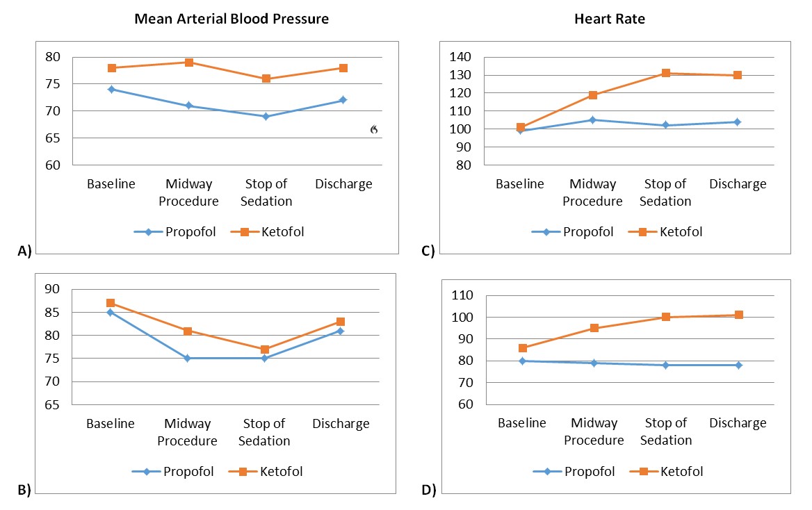
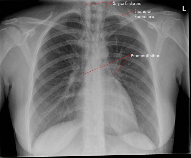
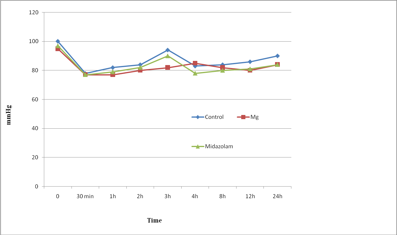
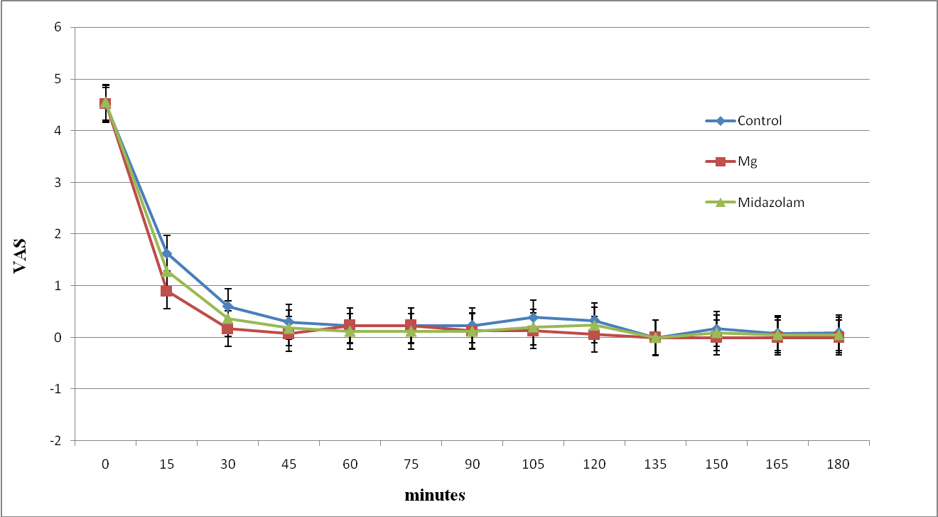
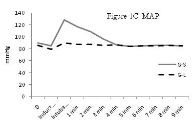
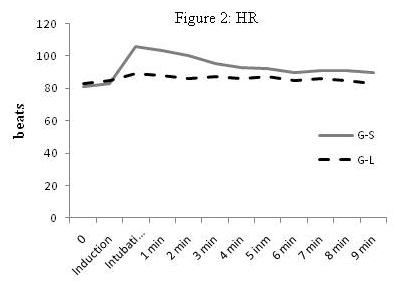
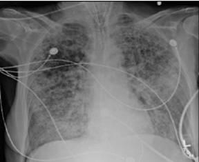
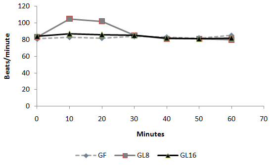
Fig 1: Intra-operative changes in heart rate in groups 
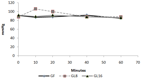
Fig 2: Intra-operative changes in MAP in groups 
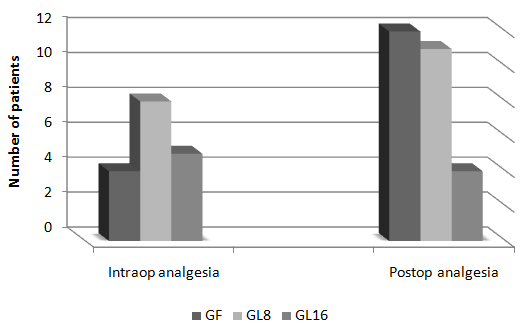
Fig 3: Number of patients requested perioperative analgesic supplementation 
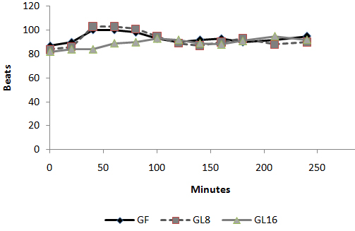
Fig 4: Changes in heart rate in PACU 
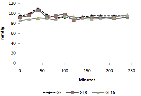
Fig 5: Changes in MAP in PACU: 
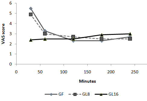
Fig 6: Changes in VAS in PACU Discussion: The use of an opioid, even a short acting one can be associated with adverse effects, which may not be acceptable for patients scheduled for day case surgery. For this reason, it was suggested to substitute an opioid with a non-opioid analgesic for postoperative pain control. The use of a NSAID is associated with adverse effects [1]. Lornoxicam has been successfully used in the prevention and treatment of postoperative pain. It has been shown to be as effective as morphine [4], meperidine [5] and tramadol [6]. To the best of our knowledge, this is the first study to compare the perioperative analgesic efficacy of lornoxicam to fentanyl in patients undergoing day case ENT surgery. We gave Lornoxicam half an hour before induction of anesthesia as the time taken to reach peak plasma concentration (Tmax) was determined to be 0.5 h [7]. During the operative procedure, HR and MAP were significantly higher in group L8 compared to group F and L16. While in PACU, patients in groups F and L8 had higher HR, MAP and VAS score in the early postoperative period compared to patients in group L16. This may be due to inadequate analgesic effect of L8 and the shorter plasma half life of fentanyl compared to L16. The analgesic efficacy of L16 might be attributable to inhibition of cyclo-oxygenase (COX1) and (COX2) activity [2], release of endogenous dynorphin and β-endorphin [5], decrease in peripheral and central prostaglandin production [8] as well as exertion of some of its analgesic activity via the central nervous system [9]. Lornoxicam has a more potent anti-inflammatory and analgesic effect than other oxicams as well as a shorter half life, which decreases the incidence of side effects of drugs with long plasma half life [10]. Arslan and colleagues reported decreased opioid need, PONV and postoperative pain scores when 16 mg of lornoxicam was administered after thyroidectomy [11]. While Xuerong and colleaguessuggested that the increase of postoperative morphine requirements induced by intra-operative administration of fentanyl could be prevented by ketamine or lornoxicam [12].  Rawal reported that NSAIDs are effective as the sole analgesic in a high proportion of cases of mild to moderate pain and it is more convenient to give these drugs by the IV route rather than by IM or rectal administration [13]. The analysis of pain intensity differences was complicated by the fact that many patients postoperatively were asleep at the time their pain assessments were due which may be attributed to effect of opioid and anesthetic medications used. To minimize any missing data we used time to the first dose rescue analgesia (based on changes in hemodynamic data) to evaluate pain intensity differences from baseline. L16 was well tolerated in this study, and was associated with a significantly lower incidence of adverse events than F and L8 which could be due to the opioid side effects in both groups. Norholt and colleaguessupported our results as they reported that, in terms of common acute adverse events, lornoxicam appeared to possess a higher benefit/risk ratio compared with morphine [4]. Zuurmond et al reported that, there is good evidence that avoidance of opioid virtually abolishes the PONV that preclude oral intake of fluids after surgery [14].  In our study, nausea developed in 25.7% of patients in group F, 20% in group L8 but only 8.6% in group L16 who received the least rescue opioid analgesia. Regarding bleeding abnormalities, Hodsman et al reported extensive bleeding required reoperation on two diclofenac group patients submitted to abdominoperitoneal resection of the rectum [15]. In our study no abnormal bleeding was reported by ENT surgeons in any of the study patients. In agreement with our results, Ilias et al[16], Trampitsch et al[17] and Karaman et al [18] used lornoxicam and they did not detect problems with surgical bleeding, bleeding time, blood transfusion requirement or postoperative bleeding.  Stroissnig et al reported that overall, in healthy adult volunteers, oral doses of lornoxicam up to 70 mg have been well tolerated, and there have been no effects on vital signs, urine analysis parameters or clinical serum biochemistry [19]. In our study, none of the patients receiving study drugs experienced severe gastric discomfort, needed rescue antiemetic medication or required admission because of poor pain control.  Previous studies used lornoxicam for reduction of postoperative opioid consumption but none of them had studied the intra-operative use of lornoxicam. So, we selected certain type of surgical procedures which might be suitable to use lornoxicam as a sole intra-operative analgesia. The adjunctive use of acetaminophen may have additive analgesic efficacy to lornoxicam because of its intrinsic opioid-sparing activity. Measurement of serum catecholamine would have been useful. These could be considered as a limitation for the present study. Conclusion:  Intravenous 16 mg lornoxicam with the present study design was comparable to 100 µg fentanyl as intra-operative analgesia but more effective than fentanyl in preventing early postoperative pain in mild to moderate ENT surgical procedures. Intravenous lornoxicam 8 mg was not satisfactory as a sole intra-operative analgesia. The overall incidence of adverse effects of lornoxicam was lower than that of fentanyl.


Acknowledgements / Conflicts / Author Details
Competing Interests: 
None Declared
Details of Authors: 
<p>MOHAMED DAABISS, M.D.Department of Anesthesia, Riyadh Military Hospital, Riyadh, Saudi Arabia. MEDHAT AL-SHERBINY,Department of Anesthesia, Riyadh Military Hospital, Riyadh, Saudi Arabia. RASHID AL-OTAIBI, RIMA AL-NIMAR, Department of Anesthesia, Department of Pharmacy, Riyadh Military Hospital, Riyadh, Saudi Arabia. RIMA AL-NIMAR, Department of Pharmacy, Riyadh Military Hospital, Riyadh, Saudi Arabia</p>
Corresponding Author Details: 
Dr. DAABISS M. Department of Anaesthesia, Riyadh Military Hospital. Personal Mail Box: D186, P.O.Box 7897, Riyadh 11159, Saudi Arabia
Corresponding Author Email: 
madaabiss@yahoo.com
References
References: 


1.        Nuutinen LS, Laitinen JO, Salomaki TE. A risk-benefit appraisal of injectable NSAIDs in the management of postoperative pain. Drug Safety 1993; 9:380-93.2.        McCorrmack K. The evolving NSAID: focus on Lornoxicam. Pain Rev 1999; 6 (4), 262-78.3.        Pruss TP, Stoissnig H, Radhofer-Welte S, et al. Overview of the pharmacological properties, pharmacokinetics and animal safety assessment of lornoxicam. Postgrad Med J 1990; 66 (suppl4):435- 50.4.        Norholt ES., Pedersen S, Larsen U. Pain control after dental surgery: a double blind, randomised trial of lornoxicam versus morphine. Pain 1996; 67: 335-43.5.        Balanika M., Tsitsika M., Wilczynski W. The use of lornoxicam-mepridine combination for postoperative analgesia. Eur J Anaesthiol 2000; 17, 771-8.6.        Staunstrup H, Ovesen J, Larsen T. Efficacy and tolerability of lornoxicam versus tramadol in postoperative pain. J Clin Pharmacol 1999; 39: 834-41.7.        Ankier SI, Brimelow AE, Crome P et al. Chlortenoxicam pharmacokinetics in young and elderly human volunteers. Postgrad Med J 1988; 64: 752–754.8.        Hitzenberger G, Radhofer-Welte S, Takacs F, et al. pharmacokinetics of lornoxicam in man. Postgrad Med J. 1990; 66(Suppl 4): S22-7.9.        Buritova J, Besson JM. Dose-related anti-inflammatory analgesic effects of lornoxicam: a spinal c-Fos protein study in the rat. Inflamm Res 1998; 47 (1), 18-25.10.      Radhofer-Welte S, Rabasseda X: Lornoxicam, a new potent NSAID with an improved tolerability profile. Drug Today 2000; 36: 55- 76.11.     Arslan M, Tuncer B, Babacan A, et al. Postoperative analgesic effects of lornoxicam after thyroidectomy: A placebo controlled randomized study. Experiment clin studies 2006; 18 (2), 27-33.12.     Xuerong Yu, Yuguang H, Xia J, et al. Ketamine and Lornoxicam for Preventing a Fentanyl-Induced Increase in Postoperative Morphine Requirement. Anesth Analg 2008; 107:2032-7.13.     Rawal N. Analgesia for day-case surgery. Br J Anaesth 2001, 87(1): 73-87.14.     Zuurmond WWA, Van Leeuwen L. Alfentanil vs. isoflurane for out-patient arthroscopy. Acta Anaesthesiol Scand 1986; 30: 329–31.15.     Hodsman NB, Burns J, Blyth A, et al. The morphine sparing effects of diclofenac sodium following abdominal surgery. Anesthesia 1987; 42: 1005-8.16.     Ilias W, Jansen M. Pain control after hysterectomy: An observerblind, randomised trial of lornoxicam versus tramadol. BJCP 1996; 50: 197-202.17.     Trampitsch E, Pipam W, Moertl M, et al. Preemptive randomized double-blind study with lornoxicam in gynecological surgery. Schemerz 2003; 17: 4-10.18.     Karaman Y, Kebapci, Gurkan A. The preemptive analgesic effect of lornoxicam in patients undergoing major abdominal surgery: A randomized controlled study. Int J Surg 2008; 6(3): 193-6.19.     Stroissnig H, Frenzel W. Lornoxicam. A novel highly potent anti-inflammatory and analgesic agent. Clinical Investigator's Brochure.6th ed. Vienna Hafslund Nycomed Pharma AG, 1992.


A case of accidental carotid artery cannulation in a patient for Hemofilter: complication and management

Authors
Sanil Nair, Harshal Wagh, Kavita Mordani and Salim Bhuiyan
Article Citation and PDF Link
BJMP 2009: 2(3) 57-58
Abstract / Summary
Keywords: 
Carotid puncture, Haemofiltration, Surgical Repair

Inadvertent carotid arterial puncture complication is reported to have an incidence of 2% - 8% 1, 2 and usually results in localised haematoma formation.

Case presentation

We present a case of a 72 year old man with acute on chronic renal failure referred for intensive care (ITU) management. History revealed dehydration and anuria for 3 days. His blood pressure was 80/40, sinus tachycardia around 130/min with cold clammy peripheries. Blood investigations revealed urea 23mmol, creatinine 800mmol, and potassium-7.7, with ECG changes of hyperkalemia. We attempted a right internal jugular (IJV) venous cannulation using landmark technique and inserted an Arrow-Howes central venous catheter with blue flexi tip, 8.5FR, 5 lumen, and 16cm length. The technique was difficult due to low blood pressure, and patient’s inability to lie flat. The ultrasound machine was unavailable at that moment.Soon after the catheter was introduced using seldinger technique, a swelling was noticed from the site of IJV cannulation .the blood sample analysis revealed arterial blood. The catheter was removed and pressure applied for 45 minutes. Thereafter, a left femoral vein catheter was inserted for central venous access and right femoral vascath –VYGON, DUALYSE Expert, 2 lumen catheter set, 12 fr, 20cm was inserted uneventfully.1 hour later the bleeding from the right carotid puncture site was examined and was found to have ceased. The patient was put on the continuous veno- venous haemofiltration (CVVHF), which clotted after 15minutes.Heparin was used as an anticoagulant for the filter. This was repeated thrice with same result. Three hours later, a haematoma was noticed in area of IJV puncture, which was gradually increasing in size. The haematoma spread to the neck and chest after which the patient complained of discomfort with breathing difficulty. In view of increasing airway compromise the patient was transferred to theatre for elective intubation and surgical intervention. Ultrasonongraphy by radiology consultant showed a large haematoma, but unable to see the damage in the vessel and a CT neck, thorax angiography was suggested.

CT angiogram revealed a leak in the bifurcation of the carotid artery. Surgical access was difficult in view of the large persistent leak. Ultrasound probe was used to make the incision superficial to the leak. A small leak was identified in the artery, which was sutured uneventfully. He was then continued on CVVHF with no further complications Discussion Complications include arterial puncture, pneumothorax, neck or mediastinal haematoma and hemothorax3, 4. The haematoma may enlarge rapidly if the patient is coagulopathic, or if a large puncture wound is produced by the introduction of the sheath itself into the carotid artery. Airway obstruction 5, 6, pseudo aneurysm 7, 8, arterio-venous fistula formation 9 and retrograde aortic dissection 10 have all been reported as a consequence of carotid puncture. In the presence of occlusive (atheromatous) carotid disease, inadvertent puncture may carry the risk of precipitating a cerebrovascular accident 11. NICE guidelines 2002 recommended use of ultrasound for central venous cannulation12. The Royal College of Radiology (RCOR) recommends practical training in vascular ultrasound should involve at least two ultrasound lists per week over less than three months up to 6 months. Meta-analysis including 18 Randomised Controlled Trials by NICE concluded that ultrasonongraphic (USG) venous catheterisation was more effective than landmark technique for all outcomes 13 Royal College Of Anaesthetists feels guidance is fair and sensible but landmark technique is still acceptable alternative whether 2D available or not. Conclusion Complications following central venous cannulation are reduced if the procedure is USG guided and therefore must be used whenever possible. But landmark technique is still an acceptable alternative.

Acknowledgements / Conflicts / Author Details
Competing Interests: 
None Declared
Details of Authors: 
<p>SANIL NAIR MBBS,DA Trust anaesthetist, Albans hospital West Herts NHS trust AL3 5PN<br /> HARSHAL WAGH MBBS,FRCA, Registrar Anaesthetics, St Albans Hospital West Herts NHS Trust, Waverley road AL3 5PN.<br /> KAVITA MORDANI MBBS,FRCA Consultant Anaesthetics St Albans Hospital West Herts NHS Trust Waverley Road AL3 5PN<br /> SALIM BHUIYAN MBBS FRCA Consultant Anaesthetics St Albans Hospital West Herts NHS Trust. Waverley road AL3 5PN</p>
Corresponding Author Details: 
SANIL NAIR MBBS,DA Trust anaesthetist, St Albans hospital West Herts NHS trust AL3 5PN
Corresponding Author Email: 
docsanil@gmail.com
References
References: 

1. Davies MJ, Cronin KD, and Domaingue CM Pulmonary artery catheterization: an assessment of risks and benefits in 220 surgical patients. Anaesth Intensive Care 10:9, 1982.
2. Patel C; Laboy V; Venus B; Mathru M; Wier D Acute complications of pulmonary artery catheter insertion in critically ill patients. Crit Care Med, 14(3):195-7 1986 Mar
3. Abbound P, Kendall J.Ultrasound guidance for vascular access.Emerg Med Clin North Am 2004; 22:749-73
4..Randolf AG,Cook DJ,Gonzales CA, et al. Ultrasound guidance for placement of central venous catheters. A meta-analysis of the literature.Cric care Med. 1996; 24:2053-8
5. Knoblanche GE Respiratory obstruction due to haematoma following internal jugular vein cannulation. Anaesth Intensive Care 7: 286, 1979
6. Kua JS; Tan IK Airway obstruction following internal jugular vein cannulation. Anaesthesia, 52:776-80, 1997
7. Shield CF, Richardson JD, Buckley CJ, et al: Pseudoaneurysm of the brachiocephalic arteries: A complication of percutaneous internal jugular vein catheterization. Surgery 78:190-193, 1975
8. Aoki H. Mizobe T. Nozuchi S. et al: Vertebral artery pseudoaneurysm: A rare complication of internal jugular vein catheterization. Anesth Analg 75:296-298, 1992
9. Gobeil F. Couture P. Girard D, Plante R: Carotid Artery-lnternal Jugular Fistula: Another Complication following Pulmonary Artery Catheterization via the Internal Jugular Venous Route. Anaesthesiology 1994, 80:23-232.
10. Applebaum RM, Adelman MA, Kanschuger MS, Jacobowitz G, Kronzon I Transesophageal echocardiographic identification of a retrograde dissection of the ascending aorta caused by inadvertent cannulation of the common carotid artery. J Am Soc Echocardiogr 1997 Sep;10(7):749-51
11. Zaidi NA, Khan M, Naqvi HI, Kamal RS Cerebral infarct following central venous cannulation. Anaesthesia, 53:186-91, 1998 Feb
12. NICE guidelines on use of ultrasound devices for placing central venous catheters. Technology appraisal guidance no-49 September 2002.
13. Hind D Calvert N, McWilliams R, et al. Ultrasound locating devices for central venous cannulation: meta-analusisi.BMJ 2003, 32:361-7. 


Anaesthetic management of a patient with Klippel-Feil syndrome for elective Caesarean section

Authors
MK Kada Venkata, Jameel Ahmed Khan, Muhammad Tayyab Qureshi, Asif Qureshi and Imtiaz Kar
Article Citation and PDF Link
BJMP 2009: 2(3) 54-56
Abstract / Summary
Abstract: 

Klippel-Feil syndrome is an inherited autosomal dominant condition and is associated with various anomalies including short neck, complete fusion of the cervical spine and severe restriction of neck movements which can cause difficulty in securing the airway. The spinal deformities pose a challenge for positioning and regional anaesthesia. We present a case of a primigravida scheduled for an elective Caesarean section successfully managed with a general anaesthetic technique using awake fibreoptic technique for endotracheal intubation.

Case report

We describe a case of a 32 year old primigravida with type III Klippel-Feil Syndrome for an elective Caesarean section. A preanaesthetic assessment at 32nd week of pregnancy revealed a history of mild asthma for which the patient was on regular salbutamol inhaler. She had no history of cardiovascular or respiratory insufficiency. There was no significant family history. Anaesthetic history included a repair of encephalomyelocele immediately after birth and a squint correction surgery at the age of 6 years under general anaesthesia which was uneventful.

 Examination revealed a woman of small stature, 137 cm in height and weighing 52 kg. Airway examination revealed Mallampati III with a normal mouth opening, a reduced thyromental distance (3 cm) and inability to flex or extend the neck (Figure 1). She had thoraco-lumbar kyphoscoliosis and examination of the respiratory and cardiovascular system was unremarkable. Electrocardiogram was within normal limits as were haemoglobin, urea and electrolyte estimation. Radiography revealed fusion of atlanto-axial joint (flexion and extension views, Figure 2 ) and also fusion of lumbar vertebrae with associated scoliosis (Figure 3Cobb’s angle 25 degree) and Cobb’s angle 25 degree Figure 1. A 32 year old parturient with Klippel-Feil syndrome with a short webbed neck and severely restricted neck movements  
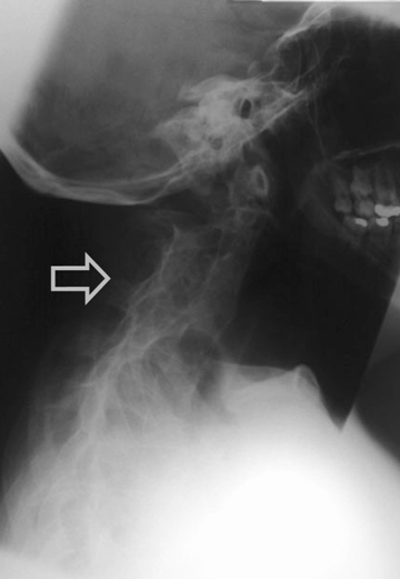
Figure 2. Radiograph of a Lateral view of cervical spine showing fusion of atlanto - occipital joint and cervical vertebrae 
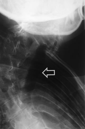
Figure 3. Antero - posterior view of dorsal spine showing deviation of trachea and marked kyphoscoliosis of thoracic spine She was scheduled to have an elective Caesarean section. The anaesthetic management options in this case included either a general anaesthetic with an anticipated difficult endotracheal intubation or a regional anaesthetic. After discussion with the patient, we planned a general anaesthetic technique with awake intubation using a fibreoptic bronchoscope (FOB) as we considered this safe. For FOB through oral route was preferred due to narrow anterior nares. Oral Ranitidine 150 mg was administered as a premedication the night before and on the morning of the planned section. After institution of standard monitoring and securing two peripheral intravenous cannulae (18G & 16 gauges), the upper airway was anaesthetised with nebulisation of 4% Lignocaine (5 ml) and 10% Lignocaine spray to the posterior pharynx. The FOB was passed through the Berman airway by using a ‘spray as you go” technique to anaesthetise the larynx and upper trachea using 4% Lignocaine and keeping well below the toxic dose (3 mg/kg). A 6.5 mm ID endotracheal tube was rail roaded over the FOB prior to its insertion and the airway was successfully secured. Once position of the endotracheal tube was confirmed, anaesthesia was then induced using intravenous Thiopentone 200 mg, Alfentanil 0.5 mg followed by Rocuronium 25 mg. Anaesthesia was maintained using oxygen with nitrous oxide (1:1 ratio) and Sevoflurane (1 MAC). Patient was ventilated with intermittent positive pressure ventilation to maintain normocapnia. The surgery lasted for 45 min and was uneventful. Using a nerve stimulator for assessing neuromuscular blockade she was reversed with Neostigmine 2.5 mg and Glycopyrrolate 0.5 mg towards the end of surgery. The patient was extubated in supine, head-low position when fully awake and in presence of protective airway reflexes. Discussion Klippel-Feil syndrome is an inherited autosomal dominant condition. In 1912, Klippel and Feil 1 first reported on a patient with a short neck, a low posterior hairline, and severe restriction of neck movements due to complete fusion of the cervical spine. These features now constitute the classic clinical triad which is the hallmark of Klippel-Feil syndrome. A great number of other anomalies associated with Klippel-Feil syndrome may pose a threat to the patient than the obvious deformity of neck. The spinal deformities may cause difficulties with both tracheal intubation and regional anaesthetic techniques. Anaesthetic management may therefore be challenging in these patients. In our case we opted for a general anaesthetic technique rather than a regional technique because of the following reasons: firstly, the patient was not keen to have regional anaesthesia, secondly, it would entail difficulty for regional anaesthesia keeping in view the spinal fusion and scoliosis and thirdly, the dose of a single bolus of spinal anaesthetic would be difficult to judge in this patient and epidural anaesthesia 2 might prove technically difficult and is associated with an increased risk of inadvertent dural puncture and poor spread within the epidural space. This patient’s abnormalities posed problems for all the commonly used anaesthetic techniques for Caesarean section. General anaesthesia could be complicated by difficult intubation. While greater use of regional anaesthesia may have reduced the number of deaths due to failed intubation in obstetric practice, several complications of epidural and spinal anaesthesia may still require intubation as part of their management. Some of these include total or high spinal anaesthesia, inadvertent intravascular injection, overdose of local anaesthetic, anaphylaxis and failure. For these reasons, the choice of regional anaesthesia for a patient with known difficult airway does not necessarily bypass the problem of unanticipated intubation. The most commonly associated anomaly in a series of 50 patients from Delaware, USA 3 was scoliosis (60% of cases), renal abnormalities (35%), Sprengel deformity (30%), deafness (30%), synkinesia (20%) and congenital heart disease (14%). The most common heart disease variant was ventricular septal defect. Less commonly associated were ptosis, lateral rectus palsy, facial nerve palsy and upper extremity anomalies. There are 3 variants of Klippel-Feil Syndrome. 4 Type I is an extensive abnormality where elements of several cervical and upper thoracic vertebrae are incorporated into a single block. In Type II variant, failure of complete segmentation occurs at one or two cervical interspaces. Type III variant includes Type I or II deformities with coexisting segmentation errors in the lower thoracic or lumbar spine. The incidence of Type II abnormalities was found to be 0.71% of Black and Caucasian skeletons that were between the ages of 17 and 102 years in a study from St Louis, Missouri, USA 5 and it is considered to be the most common form. C2-3 and C5-6 are the interspaces usually involved. It often remains unrecognised since the neck may appear normal and the patients are asymptomatic until later in life, when they present due to their increased susceptibility to cervical osteo-arthritis. Patients with Type I abnormalities are 50 times less common than Type II but are reported more frequently. This is probably because they exhibit the classic triad and thus have bizarre appearances. 6 These patients are frequently disabled by birth injuries, or have major anomalies in other organ systems. A planned elective section at term was considered as the best option for delivery of the baby by the obstetricians due to severe cephalo-pelvic disproportion and also due to anaesthetic issues regarding management. The case was managed successfully with a favourable outcome both for the mother and the baby.

Acknowledgements / Conflicts / Author Details
Competing Interests: 
None declared
Details of Authors: 
<p>MK KADA VENKATA.MD, FRCA, FFARCSI. Consultant Anaesthetist, Ulster Hospital, Belfast, UK<br /> JAMEEL AHMED KHAN. MD, FCARCSI. Specialty Registrar Anaesthetics, Royal Victoria Hospital, Belfast, UK<br /> MUHAMMAD TAYYAB QURESHI. FFARCSI. Consultant Anaesthetist, Altnagelvin Hospital, Londonderry, UK<br /> ASIF QURESHI. MBBS. Staff Grade Anaesthetist, Altnagelvin Hospital, Londonderry, UK.<br /> IMTIAZ KAR. MBBS, MD. Staff Grade Anaesthetist, Altnagelvin Hospital, Londonderry, UK</p>
Corresponding Author Details: 
JAMEEL AHMED KHAN, ST-4 Anaesthetics, Royal Victoria Hospital, Belfast, UK
Corresponding Author Email: 
drjameel@hotmail.co.uk
References
References: 

1.  Robert NH, John EL, Dean M. Klippel-Feil Syndrome: A constellation of associated anomalies. J. Bone Joint Surg. Am 1974, 56A: 1246-53
2. Dresner MR, Maclean AR. Anaesthesia for Caesarean section in a patient with Klippel-Feil syndrome. Anaesthesia 1995, 50:807-09
3. Hensinger RN, Macewen GD. Congenital anomalies of the spine. In: Rothman RH,  Simeone FA, eds. The spine. Philadelphia: W. B. Saunders, 1982: 216-33
4. Morrison SDG, Perry LW, Scott LP. Congenital brevicolis (Klippel-Feil syndrome) and cardiovascular anomalies. American Journal of Diseases of Children 1968; 115: 614-20
5. Brown MW, Templeton AW, Hodges FJ. The incidence of acquired and congenital fusions in the cervical spine. American Journal of Roentgenology, Radiotherapy and nuclear Medicine 1964; 92: 1255-9
6. Helmi C, Pruzansky S. Craniofacial and extracranial malformation in the Klippel-Feil syndrome. Cleft Palate Journal 1980; 17: 65-88

Pictorial essay: central venous catheters on chest radiographs

Authors
Krishnan Melarkode and M Y Latoo
Article Citation and PDF Link
BJMP 2009:2(2) 55-56


Chest radiographs are one of the most common radiological procedures performed in medical practice. The chest radiograph should ideally include views of the heart, lungs, trachea, mediastinum, bones of the chest and upper part of the abdomen. Chest radiographs are normally taken in the posterior-anterior (PA) view with the patient in upright / standing position but for patients admitted in the intensive care unit (ICU) or other emergency situations, this is not possible and so they are taken in the supine (anterior-posterior views) or semi-erect position.

Chest radiographs are done not only for diagnostic reasons to look for abnormalities in the lungs, soft tissues and bones but also to check the position of various invasive lines and tubes. In this article, we aim to discuss and compare the normal and abnormal positions of central venous catheter (CVC) on chest radiographs.

Indications for Central Venous Catheter (Internal Jugular Vein Cannulation)

There are many indications for central venous cannulation 1. These include:

  • Central venous pressure (CVP) monitoring
  • Pulmonary artery catheterisation and monitoring
  • Transvenous cardiac pacing
  • Administration of drugs (vasoactive drugs, chemotherapy etc)
  • Aspiration of air emboli
  • Administration of fluids (in case of difficult peripheral venous access)

Confirming the position of the central venous catheter tip:

For accurate CVP measurement, the tip of the central venous catheter (CVC) should lie within the superior vein cava (SVC), above its junction with the right atrium and parallel to the vessel walls 1. After insertion of a CVC, the position of the catheter tip must be confirmed radiologically, as catheter tips located within the heart can cause cardiac perforation and tamponade 1. Hence, optimum positioning of the CVC tip is required to prevent complications.

If the CVC tip is situated high up (above the pericardial reflection), this can cause vessel wall erosion and if they are very low (in the right atrium), they can cause arrhythmias, placement in the coronary sinus and damage to the tricuspid valve 2.

The carina is a useful radiological landmark for CVC tip position. In this edition of pictorial essay, we aim to discuss the optimum position of both the right and left sided IJV cannula on chest radiographs.


Fig: 1 (CR-1819) shows the normal position of a right sided IJV catheter. The tip of the right sided IJV cannula should ideally lie just above the level of the carina 2. This is the junction of the left and right innominate veins with the superior vena cava (SVC).


Fig: 2 (CR-1829): The optimum position of the left sided IJV cannula is at or just below the level of the carina 2. This radiograph shows the comparison between the right and left sided IJV cannula in the same patient.

The right sided IJV cannula is too low (below the level of the carina) and is probably in the right atrium while the tip of the left sided IJV cannula is optimally placed.


Fig: 3 (CR-1832): In this radiograph, the right sided IJV cannula is too high in the neck. This will not give an accurate CVP measurement. Besides, there is also a risk that the CVC might get dislodged and lead to extravasation of administered fluids and drugs.

Seldinger technique for CVC insertions:

The CVC’s are usually inserted using the Seldinger technique. The IJV can be located by using anatomical landmarks or under direct vision with the help of an ultrasound machine. In the Seldinger technique, after puncture of the IJV, a thin J-shaped guide wire is introduced through the puncture needle. The needle is then slowly withdrawn leaving the J-shaped guide wire in place. A dilator is then introduced over the guide wire to dilate the skin and the subcutaneous tissue. Next, the dilator is removed and the CVC is introduced over the guide wire. Finally, it is important that the guide wire is removed and the CVC is secured.


Fig: 4 (CR-1831). This chest radiograph shows an unusual complication where the guide wire has been left accidentally in situ on the right side. (Note the presence of the J-shaped guide wire on the right side of the neck). This can result in serious complications if the guide wire migrates distally.

Conclusion:

In this article, we have highlighted the optimum placement of central venous catheters on chest radiographs. It is imperative that after every CVC insertion (via the IJV or subclavian vein), the position of the tip be confirmed radiologically and if any re-positioning is required, it must be done. The above discussion is true for even CVC’s inserted through the subclavian veins.


Self Assessment
MCQ:

The tip of the right sided IJV cannula should be located
a. below the level of the carina
b. at the level of the clavicle
c. just above the level of the carina
d. in the right atrium


Answer: c


ACKNOWLEDGEMENTS
We wish to thank the Department of Radiology in Bedford Hospital for helping us with the chest radiographs.
COMPETING INTERESTS
None Declared
AUTHOR DETAILS
DR. KRISHNAN MELARKODE, MD DNB FRCA, Specialist Registrar in Anaesthesia, Bedford Hospital NHS Trust, UK
DR. M Y LATOO, MBBS FRCA, Consultant Anaesthetist, Bedford Hospital NHS Trust, UK
CORRESPONDENCE: Dr. Krishnan Melarkode, Specialist Registrar in Anaesthesia, Bedford Hospital, Bedford, UK
Email: drkrishnanmr@gmail.com


References

  1. Mark JB, Slaughter TF and Gerald Reves J. Cardiovascular monitoring. In: Miller RD ed. Anesthesia. 5th edition. Churchill Livingstone; 1144-51.
  2. Stonelake PA and Bodenham AR. The carina as a radiological landmark for central venous catheter tip position. British Journal of Anaesthesia 2006; 96: 335-340

Assessment of different concentration of Ketofol in procedural operation

Authors
Mohamed Daabiss, Medhat Elsherbiny and Rashed Alotibi
Article Citation and PDF Link
BJMP 2009:2(1) 27-31


Abstract

Propofol is an IV anesthetic that is often used as an adjuvant during monitored anesthesia care, the addition of ketamine to propofol may counteract the cardiorespiratory depression seen with propofol used alone. Ketofol (ketamine/propofol combination) was used for procedural sedation and analgesia. However, evaluation of the effectiveness of different concentrations of Ketofol in procedural operation regarding changes in haemodynamics, emergence phenomena, recovery time, the doses, and adverse effects was not yet studied, so this randomized, double blinded study was designed to compare the quality of analgesia and side effects of intravenous different concentrations of ketofol in hundred children of both sex undergoing procedural operation, e.g. esophgoscopy, rectoscopy, bone marrow aspiration and liver biopsy participated in this randomized, double-blinded study. Patients received an infusion of a solution containing either combination of propofol: ketamine (1:1) (Group I) or propofol: ketamine (4:1) (Group II). Subsequent infusion rates to a predetermined sedation level using Ramsay Sedation Scale. Heart rate, noninvasive arterial blood pressure (NIBP), oxygen saturation (SpO2), end tidal carbon dioxide (Etco2) and incidence of any side effects were recorded. There were no significant hemodynamic changes in both groups after induction. However, there was an increase in postoperative nausea , psychomimetic side effects, and delay in discharge times with the largest ketamine dosage (Group I). We concluded that the adjunctive use of smaller dose of ketamine in ketofol combination minimizes the psychomimetic side effects and shorten the time to discharge.

Key words: ketofol, procedural operation, psychomimetic effect.

Procedural operations, are procedures outside the operating room, which developed from a facilitation of diagnostic and therapeutic procedures into an independent subspecialty. Procedural sedation and analgesia is a minimally depressed level of consciousness that retains the patient's ability to maintain a patent airway independently and continuously(1).

Propofol is a short-acting intravenous sedative agent used for the induction and maintenance of general anesthesia for adults and children, sedation for intubated, mechanically ventilated adults in Intensive Care Units (ICU), and in procedures such as colonoscopy. It provides no analgesia(2). Ketamine is classified as an NMDA receptor antagonist and has also been found to bind to opioid receptors and sigma receptors. It induces a state referred to as "dissociative anesthesia"(3).

Ketofol (ketamine/propofol combination) was used for procedural sedation and analgesia. Ketamine and propofol are physically compatible for 1 hr at 23oC with no increase in particle content at Y site injection (4). Ketamine and propofol administered in combination have offered effective sedation for spinal anesthesia and for gynecologic, ophthalmologic, and cardiovascular procedures in all age groups. The opposing hemodynamic and respiratory effects of each drug may enhance the utility of this drug combination, increasing both safety and efficacy and allowing reduction in the dose of propofol required to achieve sedation.

However, evaluation of the effectiveness of different concentrations of Ketofol in procedural operation regarding changes in hemodynamic, emergence phenomena, recovery time, the doses, and adverse effects was not yet studied, so this randomized, double blinded study was designed to compare the quality of analgesia and side effects of intravenous different concentrations of ketofol in children scheduled for procedural operations.

Methods:

A hundred patients, American Society of Anesthesia (ASA) class I or II scheduled for procedural operation, ages 3 to 12 years were enrolled in this study. After obtaining approval from the local research ethics committee, all patients and their parents were informed about the procedure and the anesthetic technique and an informed written consent was obtained from each. Patients with clinically significant cardiovascular, respiratory, hepatic diseases or epileptic patients, longer procedures more than one hour and sensitivity to the drugs were excluded from the study.

Patients were randomized into two equal groups each of 50 patients going for procedural operation, e.g. esophgoscopy, rectoscopy, bone marrow aspiration and liver biopsy. Pre procedural visit was done to evaluate if that patient fulfils the criteria of study and for fasting instruction. In the preoperative waiting area, an IV catheter was placed after applying emla cream. Baseline measurements included Non Invasive Blood Pressure (NIBP), heart rate, respiratory rate, and pain faces scale which is recommended for children aged 3 years and older (Fig1)(5). The level of sedation was determined by Ramsay Sedation Scale(6). A separate observer who was blind to the drug combination being used assessed the depth of sedation of such patients.

*Ramsay Sedation Scale(6)

Patient is anxious and agitated or restless, or both

Patient is co-operative, oriented, and tranquil

Patient responds to commands only

Patient exhibits brisk response to light glabellar tap or loud auditory stimulus

Patient exhibits a sluggish response to light glabellar tap or loud auditory stimulus

Patient exhibits no response


Fig 1: Pain Faces Scale.

A propofol/ketamine admixture was prepared by an assistant who was not involved in the clinical management of the study patients. According to a prestudy randomization schedule of study group assignment, a ketofol (1:1): propofol 14.285 mg, ketamine 14.285 mg/ml by mixing 10 ml propofol 2% (20 mg/ml) with 4 ml ketamine (50 mg/ml) in group I, while in group II ketofol (4:1): propofol 18.1818 mg , ketamine 4.5454 mg/ml by mixing 10 ml propofol 2% (20 mg/ml) with 1 ml ketamine (50 mg/ml).

Both bolus and maintenance doses were given using syringe pump (B/Braun). Set up for delivery of Ketofol in both groups as an initial bolus of 600 g/kg IV (calculated as dose of propofol in these infusions for simplicity), followed by an initial maintenance infusion at 100 g/kg/min by anesthetist who was blinded to the identity of study infusion.The level of sedation was assessed at 1- 3 minutes intervals, and the initial infusion rate was adjusted (in 25 g/kg/min increments) to achieve Ramsay Sedation Scale of 5 before starting the procedure.

During the procedure, patients were kept on oxygen nasal prongs with a CO2 sampling port. All patients were monitored with NIBP, electrocardiography (ECG), Pulse oximetry(SpO2), heart rate (HR), and end tidal carbondioxide( EtCO2). The measurement started before commencement of the intravenous (IV) line and continued five minutes after induction. The patients were also assessed for apnoea, which was defined as the loss of respiratory efforts for more than 20 seconds or fall of SpO2 below 95%. Complaints of pain/discomfort were treated by an incremental increase in the study drug infusion rate.

The study drug infusion was discontinued at the end of the surgical procedure, and the total drug requirements were noted. After the completion of the procedure, patients were transferred to recovery room when an Aldrete score(7) 9-10 was confirmed, and kept there till ready for discharge. The recovery room nurses were blinded to the study medication received by the patients. The incidence of any episode of postoperative nausea and vomiting (PONV) or any other side effects (e.g. hallucinations, agitation or pain) was noted. The patients vital signs were assessed at 5-min intervals. Patients were considered "ready for discharge" when they had stable vital signs, oriented, able to ambulate unassisted, had no intractable nausea or vomiting, and had minimal pain. Discharge times were determined from the time the study drug infusion was discontinued. Outpatients were given written discharge instructions regarding post-procedure precautions and a telephone number to use in case of emergency.

Descriptive variables were analyzed using Students t-test and X2 test as appropriate using SPSS software statistical computer package version 15. Differences between the groups in mean blood pressure (BP),heart rate( HR), end-tidalCO2, oxygen saturation and ketofol requirements were compared using analysis of variance with repeated measures. A P value < 0.05 was considered to be statistically significant. Values are expressed as meanSD.

Results:

There were no significant differences among patients in both groups regarding number of patients, age, sex, weight, ASA physical status, and duration of ketofol infusion (table 1). There was 2 patients excluded in group I; one had a history of epilepsy and the other was acute lymphocytic leukemia on corticosteroids, while in group II only one patient was excluded due to history of epilepsy (Table 1).

Sedation scores were similar in both groups. The average ketofol initial dose in group I was 600 g /kg followed by an average infusion rate of 116 24 g/kg/min, while in group II the average ketofol initial dose was 600 g/Kg followed by an average infusion rate of 132 36 g/kg/min with a significant difference between groups (P <0.05).

There was a minimal decrease in mean arterial blood pressure (MAP) from baseline in both groups following the initial dose of ketofol. Significant hypotension was noted in 2 patients in group I (4%) and in 3 patients in group II (6%) which was corrected by a bolus of Ringers solution 10 ml/kg IV. The diffrence between the groups was statistically insignificant . All the patients had increase in pulse rate compared to the baseline. The change was least in group II (p <0.05), but no patient had severe tachycardia requiring treatment in both groups.

There was increase in Etco2 in both groups after induction with statistically insignificant difference between groups. Patients in both groups had decrease in arterial oxygen saturation (SpO2) after induction. Five patients (10%) in group I and three patients (6%) in group II had apnea and hypoxia after induction (SpO2 <95%). Excessive salivation was noted in 15 patients (31%) in group I but only two patients (4%) in group II. Eight patients (16%) in group I and two patients (4%) in group II experienced airway obstruction or apnea which required airway support. These changes were statistically significant between both groups.

Table 1. Demographic Characteristics, Intraoperative Management, and Recovery Times of Patients in the Study Groups


Group 1

Group 2

Number

50

50

Age (yr)

6.6 3.6

7 3.1

Weight (kg)

21 8

23 7.5

ASA physical status

(III)

(III)

Duration of infusion (min)

25.312.4

23.8 14.8

Average propofol concentration (mg/ml)

14.3

18.18

Average ketamine concentration (mg/ml)

14.3

4.54

ketofol bolus dose (g /kg)

600

600

Average Ketofol infusion rate (g/kg/min)

116 24

132 36*

Time to ambulation (min)

15.4 9.5

8.2 6.7*

Ready for discharge (min)

26.511.3

15.3 8.4*

Time to actual discharge (min)

38.813.5

28.2 8.9*

-Data are mean SD, median (range).

-Group I propofol/ketamine (1:1), Group II = propofol/ketamine (4:1)

-ASA = American Society of Anesthesiologists.

-Average bolus dose were calculated as dose of propofol in infusion. Average infusion rates were calculated as total drug (propofol) divided by weight and case duration.

* Significant difference (P < 0.05) versus group I.

Two patients in group II (4%) and one patient in group I (2%) had pain and discomfort during the procedure which was overcome by incremental boluses of infusions. In group I, one patient (2%) complained of postoperative nausea, four patients (8%) experienced bad dreams and hallucinations and five patients (10%) complained of agitations with no psychomimetic changes in group II

The time to ambulation in group I and II patients was 15.4 9.5 and 8.2 6.7 minutes respectively, while readiness to discharge was 26.5 11.3 in group I and 15.3 8.4 minutes in group II. The time to actual discharge was 38.8 13.5 minutes in group I and 28.2 8.9 minutes in group II. These changes in recovery timings were statistically significant.

Discussion:

The goals of procedural sedation are to provide an adequate level of sedation while minimizing pain and anxiety, maximizing amnesia, minimizing the potential for adverse drug-related events, controlling behavior, and maintaining a stable cardiovascular and respiratory status. A number of studies have demonstrated that the combination of ketamine and Propofol (ketofol) for sedation is safe and effective. The combination of the two agents appears to reduce side effects of each medication used alone, and allows for a rapid recovery time(1).

We compared the safety and efficacy of different concentrations of ketofol in procedural operations in children. The rate of ketofol infusion in group II was higher than in group I due to due to incremental doses of ketofol given to get the desired depth of sedation and abort pain sensation which was due to less ketamine content in such infusion compared to group I. Propofol in the recommended dose of 2-2.5 mg/kg almost always causes fall in blood pressure and the extent of fall depends upon the dose and adjuvant drugs used. Because we used an initial infusion dose of only 600 g/kg, the fall in MAP was mild (6%) and similar in both groups. The induction doses of propofol are reduced considerably by combination with small doses of ketamine. Ketamine had the additional advantage of better hemodynamic stability. Our results are consistent with Furuya et al and Hui et al who suggested that the minimal change observed in arterial pressure may be dose related and also because sympathomimetic actions of ketamine were effective in counter-acting the hemodynamic depression of propofol. There was a trend for pulse rate to increase after the induction in all the groups, but there was no occurrence of profound tachycardia in any group(8,9).

Akin et al published a trial of 60 patients between one month and 13 years of age undergoing cardiac catheterization who received sedation with propofol or propofol plus ketamine (3:1). They found a significant (decrease in MAP in 11 patients in the propofol monotherapy group and three patients in the ketofol group. They concluded that the addition of low-dose ketamine to propofol preserved MAP without prolonging recovery or increasing the incidence of adverse events(10). While, Goh et al published a 90 patients having a laryngeal mask airway (LMA) placed received propofol with either ketamine (5:1), fentanyl (1 g /kg), or placebo normal saline. They found the ketofol group had a significantly higher systolic blood pressure than the other two groups. They concluded that ketofol provided equivalent LMA insertion conditions while maximizing hemodynamics and minimizing apnea(11).

End-tidal CO2 increased slightly after induction in both groups. In agreement with our results, Mildh et al and Persson et al who reported that ketamine-induced sympathoadrenal activation may account for improved ventilation, also arousal secondary to the subjective side effects of ketamine (e.g., perceptual changes and anxiety) may also contribute (12,13). Also our results have confirmed the previous reports of Frey et al and Badrinath et al (14,15), suggesting that the combination of a small-dose ketamine with propofol improves ventilation during sedation.

We expect that the apnea and desaturation recorded in group I (10%) was due to the excessive salivation complicated the higher dose of ketamine in this group which led to impaired breathing and required airway support in 16% of such patients. While apnea and desaturation which happened in group II could be due to the higher infusion rate of propofol in ketofol combination.

Willman and Andolfatto published a study of 114 patients requiring procedural sedation and analgesia mainly for orthopedic procedures were given a 1:1 mixture of propofol and ketamine. Transient hypoxia occurred in 2.6% of patients, out of them one patient required bag valve mask ventilation. Three patients had an emergence reaction, one of whom received midazolam. No patient had vomiting or aspiration. Procedural success rate in this study without the use of adjunctive medications was 96.5%. Median time until recovery was 15 minutes (range 5 to 45 minutes) (16). Furthermore, Akin et al compared propofol to propofol plus ketamine (3:1) in 60 patients between one and 13 years of age undergoing auditory brainstem response testing. There were no cases of desaturation in the ketofol group, but in the propofol group 4/30 experienced desaturation and 6/30 had apnea. The authors concluded that the addition of low dose ketamine to propofol reduced the risk of respiratory depression and the need for repeat medication administration(17).

The incidence of clinically significant psychotomimetic effects was noted in the large-dose ketamine group (group I). This could be a dose-dependent interaction of the excitatory anesthetic ketamine with a pure central nervous system depressant, such as propofol (18,19). There were no post procedural psychotomimetic symptoms recorded in group II. In addition, the patients mood was significantly better in the recovery room and cognitive function recovered more rapidly in such group than those given higher dose of ketamine. Nagata et al and Mortero et al are coinciding with our results as they suggested that ketamine in sedative doses is associated with electroencephalographic activation. Furthermore, small-dose ketamine increases thalamic sensory output and arousal. Sedative effects of propofol may be partially antagonized by the arousal effects of ketamine(20,21). While Akin et al in a trial of 40 adult patients undergoing endometrial biopsy, reported that the combination of propofol (1 mg/kg) plus fentanyl (1 g/kg) was compared to the combination of propofol plus ketamine (2:1). Time to recovery was similar; however time to discharge was longer in the ketofol group secondary to the increased presence of adverse events including nausea, vertigo, and visual disturbances. These authors concluded that although both regimens seem safe, ketofol (2:1) had more adverse events leading to a longer time until discharge and had a lower overall patient satisfaction(22).

Badrinath et al, published One hundred female outpatients undergoing breast biopsy procedures under local anesthesia received an infusion of a solution containing propofol in combination with different doses of ketamine . The sedative infusion rate was varied to maintain a deep level of sedation and normal respiratory and hemodynamic functions. They reported that combination of propofol and ketamine (5:1) provides effective sedation/analgesia during monitored anesthesia care(15). Our results suggest that our combination propofol and ketamine (4:1) was more suitable in procedural operations as Badrinath et al used their preferred combination (5:1) only in monitored anesthesia care and they supplement their sedation with local anesthesia infiltration.

In conclusion, propofol combined with ketamine (4:1) infusion for procedural operations contributed adequate sedation and analgesia without hemodynamic and respiratory depression or psychotomimetic side effects and appears to be a safe and useful technique for procedural operations in the ambulatory setting.


COMPETING INTERESTS

None Declared

AUTHOR DETAILS

MOHAMED DAABISS, MEDHAT ELSHERBINY, RASHED ALOTIBI, Department of Anesthesia, Riyadh Armed forces Hospital, Kingdom of Saudi Arabia.

CORRESPONDENCE: DR MOHAMED DAABIS, P.O.Box 7897 - D186, Riyadh 11159, Saudi Arabia

Email: madaabiss@yahoo.com


References

  1. Aouad MT, Moussa AR, Dagher CM. Addition of ketamine to propofol for initiation of procedural anesthesia in children reduces propofol consumption and preserves hemodynamic stability. Acta Anaesthesiol Scand; 2008, 52 (4) : 561-5.

  2. Miner JR, Burton JH. Clinical practice advisory. Emergency department procedural sedation with propofol. Ann Emerg Med. 2007;50(2):182-7

  3. Harrison N, Simmonds M. "Quantitative studies on some antagonists of N-methyl D-aspartate in slices of rat cerebral cortex". Br J Pharmacol 1985; 84 (2): 38191.

  4. Trissl LA, Gilbert DL, and Martinez JF: compatibility of propofol injectable emulsion with selected drugs during simulated Y-site administration, Am J Health-Syst Pharm 1997;54:1287-92

  5. Wong DL, Hockenberry-Eaton M, Wilson D, Windelstein ML, Schwartz P. Wong's Essentials of Pediatric Nursing, 6th Edition.St. Louis: 2001; page 1301.

  6. Griffiths RD, Jones C. Recovery from intensive care. British Medical Journal 1999; 319: 427 9.

  7. Furuya A, Matsukawa T, Czaki M, Nishiyama T, Kume M, Kumazawa T. Intravenous ketamine attenuates arterial pressurechanges during induction of anesthesia with propofol. Eur JAnesthesiol 2001; 18: 88-92.

  8. Hui TW, Short TG, Hong W, Suen T, Gin T, Plummer J. Additive interactions between propofol and ketaminewhen used for anesthesia induction in female patients.Anesthesiology 1995; 82: 641-48.

  9. Akin A, Esmaoglu A, Guler G, et al. Propofol and propofol-ketamine in

  10. Pediatric patients undergoing cardiac catheterization. Pediatr Cardiol. 2005; 26:553-557.

  11. Goh PK, Chiu CL, Wang CY, et al. Randomized double-blind comparison of ketamine-propofol, fentanyl-propofol and propofol saline on haemodynamics and laryngeal mask airway insertion conditions. Anaesth Intensive Care. 2005; 33:223-8.

  12. Mildh L, Taittonen M, Leino K, KirvelO. The effect of low-dose ketamine on fentanyl-induced respiratory depression. Anaesthesia 1998; 53: 96570.

  13. Persson J, Scheinin H, Hellstrm G, et al. Ketamine antagonizes alfentanil-induced hypo-ventilation in healthy male volunteers. Acta Anaesthesiol Scand 1999; 43: 74452

  14. Frey K, Sukhani R, Pawlowski J, et al. Propofol versus propofol-ketamine sedation for retrobulbar nerve block: comparison of sedation quality, intraocular pressure changes, and recovery profiles. Anesth Analg 1999;89:31721.

  15. Badrinath S, Avramov MN, Shadrick M, et al. The use of a ketamine-propofol combination during monitored anesthesia care. Anesth Analg 2000; 90: 85862

  16. Willman EV, Andolfatto G. A prospective evaluation of ketofol (ketamine/propofol combination for procedural sedation and analgesia in the emergency department. Ann Emerg Med. 2007; 49:23-30.

  17. Akin A, Esmaoglu A, Tosun Z, et al. Comparison of propofol with propofol-ketamine combination in pediatric patients undergoing auditory brainstem response testing. Int J Pediatr Otorhinolaryngol. 2005; 69:1541-1545.

  18. Mori K, Kawamata M, Mitani H, et al. A neurophysiologic study of ketamine anesthesia in the cat. Anesthesiology 1971;35:37383.

  19. Tomoda K, Shingu K, Osawa M, et al. Comparison of CNS effects of propofol and thiopentone in cats. Br J Anaesth 1993;71:3837.

  20. Nagata A, Nakao S, Miyamoto E, et al. Propofol inhibits ketamine-induced expression in the rat posterior cingulate cortex. Anesth Analg1998;87:141620.

  21. Mortero RF, Clark LD, Tolan MM, et al. The Effects of Small-Dose Ketamine on Propofol Sedation: Respiration, Postoperative Mood, Perception, Cognition, and Pain. Anesth Analg 2001;92:1465-9

  22. Akin A, Guler G, Esmaoglu A, et al. A comparison of fentanyl-propofol with a ketamine-propofol combination for sedation during endometrial biopsy. J Clin Anesth. 2005; 17:187-90.

Anaestheic management of obese parturient

Authors
Nimit Shah and Yaqub Lattoo
Article Citation and PDF Link
BJMP 2008:1(1) 15-23

SUMMARY

Obesity has become a ticking time bomb. The population of obese people and so also obese pregnant patients is increasing worldwide and it won’t be long before when anaesthetists will be more commonly faced with managing obese parturients with a large spectrum of comorbidities. The last confidential enquiry into maternal and child health (CEMACH) 2002 - 2005 report stressed obesity as a major risk factor associated with maternal mortality and following suit of its recommendation, we write this review article on management of obese parturients, highlighting the problems in obese parturients and recommending guidelines for management of such patients. As the use of regional anaesthesia in obstetrics anaesthesia has increased, the trainee anaesthetists are relatively less skilled to provide general anaesthesia. General anaesthesia with all the airway management problems has been the major reason of maternal mortality in the previous CEMACH reports. An epidural block though technically difficult, provides optimal analgesia and can be extended for caesarean section if required. Hence obese parturient should be assessed and consulted by a senior anaesthetist as early as 28 weeks of gestation in the pregnancy for formulating a plan for labour analgesia and anaesthesia for caesarean section if required. Epidural analgesia should be provided in early labour prophylactically to avoid general anaesthesia. Early anaesthetic assessment, prophylactic epidural block, ensuring its effectiveness, alternative plan for failed regional block along with preparation for general anaesthetic and difficult intubation, involving senior help in the management and multidisciplinary approach are advocated to mitigate potential anaesthetic risks.

Abbreviations: BMI - Body mass index

Obesity has become a major health problem of modern society and increasing globally at nearly epidemic proportions especially in western and European countries 1,2,3,4.

DEFINING OBESITY

Obesity can be simply defined as a condition in which body fat is in excess beyond a point incompatible with physical and mental health and normal life expectancy 5 or as a metabolic disorder that is primarily induced and sustained by an over consumption or underutilization of caloric substrate. There are 2 types of obesity; Android obesity which is truncal distribution of fat associated with high incidence of cardiovascular disorders and Gynecoid obesity where fat is distributed to thighs and buttocks associated with pregnancy and not tightly linked to cardiovascular problems 6, 7.

Indices used to for obesity are Ideal body weight in kilograms (Broca’s Index), and more commonly the BMI or body mass index

( also called Quetelet’s index ).

1) Ideal body weight = height in centimeters - 100 for men ( 105 for women ). Overweightness is 20% more than ideal body weight and morbid obesity is twice the Ideal Body weight.

2) BMI = weight in kgs/ square of height in meters


Prevalence

In the US more than 60 million adults can be classified as either overweight or obese with morbid obesity affecting more than 9 million adults. Approximately 30 – 40 % of females are obese and it is estimated that 50 per cent of women will be obese by 2050. A study looking at trends in pre-partum obesity in nine states of the United States found an increase in pre-partum obesity from 13% in 1993–1994 to 22% in 2002–2003 8.

WHO classification of Obesity 9


Classification

Body mass index (kg/m2)

Associated health risks

Underweight

<18.5

Low

Normal range

18.5–24.9

Average

Overweight

>25.0


  Preobese

25.0–29.9

Increased

  Obese class I

30.0–34.9

Moderately increased

  Obese class II

35.0–39.9

Severely increased

  Obese class III

>40

Very severely increased


In the UK, 56% of all women are over the recommended BMI, with 33% of them classified as overweight (BMI > 25) and 23% obese (BMI > 30). The Health Survey of England published in 2002 gives data about the prevalence of obesity in England. Females in the reproductive age group (16 – 44 years) have shown a dramatic increase in BMI. The percentage of women with BMI above 30 increased from 12% in 1993 to 18.3% in 2002. Also alarming is that the percentage of morbidly obese women has doubled in the last decade 10. The dramatically increasing rate of obesity in the general population also extends to women of reproductive age.

Pathophysiological changes in obese pregnant patient

Obesity compounds most of the physiological changes in pregnancy

Airway - Obesity and pregnancy each increase the chance of difficult airway. Limited mouth opening and limited neck movements are common in obesity. There is narrowing of the pharyngeal opening due to excess adipose tissue and on airway examination, the airway will have more commonly high of mallampati grades. In pregnancy, particularly in pregnancy induced hypertension, the mucous membranes in the airway are oedematous and hence more prone to bleeding. Breast enlargement in pregnancy also predisposes to difficult airway.

Respiratory system – There are significant changes in an obese parturient and most of them are additive. In early pregnancy , in a non obese parturient, even before the uterus is large enough to affect respiratory function, women begin to have a sensation of dyspnea. This sensation likely occurs from the increased alveolar ventilation, probably secondary to progesterone effects on the respiratory center in the brainstem 11,12. By the fifth month of pregnancy, the growing uterus begin to cause a progressive decrease in expiratory reserve volume (ERV), residual volume (RV) and functional residual capacity (FRC), which at term are about 15–20% less than those of the non-pregnant state13. Obesity in non-pregnant subjects is associated with a decrease in expiratory reserve volume (ERV), residual volume (RV) and functional residual capacity (FRC), most likely caused by the added weight of excess fat on the chest and abdomen and decreased chest compliance13 – 16. Eng et al.13 showed, however, that obese parturients did not have a significant additional reduction in functional residual capacity (FRC) compared to normal-weight parturients, which might be partially explained by the fact that the study was performed with the parturients in the sitting position. Another possible explanation is progesterone which has a relaxing effect on smooth muscle and decreases airway resistance, thus reducing some of the negative effects of obesity on the respiratory system 12, 17.

Dempsey et al.18 have showed that excess body weight in obesity increases oxygen consumption and CO2 production in a linear fashion. The work of breathing is increased in obese parturients due to chest wall weight and they typically show a rapid and shallow breathing pattern 18. This leads in turn to a higher ventilatory requirements and work of breathing 12,19. The supine, lithotomy, induction of general anaesthesia and especially the Trendelenburg position worsen lung volumes significantly. The funtional residual capacity (FRC) is further reduced and the closing capacity (CC) encroaches on the funtional residual capacity (FRC) resulting in small airway collapse, ventilation perfusion mismatch, shunting and hypoxemia20. These physiologic changes make the obese parturient particularly prone to rapid desaturation, stressing the importance of adequate denitrogenation ('pre-oxygenation') before induction of general anaesthesia.

In non-obese parturients, physiologic changes during pregnancy are thought to protect from obstructive sleep apnea, due to high circulating levels of progesterone, which is a ventilatory stimulant 18. However, obesity increases the risk for obstructive sleep apnea significantly and this syndrome is not uncommon in the obese parturients. Obesity hypoventilation syndrome (OHS , Pickwickian syndrome ) is seen in 8% of population of obese parturients characterized by morbid obesity, alveolar hypoventilation and daytime somnolence. In response to chronic hypoventilation and hypoxemia, they develop polycythaemia, increased cardiac output, cardiomegaly, pulmonary hypertension and eventually right heart failure. There is a significant increase in morbidity and mortality. They are more to obstructive sleep apnoea. Pulmonary embolism and pneumonia are also common in these patients 25.

Cardiovascular system – Cardiac output increases in pregnancy, with a significant increase in cardiac output, becoming detectable by the third week of pregnancy and a 35 - 40% increase by the end of the first trimester. Cardiac output continues to rise throughout the second trimester until it reaches a level that is approximately 50% more than that in the non-pregnant state. For the remaining pregnancy, cardiac output remains relatively stable around that level. During labour, cardiac output increases further by approximately 10% in the early first stage, 25% in the late first stage and 40% in the second stage. Uterine contractions increase cardiac output by further 10–15% and in the immediate post-partum period the cardiac output peaks at as much as 75% above prepartum values 19. Obesity increases cardiac output even further because of extra amount of fat. Every 100 g of fat increases the cardiac output by 30–50 ml/min22. Blood volume is increased in pregnancy and even more when pregnancy is complicated by obesity. In non-obese parturients, there is a significant reduction in afterload 22. In obese pregnant parturients, however, afterload reduction may be impaired due to increased peripheral resistance and greater conduit artery stiffness 23. Additionally, obesity is associated with a higher prevalence of hypertension, diabetes mellitus, hyperlipidemia and poor cardiac function and it is one of the leading risk factors for coronary artery disease and cerebrovascular accidents 24. Due to hyperdynamic circulation, there ensues left ventricular hypertrophy and diastolic dysfunction. Systolic function might remain normal but progressively systolic dysfunction may ensue. Pulmonary blood volume increase due to increased cardiac output. Pulmonary hypertension can develop and is exacerbated by supine position, airway obstruction and hypoxemia can develop. In obesity hypoventilation syndrome, right ventricular failure can develop. Increased number of peripartum cardiomyopathy cases are seen in obese pregnant parturients but it is unclear if obesity is a risk factor 25.

The obese pregnant parturients are at an increased risk of supine hypotension syndrome (SHS) due to compression of major abdominal vessels. This is exacerbated by large panniculus which adds to the uterine compression. Tseuda et al. have reported two cases of sudden death on assuming the supine position in morbidly obese patients 26.

Gastrointestinal system – Obesity further decreases lower oesophageal tone which is already decreased in pregnancy and increase the risk of aspiration of gastric contents and Mendelson’s syndrome 27, 28. Hiatus hernia is increased in obese patients. Roberts and Shirley studied obese and non obese pregnant parturients in labour; the gastric volume in obese parturients is five times greater than in the controls 29,30,31. Obese population have a higher incidence of diabetes, which can cause delayed gastric emptying, increasing the risk for aspiration. Also, it is well known that obesity predisposes to difficult or failed intubation, both of which are associated with a higher incidence of aspiration.

Others - Gestational diabetes is common. Obesity metabolic syndrome includes dyslipidemia, impaired endothelial function, high blood pressure, increased inflammatory mediators, insulin resistance and hyperinsulinemia even in absence of diabetes 25.

Pharmacokinetics and pharmacodynamics changes

Obesity increases both fat and lean masses; however, the percentage of fat tissue increases more than does the lean mass, affecting the apparent volume of distribution of anaesthetic drugs according to their lipid solubility. Thiopental sodium and propofol dosages are calculated on total body weight (TBW). Benzodiazepine loading doses should be adjusted on actual weight, and maintenance doses should be adjusted on ideal body weight. The loading dose of lipophilic opioids is based on total body weight (TBW), whereas maintenance dosages should be cautiously reduced because of the higher sensitivity of the obese patient to their depressant effects. Pharmacokinetic parameters of muscle relaxants are minimally affected by obesity, and their dosage is based on ideal rather than total body weight (TBW). Minimum alveolar concentration is decreased. Inhalation anaesthetics with very low lipid solubility, such as sevoflurane and desflurane, allow for quick modification of the anaesthetic plan during surgery and rapid emergence at the end of surgery, hence representing very flexible anaesthetic drugs for use in this patient population. Drug dosing is generally based on the volume of distribution for the loading dose and on the clearance for maintenance. In the obese patient, the volume of distribution is increased if the drug is distributed both in lean and fat tissues whereas the anaesthetic drug clearance is usually normal or increased 32. Albumin binding of drugs is unchanged in the obese, but levels of fatty acids, triglycerides, and a1-acid glycoprotein are increased and may influence plasma protein binding. In pregnancy, the volume of distribution is increased, albumin concentration decreased and the renal clearance is increased. Net effect is unpredictable. Pseudocholinesterase levels are decreased in pregnancy.

Local anaesthetic requirements

Lower dose of local anaesthetic is required (less by 25%) when injected neuraxially. Proposed mechanisms are pregnancy induced hormone related changes in the action of spinal cord neurotransmitters, potentiation of the analgesic effect of the endogeneous analgesic systems, increased permeability of the neural sheath 25 and decreased dilution by decreased volume of cerebrospinal fluid (CSF) Hodgkinson et al 33 have shown an increased cephalad spread of local anesthetics in obese patients. Hogan et al.34 found a lower average cerebrospinal fluid (CSF) volume in obese subjectsI, which could explain the decreased local anesthetic dose requirements due to decreased anaesthetic dilution. Since similar changes were noticed with external abdominal compression and abdominal pressure increases linearly with increased body weight 35, increased abdominal pressure is probably the cause. Some 36 have also attributed the decrease in cerebrospinal fluid (CSF) volume to compression of the dural sac due to engorgement of the epidural venous plexus and increased epidural space pressure, resulting from compression of the inferior vena cava by gravid uterus with redistribution of the venous return from the lower limbs and pelvis. Hogan et al.34, however, suggested that the mechanism by which increased abdominal pressure decreases the CSF volume is probably inward movement of soft tissue (mostly fat) in the intervertebral foramen, which displaces CSF. This hypothesis is based on their findings that the greatest change in CSF volume during abdominal compression was found at sites in which intervertebral foramina were present. Greene suggests that larger buttocks of obese patients place the vertebral coloumn in the Trendelenburg position, exaggerating the cephalad spread of the local anaesthetic 25, 37

Consequences of obesity in pregnancy

Obesity severely complicates pregnancy. It affects both the mother and foetus

Maternal consequences

There is increased risk of gestational diabetes and type 2 Diabetes. There is 3 - 10 fold higher risk of gestational diabetes (type 1 insulin dependent diabetes mellitus)38. Studies have shown when pregnancy is complicated by gestational diabetes, there is a higher risk of developing type 2 diabetes mellitus in later life 39. There is a 2 – 4 fold increased risk of preeclampsia. The risk is almost 5 times greater in the morbidly obese group; typically a BMI > 35 38, 40. But there is no increased risk of HELLP (haemolysis, elevated liver enzymes, low platelets) syndrome 31. Obesity is an independent risk factor for hypertension 41.

It is reported 42 that there is a higher chance of failure to progress, prolonged second stage of labour and a failed induction of labour in obese compared to non obese parturients and this is secondary to soft tissue dystocia. There is a higher risk of instrumental delivery of up to 18% in women with a BMI between 35 and 40 and up to 34% in patients with BMI greater than 40. Also there is an increased risk of failed instrumental delivery leading to caesarean section. There is 3 times higher risk of caesarean section in a obese parturient. This is due to fetal macrosomia, higher risk of shoulder dystocia and/or failed cervical dilatation 38, 43, 44. About two thirds present as emergency caesarean section 45. The obese parturient is at a higher risk of having a prolonged incision to delivery time, blood loss greater than 1000 ml and prolonged operative times. There is an increased risk of wound infections and endometritis and dehiscence 46, 47. There is an increased risk of major postpartum haemorrhage. The risk of postpartum haemorrhage rises with increasing BMI and is about 30% more frequent for a moderately raised BMI and about 70% more frequent for a highly raised BMI compared with the normal BMI group 45,48. There is an increased risk of thromboembolism – obesity and pregnancy are each independent risk factors for deep vein thrombosis. Both pharmacological and mechanical methods should be used for thromboprophylaxis.

Obese women spend an average of 5 more days in hospital resulting in 5 fold increase in cost of care due to potential complications such as wound infections and postpartum haemorrhage 49.

Fetal consequences

Maternal obesity is associated with large for gestational age infants. There is increased risk of a macrosomic foetus, independent of maternal diabetes 43, 50. There is an increased risk of shoulder dystocia upto three times more common in the morbidly obese parturients. The risk of foetal macrosomia and shoulder dystocia increases with increase in BMI 44.

There is an increased risk of Infant birth defects. Since 1994 a number of studies have shown an association between maternal obesity and infant birth defects. Anomalies include neural tube defects such as anencephaly, anomalies of the heart and intestinal tract, omphaloceles, orofacial clefts, and multiple congential anomalies of the central nervous system ( 43, 51, 52 ).

There is an increased risk of stillbirth, a three times increase in antepartum stillbirth was found in morbidly obese parturients compared with women of normal BMI 52, 53.

Due to the depth of maternal adipose, foetal monitoring by intermittent or continuous Electronic Foetal Monitoring using external transducers may be technically difficult. The use of fetal scalp electrodes and intrauterine pressure catheters to ensure an acceptable standard of fetal monitoring may be needed.

In a study ‘Maternal obesity and pregnancy outcome: a study of 287213 pregnancies in London’ (N J Sebire, et all, International journal of obesity (2001) 25, 1175 - 82 ) complications such as gestational diabetes mellitus proteinuric pre-eclampsia; induction of labour; delivery by emergency caesarean section; postpartum haemorrhage; genital tract infection; urinary tract infection, wound infection; birth weight above the 90th centile, and intrauterine death were significantly higher in obese pregnant parturients than non obese pregnant parturients. However, delivery before 32 weeks gestation and breastfeeding at discharge were significantly less likely in the overweight groups. In all cases, increasing maternal BMI was associated with increased magnitude of risk 38. Weiss et al.54 found for nulliparous patients a caesarean delivery rate of 20.7% in the control group compared with 33.8% in the obese and 47.4% in the morbidly obese group.

The Confidential Enquiry into Maternal and Child Health 2004 reported that 35% of all maternal deaths occurring in the triennium 2000–2002 were in obese women with BMI > 30. The most recent CEMACH reports in the United Kingdom reported that obesity was a cofactor in a significant number of the maternal deaths between 2003 and 2005. Twenty seven percent of the women who died had BMI > 30. Of these women, 12% had a BMI between 30 and 34, 7% had values between 35 and 39 and 8% had a BMI of 40 or more. 295 women who died 119 were overweight and 64 of those were morbidly or super-morbidly obese. In 30 per cent of women who experienced a stillbirth or perinatal death, the maternal BMI was recorded at more than 30.

Furthermore, Cedergren et al found a 3 fold increased rate of stillbirths, 5 fold increased risk of preeclampsia and a 3 fold increased risk of caesarean section. The success rate for a vaginal delivery in obese parturient with a previous caesarean section is less than 15% 38.

Anaesthetic management

Obese parturients have severely limited physiological reserve and a higher risk of emergency surgical intervention. Hence the anaesthetic risks increase greatly. Obesity and pregnancy each has multisystem effects, many of which are additive. A thorough understanding of the physiology, associated conditions and morbidity, available options for anaesthesia and possible complications is important.

Senior anaesthetist must be involved early in multidisciplinary approach for patient care as early as 28 weeks of gestation. The preoperative assessment include evaluation of airway, respiratory and cardiovascular system and pregnancy associated problems such as pregnancy induced hypertension, gestational diabetes etc and also should include patient education. An examination of the back should be done.

Airway

The obese parturients need thorough pre-operative assessment for difficult airway as incidence of failed intubation is 8 times higher than non obese patients. In the obstetric population, between one in 280 and one in 750 attempted tracheal intubations fail 45, compared to one in 2230 in the general population 17, 55,56. In contrast, the incidence of difficult intubation in obese population, is as high as 15.5% 57. Dewan 58 found the incidence as high as 33% in morbidly obese parturients. A 6-year review of failed intubations in parturients in a United Kiingdom region reported 36 cases of failed intubation and it was found that the average BMI of these women was 33 57. So it is evident that incidence of difficult or failed tracheal intubation in obese parturients is very high and emphasizes optimal assessment and management of the airway. An airway assessment should include mallampati classification, thyromental distance, neck extension (atlanto-occipital joint extension), mouth opening (vertical dimension). The combination of two tests (mallampatti and thyromental distance), though in a small study of 80 parturients receiving general anaesthesia, has been shown to be 100% sensitive with 70% positive predictor value 59. These tests can be done in less than 1 minute; hence they are also useful in an emergency scenario. Other features shown to be of significance are short neck, receding mandible and protruding incisors 60. It is of interest to note that neck circumference, not BMI, is more predictive of a difficult intubation in morbidly obese patients 61. A study has shown a gestational weight gain of more than 15 kgs is associated with three times increase in suboptimal layngoscopic view as compared to that in non obese parturients of the same age 57, 62. This means weight gain in pregnancy should be limited in obese parturients and if an obese parturient presents who has gained more than 15 kgs of weight in pregnancy, she will be more likely to have a difficult airway 62. A plan of airway management should be formulated in case of an emergency for all women regardless of the primary obstetric and anaesthetic plan. Although rapid sequence intubation with proper positioning and back up equipment may be adequate for most women, an alternative airway plan should be considered. A history of snoring, diagnosis of sleep apnoea, lack of teeth, and large breasts all increase risk of difficult intubation and awake fibreoptic intubation should be considered in all patients with limited range of neck, head or jaw movements, short neck, neck circumference of 15 inches and above, and mallampati score of 3 and above 25

Respiratory System

Usually a complete history and chest examination and routine investigations including an ECG is adequate for a preoperative anaesthetic fitness. However chest X ray, arterial blood gas, pulmonary function tests can be done to aid further evaluation of respiratory reserve. Measurement of oxygen saturation by pulse oximetry in sitting and then supine can provide evidence of airway closure during normal tidal volume ventilation, thereby identifying candidates for post operative oxygen administration 17.

Women with obesity are more likely to have obstructive sleep apnoea but the prevalence is unknown in pregnancy. Sleep disturbances and day time fatigue are normal at the end of pregnancy and so obstructive sleep apnoea may go undiagnosed. J Mhyre 25 has suggested women with a BMI > 35, neck circumference of greater than 16 inches, symptoms of suspected airway obstruction during sleep ( include frequent or loud snoring, observed pauses in breathing during sleep, frequent arousals from sleep or arousal with a choking sensation ) should be screened by polysomnography for obstructive sleep apnoea and advised continuous positive airway pressure (CPAP) if required.

If obesity hypoventilation syndrome is suspected arterial blood gas is useful to screen hypoxia, hypercarbia and acidosis and echocardiogram should be done to evaluate cardiac function and patient should be referred to cardiologist 25,63

Cardiovascular system

Cardiovascular co-morbidities such as hypertension, ischaemic heart disease and heart failure can co-exist in obese parturients. Nearly 40% of the obese population experience angina without demonstrable coronary artery disease 64. Pulmonary hypertension can be present. Hence cardiologists should be involved early in the care of symptomatic morbidly obese parturients to investigate and optimise the disease status wherever appropriate 17. Echocardiogram may be useful.

The obese parturients cannot be accurately stratified for perioperative risk using the usual screening indices such as Goldman’s index etc as obesity and pregnancy are not included as risk factors in these indices and they might be classed in the lower risk group despite having significantly increased risk.

Others

Patients should be assessed for pregnancy associated problems such as pregnancy induced hypertension and gestational diabetes mellitus etc.

Peri-operative issues such as transfers, beds, intravenous access, central venous access, difficulty in measuring non invasive blood pressure, arterial cannulation, different size regional anaesthesia kit should be anticipated, discussed and planned for.

Post operative intensive care management /high dependency care should be sought for. Deep vein thrombosis prophylaxis must be put in place. The management plan should be liaised with whole team including consultant anaesthetists, consultant obstetricians, consultant intensivists, midwives, operating department practioners (ODP’s) and physiotherapists

Analgesia for labour

Each of the risk factors of fetal macrosomia and shoulder dystocia which are increased in obese parturient result in more painful contractions and complicated labour 65. Although there are various modalities of pain relief, analgesia using neuroaxial blockade has been shown to be the most effective 66. The anticipated technical difficulties should not preclude the use of epidural analgesia in obese parturients. It is been shown effective pain relief during labour can improve maternal respiratory function and attenuate sympathetically mediated cardiovascular responses 67, 68. Available evidence shows that the rate of caesarean delivery does not increase with epidural analgesia during labour 66, though obesity increases the need for caesarean section. Hence, placing a functional epidural catheter is advantageous should any operative intervention be required. In addition, epidural analgesia can be extended into the postoperative period where adequate pain relief can optimise care.

The challenges for the anaesthetist should not be underestimated. Technical problems include appropriate positioning of the patient, identification of the midline and epidural space, and dislodgement of catheters 45, 69, 70. The initial failure rate for epidural catheter placement can be very high (42%) 45 and multiple attempts of catheter placement are common. Jordan et al. noted 74.4% of massively obese parturients needed more than a single attempt and 14% needed more than three attempts for successful epidural placement 71. The knee–chest position required for doing epidural in the lateral position is difficult to obtain in the obese. One study has shown that cardiac output decreased more in the lateral decubitus position with maximal lumbar flexion compared with the sitting position 72. Moreover, in the lateral position, gravity can drag down the pad of fat obscuring the midline. Another study found the depth of the epidural space from skin to be greater in patients where the epidural was inserted in the lateral decubitus position 73. Overall, the sitting position is preferable and should be used.

Steps and caveats

Early placement and confirmation of optimal epidural analgesia even before onset of labour ( when a term patient presents before labour ) is prudent. This allows sufficient time to manage a failed epidural block ( because not only the incidence of failed initial epidural catheter placement is high in obese parturients, but the incidence of failed epidural during labour due to migration of epidural catheter in the fatty subcutaneous tissues is also high )17, 74, 75. Re-evaluate the airway and cardiorespiratory status. Senior anaesthetist preferably a consultant anaesthetist should be involved. Ensure wide intravenous cannula (preferably 14 or 16 gauge) in place. In case of problems with blood pressure cuff not measuring, cuff can be placed on calf/forearm; will help in getting the trends if no accurate reading. Invasive blood pressure monitoring might be needed. Ensure pulse oximetry monitoring and supplement oxygen by mask if required.

Perform in sitting position. Ensure midline position as even if slight deviation of the midline will lead to exaggerated directional errors due to increased length of epidural space from the skin and hence failure of epidural. Midline might be not possible to palpate, in this case drop a line from C1 spinous process to lower skin crease and this may be guide as a midline. Strapping excess fat away from the midline might be necessary.

If highest points of iliac crests are palpated for the Tuffier’s line then because of fat pads on the sides, higher spaces might be inadvertently selected and increased chance of spinal cord damage. In case of difficulty, lower thoracic space may be selected.

A recent study in pregnant patients has shown a positive correlation between BMI and the distance to skin to the lumbar puncture 76. Although the epidural space may be deeper in overweight people, the majority of studies report that only a few have an epidural space deeper than 8 cms 73, 77. Hence it seems appropriate to use a standard needle to identify the epidural space on the first attempt. In morbidly obese patient ultrasound technique has been found valuable in establishing epidural 78, 79

In case of difficulty in insertion, a deliberate spinal with 25 guage needle might be performed ( no injection of drugs ) to assess the midline and depth of epidural space. There is an increased risk of dural tap 25, but decreased risk of postdural puncture headache 75. In case of dural tap, epidural can be converted to continuous spinal catheter analgesia with extreme caution. Also there is an increased risk of Intravenous placement of epidural catheter due to engorged epidural veins and decrease in epidural space. The meniscus drop ( negative pressure ) test is not reliable as epidural pressure may be high 25. Minimum 5 cms of catheter in space should be left. To minimize catheter displacement, it should be secured on assumption of upright or preferably lateral position from the initial flexed position. The epidural should be checked with a test dose and a functioning epidural should be ensured. Sometimes a longer epidural needle might be required. There is an advantage to titrate block height. Minimum local anaesthetic concentration ( MLAC ) is lower in obese pregnant patients compared to non pregnant patients 80

If epidural is contraindicated or impossible to site then entonox is an useful adjunct. Intramuscular opioids are not reliable. Patient controlled analgesia can be used but cautiously as increased chance of sedation and respiratory depression. Remifentanyl, an ultra short acting opioid, has favourable pharmacokinetics to be used as an opioid for patient controlled analgesia but not enough data is available for its use in obese parturients. It is metabolized by red blood cells and tissue esterases both in mother and foetus and hence does not accumulate and is easily antagonized if required. However it is a potent respiratory depressant and hence should be used very cautiously in obese parturients who would be susceptible to its sedative side effects and hence they should be managed in high dependency unit with appropriate monitoring and one to one nursing by skilled midwife and under observation of a highly skilled anaesthetist. The dosage should be carefully titrated individually and naloxone and difficult airway trolley ready. Patients with obstructive sleep apnoea would be very susceptible to its sedative side effects and hence should be avoided. Proper training of patients is required as its peak effect is 2 -3 minutes and if the button of patient controlled analgesia is pressed at the onset of contraction it would be less effective. The duration of its use should be minimized as much as possible.

Analgesia/anaesthesia for Caesarean section

Obesity and Caesarean section have been identified as independent risk factors for maternal morbidity and mortality 44. Analysis of direct maternal deaths due to anaesthesia, in the confidential enquiries report on maternal mortality in the United Kingdom from 1979 to 2005, reveals that the majority of deaths occurred under general anaesthesia, compared with regional anaesthesia 17. Most parturients who die of complications of general anaesthesia die of airway management problems, including aspiration, failed intubation, inadequate ventilation, and respiratory failure.54, 55

Factors that play a role in general anaesthesia being more likely to be associated with maternal mortality than regional anaesthesia are unexpected airway difficulties, pulmonary aspiration of gastric contents, emergency general anaesthesia (including conversion of a failed regional), peripartum haemorrhage, and embolism necessitating general anaesthesia, and resident lack of experience in general anaesthesia for caesarean section 28 , 81.

In Why Mothers Die 2000–02, 35% of all the women who died were obese, 50% more than in the general population 82.

Direct maternal deaths due to anaesthesia by types of anaesthesia in United Kingdom 1979–2005. Derived from CEMD reports. Since 1979, maternal deaths are reported as direct and indirect.


Year

Total(n)

GA(n)

RA(n)

Other (n)

2002-05

6

4

1

1

2000-02

6

6

0

0

1997-99

3

2

1

0

1994-96

1

0

1

0

1991-93

8

7

1

0

1988-90

4

3

1

0

1985-87

8

7

1

0

1982-84

18

17

1

0

1979-81

22

22

0

0

GA, general anaesthesia; RA, regional anaesthesia; Other – eg Central VP line insertion

Hence regional anaesthesia preferably epidural should be opted unless contraindicated or difficult.


Regional anaesthesia for Caesarean section

Different techniques can be used. Epidurals are reliable but have high failure rate, spinal is a familiar technique while combined spinal epidural has minimal side effects such as headache, high block , hypotension and can be used post operatively as well as for redo surgery.

Use 25% less local anaesthetic dose compared to non obese patient due to altered neuro-axial physiology and anatomy.

Epidural

A working epidural as above can be continued for caesarean section and it also provides post operative pain relief. However it may be inadequate in more than 25% of these patients, mainly because of difficulty in blocking the sacral roots, resulting in visceral pain upon stimulation of the bladder 83.

Spinal

An obese woman is a candidate for spinal anaesthesia if the airway examination is normal, cardiopulmonary derangements are minimal and the obstetricians aim to complete the surgery within 90 minutes 25. In obese parturients spinals can be technically difficult requiring varied needle lengths and being unable to titrate to block for surgery and surgical duration. If the spinal wears off, general anaesthesia with all its inherent risks, will be required. Tuohy needle can be used as an introducer for the spinal needle 25. Spinal opioids can provide post operative analgesia but respiratory monitoring becomes essential.

Combined Spinal Epidural

Success of combined spinal epidural will depend on familiarity with technique. It is more versatile to titrate the block and dose and also a faster onset compared to epidural alone. This technique can be useful for post operative analgesia and re-operative anaesthesia 84. There is higher rate of success for surgical anaesthesia compared to spinal or epidural alone. Several studies have shown that catheters inserted as a part of combined spinal epidural technique produce anaesthesia /analgesia more reliably than those inserted via a standard epidural technique 85 – 88. The appearance of cerebrospinal fluid indirectly confirms correct epidural needle placement and increase the chance of functional epidural catheter. There is a possible flaw when spinal injection alone produces the desired block and epidural remains untested; when epidural is required and fails, general anaesthetic might be needed 25 and hence a small dose intrathecally might be used to establish the analgesia to make mother pain free (which therefore also decreases the risk of hypotension) and then epidural should be used to make sure it is working for the complete surgical anaesthesia.

Continious Spinal anaesthesia

Operators need to be familiar with technique. Continuous spinal anaesthesia must be done always with consultant anaesthetist. It is occasionally used in patients who have accidental dural puncture. It may be used when epidural is indicated and difficult to site. It provides reliable and predictable block and allows to titrate the block to desired level and duration. It provides surgical anaesthetic level within minutes in emergency situations with incremental doses. It is important to flush the catheter before placement to avoid introducing air into the spinal space which could cause pneumoencephalus headache 25. It is also very important to mark it as an intrathecal catheter and to be used by anaesthetist only. This can be used for analgesia as well as anaesthesia.

Incidence of headache and infection is higher with this technique compared to other regional techniques but overall incidence of post dural puncture headache in obese parturients is lower 89, 90. Final density and level are proportional to the dose in mgs, not the volume delivered

General anaesthesia for Caesarean

Consultant anaesthetist should be involved as early as possible. Strategy should be to avoid need for emergency general anaesthesia by being proactive and establishing effective regional analgesia and anaesthesia as early as possible. Airway assessment regarding difficult airway must be done. Preparation for general anaesthesia and difficult intubation (ensure lower sized endotracheal tube and a laryngeal mass airway) must be in place including awake fibre optic laryngoscope. Anti-aspiration prophylaxis must be given before conduct of anaesthesia

Collins et al. 91 investigated the effect of the position of the patient on the view obtained during laryngoscopy in 60 morbidly obese patients. They found that the 'ramped' position, accomplished by arranging blankets underneath the patient's upper body and head until horizontal alignment is achieved between the external auditory meatus and the sternal notch, clearly improves the laryngeal view when compared with the standard 'sniff' position. HELP (Head elevated laryngoscopy position) should be given to make sure that airway is in alignment.

After all monitoring including foetal monitoring is in place, patient must be prepared awake and draped. Adequate preoxygenation { 8 vital capacity breaths of 100% oxygen 92} is ensured as otherwise they rapidly desaturate. Baraka et al. 92 showed that pre-oxygenation achieved by eight vital capacity breaths within 60 s at an oxygen flow of 10 liters/min not only results in a higher partial pressure of arterial oxygen (PaO2), but also in a slower hemoglobin desaturation when compared with the four deep breaths technique. Use standard rapid sequence induction with cricoid pressure and left lateral tilt in patients with no anticipated difficult airway. For general anaesthesia make sure drug doses for (thiopentone, suxamethonium, atracuruim) are calculated before hand keeping in view altered distribution and elimination in obese patients. Dewan suggests that at least 4 mgs/kg of thiopentone (up to a maximum dose of 500 mgs) should be used if chosen, to avoid the risk of maternal awareness, hypertension and decreased uterine blood flow during light anaesthesia 93. Administration of a larger dose may be associated with delayed arousal in the event of failed intubation. For suxamethonium, dose based on 1 - 2 mgs/kg of actual bodyweight up to maximum of 200 mgs 93.

Tracheal intubation should be confirmed by capnography in addition to auscultation. Endobronchial intubation should be promptly recognized and managed. In the event of failure to intubate the trachea after rapid sequence induction, it is imperative to institute a failed intubation drill without delay. Repeated attempts and a second dose of suxamethonium are seldom beneficial and often detrimental. The primary objective in the management of failed intubation is to ensure adequate maternal oxygenation despite the concerns of foetal wellbeing or risk of regurgitation 17.

Patients will need suitable ventilators for adequate ventilation. They need large tidal volumes of 10–12 mls/kg and positive end expiratory pressure (PEEP) may be avoided 25, as though it increases partial pressure of oxygen in blood ( PaO2 ) 25, it might decrease cardiac output and oxygen delivery to foetus 25. Extubation must be done when awake in left lateral position or semi upright position after adequate reversal of muscle relaxant

Antibiotic prophylaxis is a must as high incidence of wound infection in these patients 46, 47. There is increased risk of post operative respiratory failure and hence morbidly obese parturients are best managed in intensive care management or high dependency care post operatively after general anaesthesia 75. Adequate pain control (Patient controlled analgesia / patient controlled epidural analgesia ( PCA/PCEA) to assure post op deep breathing. Infiltrative analgesia at the end of surgery can be carefully used to decrease requirement of post op analgesia. Post operative oxygen should be given and continuous positive airway pressure if required.

Thromboprophylaxis should be given after liasing with the obstetricians as to the dose and frequency required. Both pharmacological and mechanical methods and early mobilization should be used for thromboprophylaxis. It has been suggested that low molecular weight heparin (LMWH) dosing should be based on actual body weight 94.

The anticoagulation status of the patient becomes particularly important for the anesthesiologist when the patient has a spinal or an epidural catheter. According to European guidelines (when a single daily dosing of low molecular weight heparin (LMWH's) is used), catheters can be removed 10–12 hrs after the last dose of low molecular weight heparin (LMWH) and 4 hrs before the next dose.

Subcutaneous and Intramuscular routes of drug administration should be avoided as they are less reliable.

CONFLICT OF INTERESTS
None declared

AUTHOR DETAILS
NIMIT SHAH, FRCA, FFARCSI, DNB. Specialist registrar, Norfolk and Norwich hospital, Norwich NR4 7UY.
YAQUB LATOO FRCA. Consultant Anaesthetist, Bedford hospital, Bedford, MK42 9DJ.
CORRESPONDENCE: Dr Y Latoo, Consultant Anaesthetist, Bedford hospital, Kempston Road, Bedford, MK42 9DJ.
Email: yaqublatoo@aol.com

REFERENCES

  1. Seidell JC. Epidemiology of obesity. Semin Vasc Med 2005; (1):3–14.

  2. Friedman N et al. Overweight and obesity: an overview of prevalence, clinical impact, and economic impact. Dis Manag 2004; 7 (Suppl 1):S1–S6.

  3. Malecka-Tendera E et al. Childhood obesity: a pandemic of the twentyfirst century. Int J Obes 2006; 30 (Suppl 2):S1–S3.

  4. Deitel M. Some consequences of the global obesity epidemic. Obes Surg 2005; 15:1–2.

  5. National Institutes of Health Consensus Development Conference Statement. Health implications of obesity. Ann Int Med 1985; 103: 147–51

  6. Bray GA ( 1985 ) complications of obesity. Ann Intern Med 103:1052 – 1062

  7. M Tanaka et al – Anesthetic management in obese parturient, J of Anesthesia (1999), 13:217 - 229

  8. Kim SY, Dietz PM, England L et al. Trends in pre-pregnancy obesity in nine states, 1993–2003. Obesity (Silver Spring) 2007; 15: 986–93.

  9. World Health Organization. Obesity: Preventing and Managing the Global Epidemic. Report on a WHO consultation, WHO Technical Report Series 894. Geneva: WHO, 2000

  10. Department of Health, London. Health survey for England 2002 – trends [WWW document].

  11. Naimark A, Cherniak R. Compliance of the respiratory system and its components in health and obesity. J Appl Physiol 1960; 15: 377–82.

  12. Unterborn J. Pulmonary function testing in obesity, pregnancy, and extremes of body habitus. Clin Chest Med 2001; 22: 759–67.

  13. Eng M, Butler J, Bonica JJ. Respiratory function in pregnant obese women. Am J Obstet Gynecol 1975; 123: 241–5.

  14. Cullen JH, Formel PF. The respiratory defects in extreme obesity. Am J Med 1962; 32: 525–31.

  15. Lee JJ, Larsen RH, Buckley JJ et al. Pulmonary function and its correlation to the degree of obesity in 294 patients. Anesth Rev 1981; 8: 28–32.

  16. Vaughan RW, Cork RC, Hollander D. The effect of massive weight loss on arterial oxygenation and pulmonary function tests. Anesthesiology 1981; 54: 325–8.

  17. Saravanakumar K etal. Obesity and obstetric anaesthesia. Anaesthesia 2006; 61:36–48.

  18. Lefcourt LA, Rodis JF. Obstructive sleep apnea in pregnancy. Obst Gynecol Surv 1996; 51: 503–6

  19. Chang AB. Physiologic changes of pregnancy. In: Chestnut DH, ed. Obstetric anesthesia: principles and practice, 3rd edn. Philadelphia: Elsevier Mosby, 2004: 15–36.

  20. Øberg B, Poulsen TD. Obesity: an anaesthetic challenge. Acta Anaesthesiol Scand 1996; 40: 191–200.

  21. Stephansson O et al. Maternal weight, pregnancy weight gain and the risk of antepartum stillbirth. American Journal of Obstetrics and Gynecology 2001; 184: 463–9.

  22. Veille JC, Hanson R. Obesity, pregnancy, and left ventricular functioning during the third trimester. Am J Obstet Gynecol 1994; 171: 980–3.

  23. Vasan RS. Cardiac function and obesity. Heart 2003; 89: 1127–9.

  24. Tomoda S, Tamura T, Sudo Y et al. Effects of obesity on pregnant women: maternal hemodynamic change. Am J Perinat 1996; 13: 73–8.

  25. J Mhyre – anaesthetic management of morbidly obese pts

  26. Tsueda K et al, Obesity supine death syndrome: reports of two morbidly obese patients. Anesthesia and Analgesia 1979; 58: 345–7.

  27. Mendelson CL. The aspiration of stomach contents into the lungs during obstetric anesthesia. Am J Obstet Gynaecol 1945; 52: 191–204.

  28. Olsson GL, Hallen B, Hambraeus-Jonzon K. Aspiration during anaesthesia: a computer-aided study of 185358 anaesthestics. Acta Anaesthesiol Scand 1986; 30: 84–92.

  29. Brock Utne JG, Dow TG et al gastric and lower oesophageal pressure in early pregnancy. Br J Anaestheisa 1981; 53: 381 -384

  30. Obrien TG Jr; LOS pressure and oesophageal function in obese humans J clin gastroenterology. 1980; 2:145 -148

  31. Roberts RB, Shirley MA – reducing the risk of acid aspiration during caesarean section, anaesthe analgesia 1974; 53: 859 - 868

  32. A . Casati , M . Putzu, Anesthesia in the obese patient: Pharmacokinetic considerations . Journal of Clinical Anesthesia , Volume 17 , Issue 2 , Pages 134 - 145

  33. Hodgkinson R, Husain FJ. Obesity and the cephalad spread of analgesia following epidural administration of bupivacaine for cesarean section. Anesth Analg 1980; 59: 89–92.

  34. Hogan QH, Prost R, Kulier A et al. Magnetic resonance imaging of cerebrospinal fluid volume and the influence of body habitus and abdominal pressure. Anesthesiology 1996; 84: 1341–9.

  35. Hackney JD, Crane MG, Collier CC et al. Syndrome of extreme obesity and hypoventilation: studies of etiology. Ann Int Med 1960; 51: 541–52.

  36. Greene NM. Distribution of local anesthetic solutions within the subarachnoid space. Anesth Analg 1985; 64: 715–30.

  37. Greene NM Distribution of local anesthetics within the subarachnoid space. Anesthesia and Analgesia 1985;64:715 -30

  38. Maternal obesity and pregnancy outcome: a study of 287 213 pregnancies in London, N J Sebire et al International Journal of Obesity (2001) 25, 1175 - 82

  39. Gabbe SG : Gestational Diabetes mellitus NEJM 315 ( 1986 ) 1025

  40. Kumar AS – Pregnancy outcome in women with morbid obesity J Gynaecology Obstetric 97 (2001) 101

  41. Gross T etall - Obesity in pregnancy : risks and outcome. Obstet Gynaecol 56 ( 1980 ) 446

  42. Johnson SR et al. Maternal obesity and pregnancy. Surg Gynaecol Obstet 164: 431 -7

  43. Ehrenburg HM et al. The influence of obesity and diabetes on the prevalence of macrosomia. American Journal of Obstetrics and Gynecology 2004; 191: 964–8.

  44. Weiss et all – Obesity, obstret complications and caesarean delivery rate American J Obstetr GYnaec 190 ( 2004 ) 1091 -7

  45. Hood DD et al. Anesthetic and obstetric outcome in morbidly obese parturients. Anethesiology 1993; 79:1210–1218.

  46. Usha Kiran TS et al. Outcome of pregnancy in a woman with an increased body mass index. British Journal of Obstetrics and Gynaecology 2005; 112: 768–72.

  47. Myles TD et al. Obesity as an independent risk factor for infectious morbidity in patients who undergo cesarean delivery. Obstetrics and Gynecology 2002; 100: 959– 64.

  48. Andreasen KR et al. Obesity and pregnancy. Acta Obstetricia et Gynecologica Scandinavica 2004; 83: 1022–9.

  49. Galtier-Dereure F et al. Obesity and pregnancy: complications and cost. American Journal of Clinical Nutrition 2000; 71 (Suppl. ): S1242–8.

  50. Obesity in pregnancy. Yu CK, Teoh TG, Robinson S BJOG. 2006 Oct;113(10):1117-25. Epub 2006 Aug 10

  51. Watkins ML et al. Maternal obesity and risk for birth defects. Pediatrics 2003; 111: 1152–8.

  52. Stephansson O et al. Maternal weight, pregnancy weight gain and the risk of antepartum stillbirth. American Journal of Obstetrics and Gynecology 2001; 184: 463–9.

  53. Cedergren Mi : Maternal morbid obesity and risk of adverse pregnancy outcome Obstetr Gynecol 103 (2004) 219

  54. Weiss JL, Malone FD, Emig D et al. Obesity, obstetric complications and cesarean delivery rate – A population-based screening study. Am J Obstet Gynecol 2004; 190: 1091–7.

  55. Munnur U et al , Airway problems in pregnancy. Crit Care Med 2005; 33 (10 Suppl):S259–S268.

  56. Schneider MC. Anaesthetic management of high-risk obstetric patients. Acta Anaesthesiol Scand 1997; 41 (1 Suppl 111):163–165.

  57. Juvin P, et al. Difficult tracheal intubation is more common in obese than lean patients. Anesthesia and Analgesia 2003; 97: 595 – 600.

  58. D’Angelo R, Dewan DD. Obesity. In: Chestnut DH, ed. Obstetric Anesthesia: Principles and Practice, 2004: 893–903

  59. Merah NA et al. Prediction of difficult laryngoscopy in a population of Nigerian obstetric patients. West African Journal of Medicine 2004; 23: 38–4.

  60. Rocke DA et al,. Relative risk analysis of factors associated with difficult intubation in obstetric anesthesia. anesthesiology 1992; 77: 67–73.

  61. Bell RL et al. Postoperative considerations for patients with obesity and sleep apnea. anaesthesiol clin N Am 2005; 23:493–500

  62. Shankar KB et all – airway changes during pregnancy 1997 – anaesthesiology 87: A895

  63. ACOG committee opinion number 315. Obesity in pregnancy – Obstetr Gynaecol 2005; 106:671 -75

  64. Lean ME. Obesity and cardiovascular disease: the waisted years. British Journal of Cardiology 1999; 6: 269–73.

  65. Hess PE, Pratt SD, Lucas TP et al. Predictors of breakthrough pain during labor epidural analgesia. Anesth Analg 2001; 93: 414–8.

  66. Howell CJ. Epidural versus non-epidural analgesia for pain relief in labour (Cochrane Review). In: The Cochrane Library, Issue 4. Chichester: John Wiley & Sons, Ltd., 2004.

  67. Von Ungern-Sternberg BS et al, The effect of epidural analgesia in labour on maternal respiratory function. Anaesthesia 2004; 59: 350–3.

  68. Cascio M, et al. Labour analgesia with intrathecal fentanyl decreases maternal stress. Canadian Journal of Anaesthesia 1997; 44: 605–9.

  69. Patel J. Anaesthesia for LSCS in a morbidly obese patient. Anaesthesia and Intensive Care 1999; 27: 216–9.

  70. Chetty V et al - Difficult spinal anaesthesia for caesarean section in two obese pregnant patients. West African Journal of Medicine 2001; 20: 274–6.

  71. Jordan H, Perlow MD, Mark A, Morgan MD. Massive maternal obesity and perioperative cesarean morbidity. American Journal of Obstetrics and Gynecology 1994; 170: 560–5.

  72. Vincent RD, Chestnut DH. Which position is more comfortable for the parturient during identification of the epidural space? International Journal of Obstetric Anesthesia 1991; 1: 9–11.

  73. Hamza J et al Parturient’s posture during epidural puncture affects the distance from skin to epidural space. Journal of Clinical Anesthesia 1995; 7: 1–4.

  74. Robinson HE, O’Connell CM etall: Maternal outcomes in pregnancies complicated by obesity. Obstet Gynecol 2005; 106: 1357 - 64

  75. Manuel C Vallejo – Anaesthetic management of the morbidly obese parturient; Current opinion in Anaesthesiology 2007, 20: 175 - 180

  76. Clinkscales CP et all An observational study of the relationship between lumbar epidural space depth and BMI in Michigan parturients IJOA 2007;16:323-7

  77. Watts RW. The influence of obesity on the relationship between body mass index and the distance to the epidural space from the skin. Anaesthesia and Intensive Care 1993; 21: 309–10.

  78. Wallace DH, Currie JM. Indirect sonographic guidance for epidural anesthesia in obese pregnant patients. Regional Anesthesia 1992; 17: 233–6.

  79. Grau T, et Ultrasound imaging improves learning curves in obstetric epidural anesthesia: a preliminary study. Canadian Journal of Anesthesia 2003; 50: 1047–50.

  80. Melzack R- Severity of labour pain : influence of physical as w ell psychological variables Can Med Asso J 1984; 130:579 -584

  81. Hawkins JL. Maternal mortality: anesthetic implications. Int Anesthesiol Clin 2002; 40:1–11.

  82. Lewis G (ed). Confidential Enquiries into Maternal and Child Health. Why Mothers Die. The Sixth Report of the United Kingdom Confidential Enquiries into Maternal Deaths in the United Kingdom. London. RCOG Press. 2004. www.cemach.org.uk

  83. Rawal N, Holmstrom B, Crowhurst JA et al. The combined spinal–epidural technique. Anesth Clin North Am 2000; 18: 267–95.

  84. Kuczkowski KM, Benumof JL. Repeat Caesarean section in a morbidly obese parturient: a new anaesthetic option. Acta Anaesthesiologica Scandinavia 2002: 46: 753 – 4

  85. van de Velde M, Teunkens A, Hanssens M et al. Post dural puncture headache following combined spinal epidural or epidural anesthesia in obstetric patients. Anaesth Intensive Care 2001; 29: 595–9.

  86. Pan PH, Bogard TD, Owen MD. Incidence and characteristics of failures in obstetric neuraxial analgesia and anesthesia: a retrospective analysis of 19,259 deliveries. Int J Obstet Anesth 2004; 13: 227–33.

  87. Norris MC. Are combined spinal–epidural catheters reliable? Int J Obstet Anesth 2000; 9: 3–6.

  88. Eappen S, Blinn A, Segal S. Incidence of epidural catheter replacement in parturients: a retrospective chart review. Int J Obstet Anesth 1998;7: 220–5.

  89. Coker LL Continuous spinal anaesthesia for Caesarean section for a morbidly obese patient: a case report. AANA J 2002;70: 189 -192

  90. Faure E et al Incidence of postdural puncture headache in morbidly obese parturients. Reg Anesth 1994; 19: 361-363

  91. Collins JS, Lemmens HJ, Brodsky JB et al. Laryngoscopy and morbid obesity: a comparison of the 'sniff' and 'ramped' positions. Obes Surg 2004; 14: 1171–5.

  92. Baraka AS, Taha SK, Aouad MT et al. Preoxygenation – comparison of maximal breathing and tidal volume breathing techniques. Anesthesiology 1999; 91: 612–6.

  93. Bray GA ( 1985 ) complications of obesity. Ann Intern Med 103:1052 – 1062

  94. Michota F, Merli G. Anticoagulation in special patient populations: are special dosing considerations required? Clev Clin J Med 2005; 72 (Suppl. 1): S37–42.

share
Syndicate content