


ISSN 1757-8515
Spotted Bone - A Spot Diagnosis
Abdul Rehman Arshad, Asif Rahman, Shafqat Hussain
Cite this article as: BJMP 2014;7(2):a713
|
|
Abstract A young male was incidentally diagnosed to have a rare disorder on X rays done to rule out a bony injury after trauma to his left wrist. The radiological findings and differential diagnosis are briefly discussed. Keywords: bone dysplasia, osteosclerosis, osteopoikilosis |
Clinical Scenario / Question
A 26-year-old previously healthy male presented with a two day history of pain in his left wrist following trauma inflicted while playing volleyball. It was aggravated by movements around the affected joint. Clinical examination revealed mild tenderness over the left wrist with full range of movements and absence of any swelling. Distal neurovascular status was intact. X-ray of the left hand and wrist was done to rule out an injury to the bones (Fig. 1).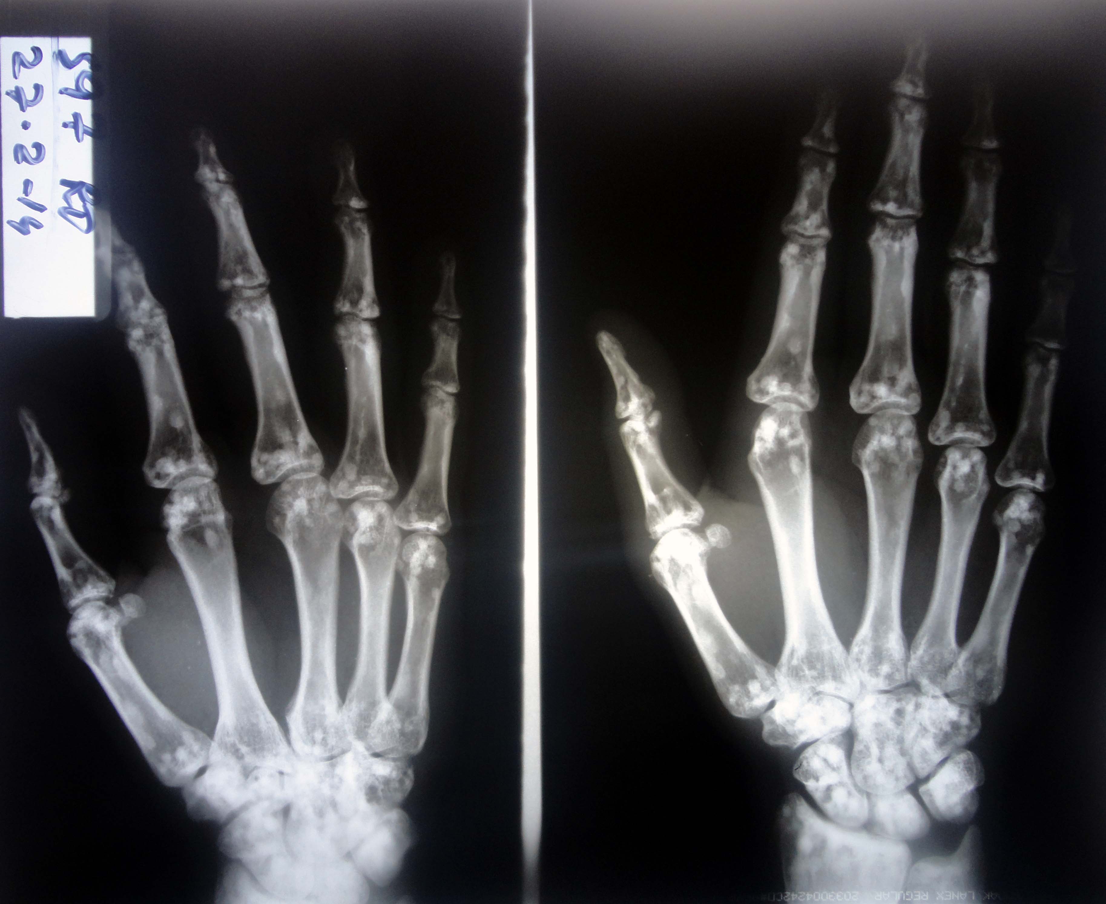
Fig. 1: X-Rays of left hand and wrist
What diagnosis does the X-ray findings indicate?
- Fracture of left scaphoid bone
- Osteoblastic metastases
- Osteopathia striata
- Tuberous sclerosis
- Osteopoikilosis
Answer / Discussion
The X-ray in Fig. 1 shows multiple small hyperdense oval and circular lesions scattered in all small bones of the left hand, with preservation of cortical thickness. These findings are suggestive of osteopoikilosis. Similar lesions were also present in the contralateral hand and wrist, as well as the pelvis (Fig. 2), on X-rays done subsequently.
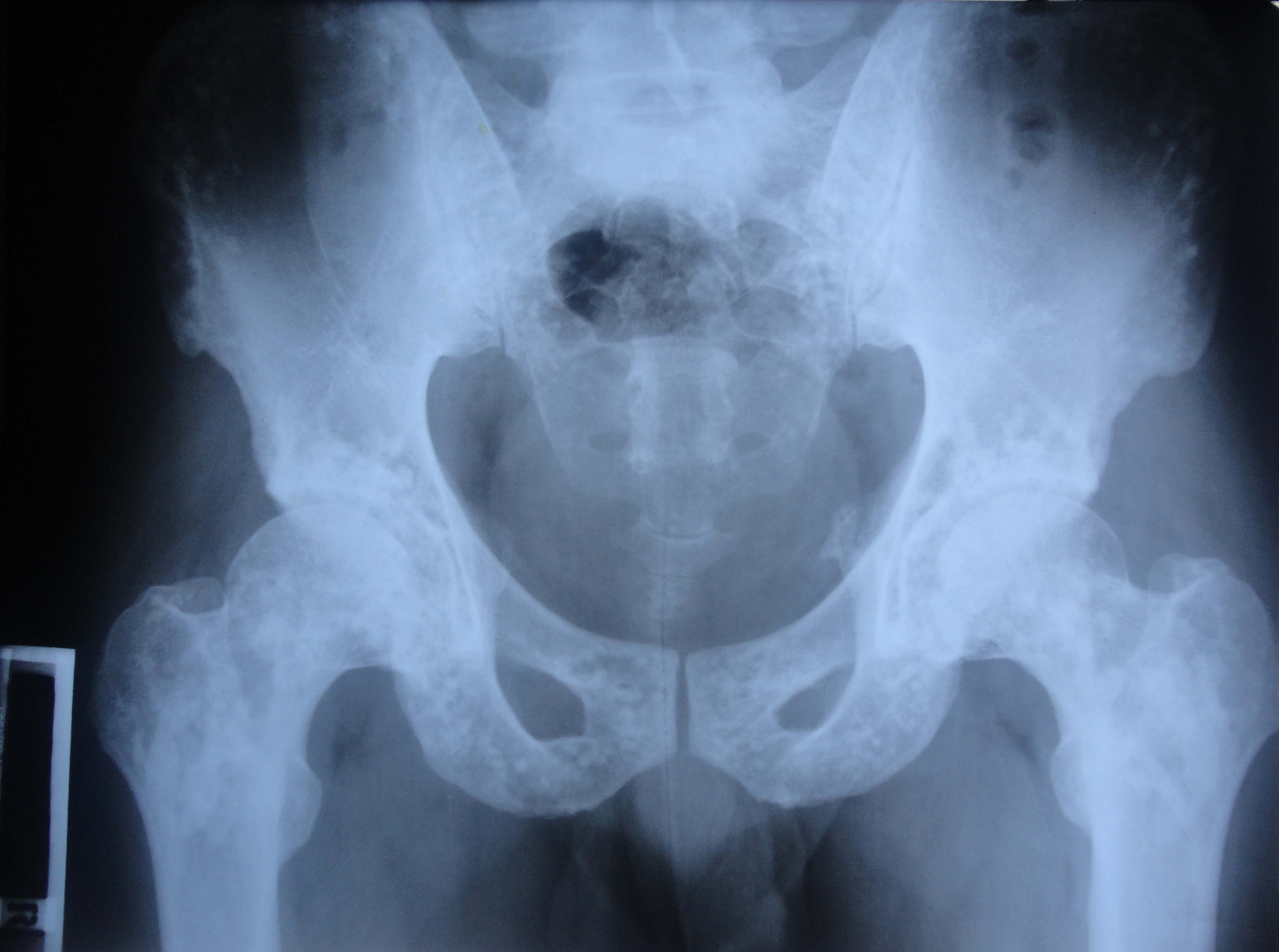
Fig. 2: X-Ray Pelvis showing bone islands
Patient was counselled and reassured about the radiological findings. He was prescribed oral Paracetamol and topical Piroxicam for three days and asked to rest the affected joint. Osteopoikilosis (also called spotted bone) is a benign, possibly autosomal dominant dysplasia of bones, occurring in 1 per 50,000 people.1 Small bones of hand and feet, long tubular bones and pelvis are most frequently affected. The condition is asymptomatic and is diagnosed incidentally on radiographs taken for other problems. The diagnosis is straightforward, based on the typical radiological appearances of small (up to 10mm) hyperdense opacities distributed symmetrically. No further investigations or any specific treatment are indicated. Patients need to be reassured about the benign nature of radiological findings.
Osteoblastic metastases occur in the older age group, are generally larger in size and do not have such a uniformly symmetric distribution. Osteopathia striata is another rare bone dysplasia, characterized by long hyperdense striations mainly in the metaphyses of long bones and pelvis.2 Sclerotic bone lesions in tuberous sclerosis are frequently seen in the axial skeleton especially calvarias and spine, are at times distributed focally and have irregular borders and variable size.3 Subperiosteal new bone formation may be present and other clinical features like epilepsy may also provide a clue. As seen in Fig. 1, there is no break in the continuity of scaphoid bone, thus ruling out a fracture.
|
Competing Interests None declared Author Details ABDUL REHMAN ARSHAD, MCPS, FCPS(Pak). 1 Mountain Medical Battalion, Bagh, AJK Pakistan. ASIF RAHMAN, MCPS, FCPS(Pak). Combined Military Hospital Bannu, Pakistan. SHAFQAT HUSSAIN, MBBS. 1 Mountain Medical Battalion, Bagh, AJK Pakistan. CORRESPONDENCE: Dr ABDUL REHMAN ARSHAD, Classified Specialist in Medicine, 1 Mountain Medical Battalion (MDS), Bagh 12500, Azad Kashmir, Pakistan. Email: maj.abdulrehman@gmail.com |
References
- Meena S, Saini P, Chowdhary B. Multiple spots on bone: diagnostic challenge or spot diagnosis? Osteopoikilosis. Neth J Med 2013; 71(7):372, 376.
- Perdu B, de Freitas F, Frints SG, et al. Osteopathia striata with cranial sclerosis owing to WTX gene defect. J Bone Miner Res 2010; 25(1):82-90. doi: 10.1359/jbmr.090707.
- Umeoka S, Koyama T, Miki Y, et al. Pictorial review of tuberous sclerosis in various organs. Radiographics 2008; 28(7):e32. doi: 10.1148/rg.e32. Epub 2008 Sep 4.

The above article is licensed under a Creative Commons Attribution-NonCommercial-NoDerivatives 4.0 International License.