


ISSN 1757-8515
Portal vein air embolism
Suhail Y Hakim, Gursimran Singh Kundan and Sofia Gani Rah
Cite this article as: BJMP 2010;3(4):a348
|
A 45- year -old man hit by a speeding vehicle presented with chest and abdominal pains, along with dyspnea and vomiting. On examination, vitals signs were pulse rate of 122 per minute, blood pressure of 100/60 mm of Hg and respiratory rate of 28 per minute. Chest examination showed tenderness on the lower ribs. The patient had ecchymotic patches and abrasions on his lower chest and abdomen. Abdominal examination showed diffuse tenderness in all quadrants. The patient also had an open fracture of right femur. Arterial blood gas analysis showed hypoxia. Liver enzymes were normal. Chest X-ray and FAST (Focused Assessment with Sonography for Trauma) did not reveal any obvious pathology. CT scan of the chest showed small contused areas of lungs on both sides. Contrast CT abdomen showed the following picture.
Medicine in pictures
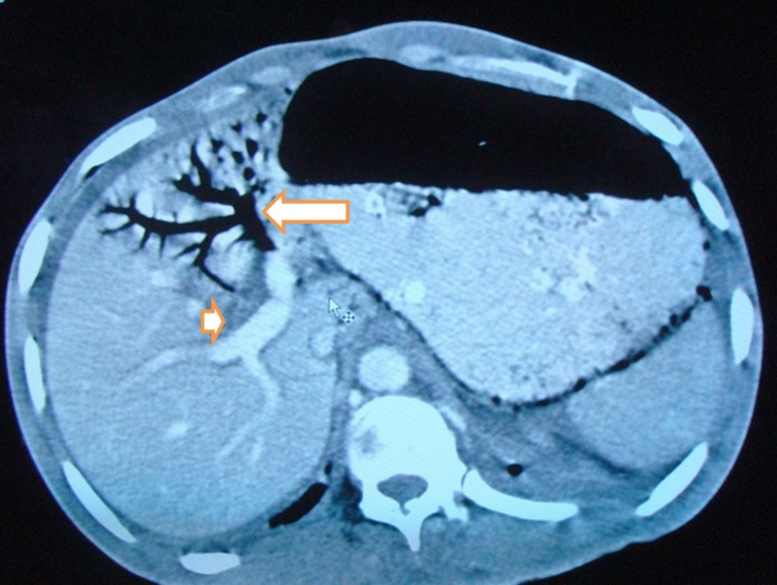
CECT (Contrast Enhanced Computed Tomography) of Abdomen.
Question: What is this radiological finding?
Answers:
1. Pneumobilia
2. Liver Laceration
3. Portal vein air embolism
4. Oriental cholangiohepatitis.
Correct answer and description – at the end of the article
Differential diagnosis:
Air in the portal vein has many causes including Necrotizing entero-colitis, Inflammatory bowel disease, Pneumatosis intestinalis, Mesenteric ischemia, Perforated peptic ulcer, Trauma etc.
Explanation:
Pneumobilia means air in the biliary tree. This condition refers to central location of the air which does not extend to within 2 cm of the liver capsule. It is most commonly seen in patients following surgery in which a biliary-enteric anastomosis has been created or a sphincterotomy (sphincter of Oddi) has been performed.
Liver laceration is seen as a non-enhancing region, linear or branching, hypo-dense wedge lesion extending to liver surface.
The CT findings in oriental cholangiohepatitis can present as intra- or extra hepatic duct stones, dilatation of the extra hepatic duct with relatively mild or no dilatation of the intrahepatic ducts or as localized dilatation of the lobar or segmental bile ducts.
Discussion:
Gas in the portal vein is a rare and usually fatal condition, and its presence in trauma is a rare occurrence. Various terms are used to describe the condition like hepatic portal vein gas (HPVG), pneumoportogram, gas embolism of the portal vein etc. It has to be differentiated from air in the biliary radicals. Portal venous gas manifests on CT as small, tubular air densities in the peripheral regions of the liver, predominantly in the left hepatic lobe in the left portal vein as it is more anterior. Due to the centrifugal flow of blood in the portal venous system, air bubbles appear to extend within 2 cm of the liver capsule 1, 2 . Susman and Senturia state that air in the biliary radicals is more centrally placed and extends up to the hilum and into the common hepatic duct due to the centripetal flow of bile 3 . In the pediatric age group the commonest cause attributable is necrotizing entero-colitis, and along with pneumatosis intestinalis it is in fact pathognomic of the condition4 . The pathophysiology behind air entering portal vein is intestinal mucosal damage leading to air entering the venules that connect into the portal vein. This was demonstrated in an experiment where hydrogen enema was given to a dog and subsequent mesenteric venous gas was noted5. Outcomes are poor in non-trauma cases. In the absence of CT findings associated with bowel ischemia, portal venous gas due to trauma or iatrogenic causes may be treated conservatively 6 . However, blunt trauma can be varying in severity and intensity, and the likelihood of polytrauma should be considered by the treating physician.
Correct answer is option 3 - Portal vein air embolism. Portal Vein Air embolization also called HPVG ( hepatic portal vein Gas).Long arrow showing air in the left portal vein and the arrow head showing the contrast filled right portal vein.
|
Competing Interests None declared Author Details SUHAIL Y HAKIM, MS,FNB (Trauma Care)Trauma Surgeon, Department of General Surgery & Trauma, Lokmanya Tilak Municipal Medical College and Hospital, Mumbai, India. GURSIMRAN SINGH KUNDAN, MS, Senior Resident, Department of General Surgery & Trauma, Lokmanya Tilak Municipal Medical College and Hospital,Mumbai, India. SOFIA GANI RAH Resident, Department of General Surgery & Trauma, Lokmanya Tilak Municipal Medical College and Hospital, Mumbai, India. CORRESPONDENCE: SUHAIL Y HAKIM Suhail MS,FNB (Trauma Care),Trauma Surgeon, Department of General Surgery & Trauma, Lokmanya Tilak Municipal Medical College and Hospital, Mumbai, India. Presently Trauma Specialist with DHS Kashmir. Email: E Mail: dryaqoob@rediffmail.com |
References
1. Sebastia C, Quiroga S, Espin E et al. Portomesenteric vein gas: pathologic mechanisms, CT findings, and prognosis. Radiographics 2000; 20:1213-1224. (s)
2. Hussain A, Mahmood H, El-Hasani S. Portal vein gas in emergency surgery. World J Emerg Surg 2008 ;3: 21. (s)
3. Susman N and Senturia H. R. Gas embolization of the portal venous system. Am J Roentgenol Radium Ther Nucl Med 1960; 83: 847.
4. Ergaz Z, Arad I, Simanovsky N. Portal venous gas following trauma in a preterm infant. Fetal Pediatr Pathol 2006 ;25 :147-50
5. Shaw A, Cooperman A and Fusco J. Gas embolism produced by Hydrogen Peroxide. N Engl J Med 1967; 277: 238.
6. Pate A, Amin F, Nuqui M et al. Intrahepatic air pneumobilia vs. Portal venous gas. The Internet Journal of Surgery 2009,Volume 21 Number 2.

The above article is licensed under a Creative Commons Attribution-NonCommercial-NoDerivatives 4.0 International License.