


ISSN 1757-8515
Photo Quiz: Palatal swelling with pain
Segun Adeoye, Sylvester Sarfraz and Kiranmayi Korimerla
Cite this article as: BJMP 2014;7(1):a704
|
|
Abstract A 32-year old female diabetic patient presents with 2-day history of pain in her mouth. A complete history, physical findings, picture of lesion, differential diagnoses and management is detailed. Keywords: palatal swelling, palatal pain, torus palatinus, palatal abscess, pleomorphic adenoma of salivary gland, hyperplastic candidiasis, median palatal cyst.Abbreviations: STD- Sexually transmitted disease, HIV- Human immunodeficiency virus |
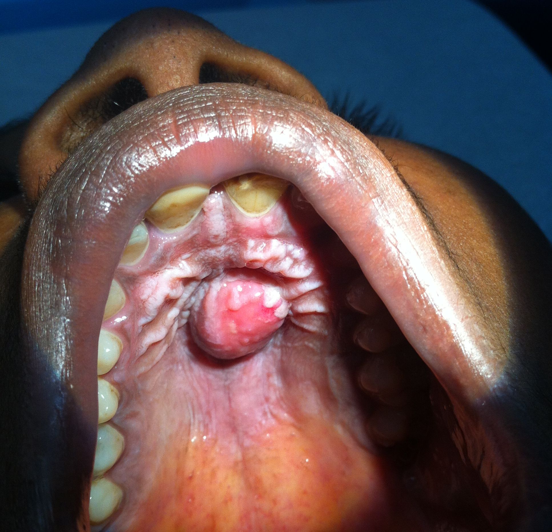
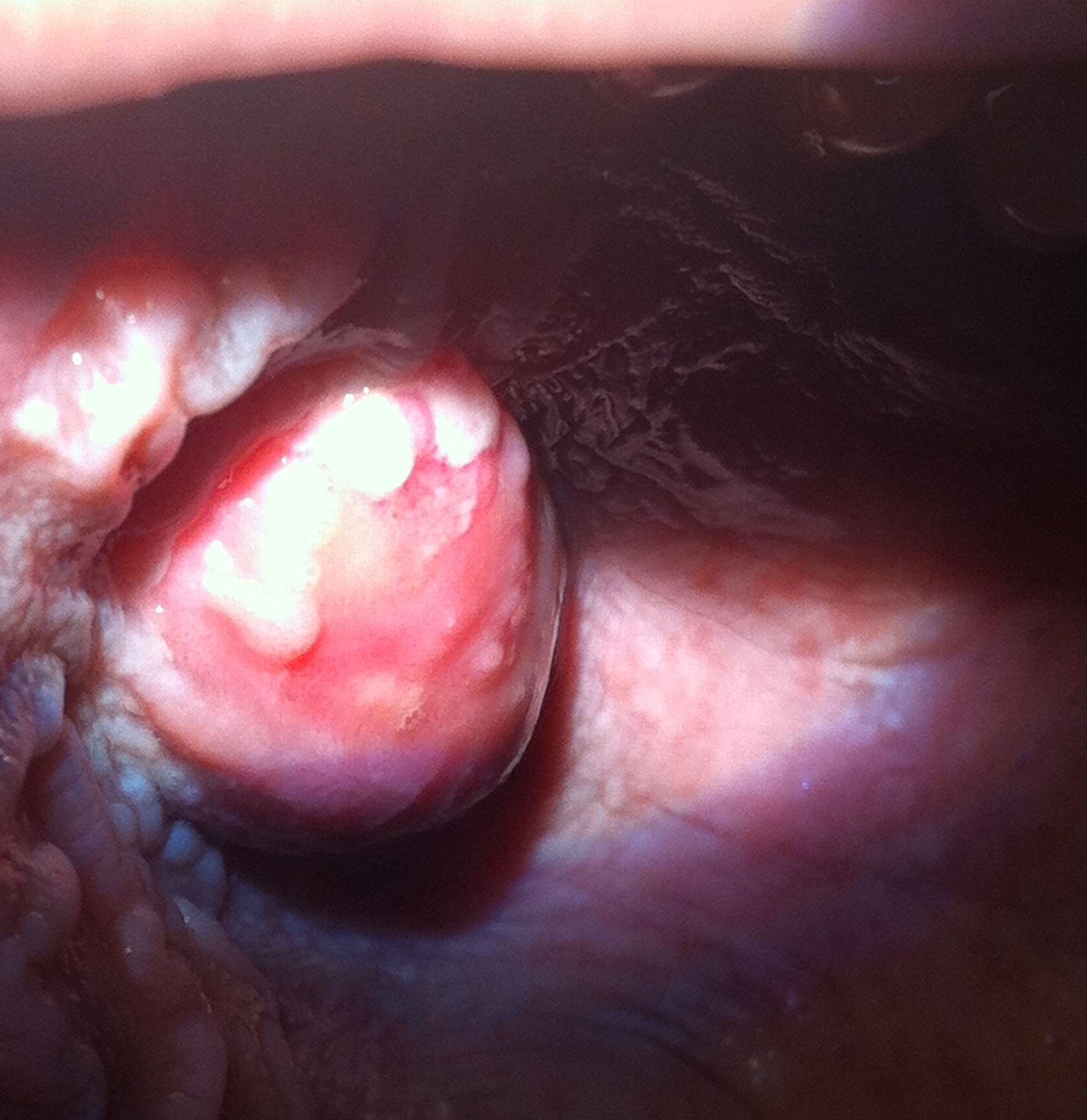
A 32-year old female with medical history of Diabetes Mellitus (type 2) presents to the outpatient clinic with a 2-day history of pain in the roof of her mouth. She described the pain as severe, throbbing, non-radiating, and unrelieved by analgesics. Fever was absent and there were no symptoms suggestive of an antecedent respiratory tract infection (RTI). She had no history of oral mucosa trauma or burns. She gave a remote history of herpes oralis and aphthous ulcers. Her diabetes is well controlled with Sitagliptan-Metformin and Lantus. An annual dental examination done in the past year was described as “normal” by the patient. She maintains oral hygiene with daily teeth brushing without flossing; she had never used dentures. Further review of systems was negative. On examination she was not in distress and vital signs were normal. No external orofacial or neck swelling was observed. Oral examination revealed dental plagues and periodontal lesions. A 2x2cm tender paramedian mass with a small central ulcer was seen and felt on the hard palate anteriorly (see Figure 1 and 2). There were no pharyngotonsillar lesions or regional lymphadenopathy. Tongue and deglutitive movements were normal. Systemic examination was normal. STD/HIV screening was negative. The clinical picture is most consistent with:
- Torus palatinus with aphtous ulceration
- Hyperplastic candidiasis
- Palatal pleomorphic adenoma
- Median palatine cyst
- Palatal abscess
Answer
The clinical picture is most consistent with a palatal abscess. Palatal abscess is a pyogenic collection representing a palatally directed drainage of infective pulpal, pericoronal or periodontal process.1, 2 The most common origin is from an infection of the palatal root of maxillary premolars or molars.3 It presents as a very painful, fluctuant swelling, with lateral or paramedian localization. The surrounding edema may give an impression of midline involvement or contralateral extension.4 The prevailing dental plagues and periodontitis present in this patient (her diabetic state abetting), creates a rich source of oral aerobes and anaerobes as well as the environment in which they thrive. An antecedent herpetic or aphthuos ulcer may also be portal of entry for causative microbes. The patient’s oral hygiene status, her diabetic state, the acuity of symptoms and markedly painful presentation are consistent with acute palatal abscess. The absence of fever in this patient does not preclude this diagnosis.
Hyperplastic candidiasis is the result of chronic colonization and superficial oral mucosa invasion by Candida sp, causing chronic inflammatory changes with edema and epithelial proliferation.2 The result of these reactive responses is a raised pebbled-like –surfaced lesion. It is most commonly seen under denture sites in denture wearers.5,6 The lesion depicted in the picture above is not typical for hyperplastic candidiasis, more so, though not an absolute discriminant, the patient had never used dentures.
Torus palatinus is a wide-based, smooth surfaced, bony protrusion in midline of the hard palate caused by cortical bone growth with a thin, poorly vascularized mucosa lining. The etiology is unclear, but is thought to be multifactorial; genetics (autosomal dominant trait) and recurrent superficial palatal injuries most often implicated.1 Torus palatinus is often an incidental finding, though some affected persons may present out of concern for its increasing size or interval development of ulceration or pain in the area of the torus. Pleomorphic adenoma is the most common neoplasm of salivary glands. Though it may occur at any age, pleomorphic adenoma of salivary glands has peak incidence in the fourth to sixth decade of life. Palatal pleomorphic adenoma often presents as a painless, slow-growing tumor. Median palatine cyst is a rare, non-odontogenic lesion of the hard palate that usually presents as a painless, fluctuant swelling. They are composed histologically of a fibrous collagenous tissue wall, with infiltration of chronic inflammatory cells, and lined by stratified squamous and/or respiratory epithelium. Pain is unusual in the above three oral diagnostic entities, when present it arises from ulcerative, hemorrhagic or infective complications. A detailed history eliciting the chronicity of a preceding midline palatal swelling is often helpful. The patient reported normal palatal examination a year earlier, the relative short history, and the acuity of presentation (severity of pain) make torus palatinus, median palatine cyst or palatal pleomorphic adenoma unlikely. See Table 1 for discriminants and differential diagnoses.
| Table 1: Differential Diagnoses of Palatal Swelling | |
| Onset and course | |
| Acute | Palatal abscess |
| Chronic | Torus palatinus, median palatal cyst, pleomorphic adenoma, hyperplastic candidiasis |
| Shape | |
| Globular | Palatal abscess, torus palatinus, median palatal cyst, pleomorphic adenoma |
| Peppled | Hyperplastic candidiasis |
| Consistency | |
| Fluctuant/tense | Palatal abscess, median palatal cyst |
| Rubbery/firm | Torus palatinus, pleomorphic adenoma, hyperplastic candidiasis |
| Associated pain | |
| Yes | Palatal abscess, hyperplastic candidiasis, infective or traumatic complications of: torus palatinus, median palatal cyst, pleomorphic adenoma |
| No | Uncomplicated: torus palatinus, median palatal cyst, pleomorphic adenoma |
| Associated fever | |
| Yes | Palatal abscess (but may not be present) |
| No | Hyperplastic candidiasis, uncomplicated: torus palatinus, median palatal cyst, pleomorphic adenoma |
| Patient attributes | |
| Poor oral hygiene/caries | Palatal abscess |
| Diabetes/HIV | Palatal abscess |
| Denture wearer | Hyperplastic candidiasis |
The patient underwent definitive treatment with incision and drainage of abscess as well as extraction of her upper left second molar by a dental surgeon. She completed a course of Clindamycin as well as multiple scaling and polishing sessions by a dental hygienist. Maintaining oral hygiene by daily teeth brushing, and flossing, use of mouth antiseptic, as well as a biannual visit to her dentist was recommended.
|
Competing Interests None declared Author Details SEGUN ADEOYE, MD, MS, Hospitalist, University of Pittsburg Medical Center (Horizon), Greenville, PA. SYLVESTER SARFARAZ, MD, Fellow, Geriatric Medicine, Brown University/Rhode Island Hospital, Providence, RI. KIRANMAYI KORIMERLA, MD, Resident, Department of Family Medicine, The Brooklyn Hospital Center, NY. CORRESPONDENCE: Segun Adeoye, MD, University of Pittsburgh Medical Center, Horizon. 110 N Main Street, Greenville, PA 16125. Email: adeoye.segun@yahoo.com |
References
- Hargreaves KM, Goodis HE. Seltzer and Bender’s dental pulp. 3rd. Quintessence Publishing; 1984.
- Mitchell CS, Nelson MD., Jr Orofacial abscess of odontogenic origin in the pediatric patient: Report of two cases. Pediatr Radiol. 1993;23:432–434.
- Jimenez Y, Bagan JV, Murillo J, et al. Odontogenic infections. Complications. Systemic manifestations. Med Oral Patol Oral C r Bucal. 2004;9:139–147.
- Houston GD, Brown FH. Differential diagnosis of the palatal mass. Compendium. 1993;14:1222–1224.
- Jainkittivong A, Aneksuk V, Langlais R. Oral mucosal lesions in denture wearers. Gerontology 2010 March 27(1):26-32.
- Odell EW. Clinical Problem solving in dentistry. 2nd ed. Elsevier Science; 2004. pp. 223–226.

The above article is licensed under a Creative Commons Attribution-NonCommercial-NoDerivatives 4.0 International License.