


ISSN 1757-8515
Phenobarbital induced Pellagra
Youssef kort, Naziha Khamassi, Heykel Abdelhedi and Ouahida Cherif
Cite this article as: BJMP 2015;8(2):a814
|
|
Abstract Background: Pellagra is a nutritional disorder due to an insufficiency in vitamin B3 also called vitamin PP for “pellagra preventing” vitamin. The disease was characterized by the “3D” consisting in dermatitis, dementia and diarrhea. The insufficiency can be due to drug use as anti epileptics. We describe a case of Phenobarbital-induced pellagra. |
Introduction
Pellagra is a nutritional disorder due to an insufficiency in vitamin B3 also called vitamin PP (‘Pellagra preventing’). The disease was characterized by the following ‘3 D’s’: ‘Dermatitis’, ‘Dementia’ and ‘Diarrhoea’. When it is misdiagnosed, it can lead to the fourth ‘D’ which is ‘Death’.1 It was very common in past centuries, particularly in populations that had an exclusively maize diet. Nowadays, this diet problem is rare in developed countries, so the disease is less frequent. However, many recent studies suggest that the disease has not been eradicated and can be under-diagnosed. Alcohol, drugs and malabsorption seem to be the new aetiologies of the disease. So, it is important to recognize the ‘3 D’s’ triad in such situations to avoid fatalities.
Observation
A 61-year-old patient presented to the Internal Medicine Department with an 8-month history of deterioration in her general state. She had a medical history of Epilepsy treated by Phenobarbital since she was 16.
Review of systems revealed gastrointestinal symptomatology consisting of intermittent diarrhoea (6-7 watery stools a day without blood), dysphagia and diffuse abdominal pain. The patient also reported a skin photosensitive eruption affecting her hands and feet.
Physical examination showed a listless patient with a low Body Mass Index (17 kg/m²).
On dermatological examination, symmetric, well-defined, brown-coloured and scaly eruption was observed on the dorsa of her feet (Picture 1) and hands (Picture 2). The mucous examination showed a commissural Cheilitis and Glossitis.
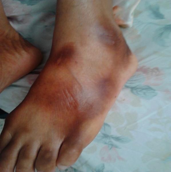
Picture 1: Brown well-defined patches on feet.
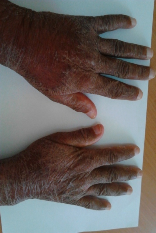
Picture 2: Brown pigmentation and scales of hands.
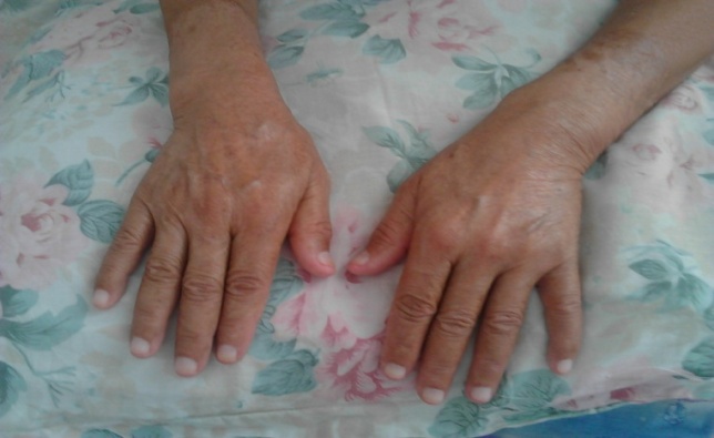
Picture 3: Hands aspect after treatment.
The nervous system examination revealed a pyramidal and cerebellar syndrome. The response to neurocognitive tests was altered suggesting Dementia.
The rest of physical examination was normal. In particular, there were neither peripheral lymph nodes, nor spleen or liver enlargements, nor abdominal mass.
The laboratory tests (glucose, calcaemia, creatinine, liver function tests, urine analysis, haemoglobin, hematocrit, sedimentation rate, C-reactive protein and protein electrophoresis) were within normal limits.
Oesogastroduodenal endoscopy was normal. The cranial, thoracic and abdominal CT scans were normal.
The diagnosis of Pellagra was made on dermatological, abdominal and neurological signs. The patient was treated by Niacin (1000 mg/day) and multivitamin complex. Phenobarbital was discontinued and switched to Clobazam. The patient’s symptoms started to improve quickly. Ten days after the treatment began the skin lesions (Picture 3) and gastrointestinal signs completely disappeared.
Discussion
Pellagra was diagnosed clinically in our patient based on the skin aspects. The skin lesions have been described since 1771 by Frapolli whose name was given to the disease: Pellagra which means rough skin in Italian.1 The typical lesions consist of a brown pigmentation and scales with a photosensitive distribution and well-defined borders as seen in our patient. The face, the neck and the dorsa of the hands are the preferential locations.2 The skin lesions are not always found, and cases of Pellagra Sine Pellagra have been described.3 The extra-cutaneous manifestations are less specific, but their association with pellagrous skin lesions are sufficient to reach a diagnosis. The neurological involvement is classically a Dementia syndrome, but ‘Pellagrous Encephalopathy’ can also consist of delirium, insomnia, depression, cerebellar and extrapyramidal syndrome.4 The gastrointestinal signs are non-specific; they can be Glossitis, dysphagia, nausea, vomiting and abdominal pain. An intractable diarrhoea may occur in advanced stages of disease and can quickly lead to death.5
In 1929, Goldberger attributed such clinical manifestations to a Niacin (vitamin B3 or PP) deficiency. Niacin is a precursor for two important coenzymes namely ‘Nicotinamide Adenine Dinucleotide (NAD)’ and ‘NAD-Phosphate’ which are essential for many oxidative reactions. This probably explains why Pellagra affects tissues with a high rate of cell turnover such as the skin and digestive tract.4
Niacin can be directly absorbed by the gastrointestinal tract or synthesized from Tryptophan.
Primary Pellagra occurs when the diet is deficient in Niacin or Tryptophan as in poor populations with an exclusive maize diet (which contain Niacin but in an indigestible form).
Despite sufficient dietary Niacin, Secondary Pellagra may be caused by a problem in Niacin absorption or metabolism. Many causes of Secondary Pellagra were identified as alcohol, intestinal malabsorption, carcinoid tumours, Hartnup’s Syndrome, Anorexia Nervosa and drugs (Table 1).5 Our patient did not consume alcohol and had no biological signs of malabsorption, but she was taking Phenobarbital for about 45 years. In the English literature, we found only two cases of Phenobarbital-induced Pellagra, and with a fatality in one case.6, 7 The underlying mechanism of Pellagra caused by Phenobarbital, or other antiepileptic drugs, is an alteration in Niacinamide synthesis due to an enzymatic induction.5
Table 1: List of drugs predisposing to Pellagra.
| Predisposing Drugs |
| Antituberculosis agents: Isoniazid Pyrazinamide |
| Antiepileptic drugs: Hydantoins Ethionamide Phenobarbital Diazepam |
| Chemotherapy and immunosuppressive drugs 6-Mercaptopurine 5-Fluorouracil Chlorambucil Azathioprine Chloramphenicol |
The treatment is first based on correction of predisposing factors. In our patient, it consisted of using another antiepileptic drug instead of Phenobarbital. Second-line treatment is a vitamin therapy based on Niacin. There is no consensus on the doses, form and duration of the treatment, but the minimal dose is 300 mg of Niacin/day. A multivitamin complex containing other B-vitamins is often necessary because of the frequency of other deficits in such patients.2 With treatment, the skin lesions and gastrointestinal symptoms generally disappear within a few hours or days, as in the case of our patient, and it is a good argument for a retrospective diagnosis of pellagra.1 Testing for Niacin levels or urinary metabolites is not frequently available and it’s not necessary for the diagnosis.
Conclusion
We described a typical case of Pellagra in which the ‘3 D’s’ were present. In such a case, we should begin the treatment before the results of the laboratory investigations are known. The improvement of all symptoms within a few days is sufficient to confirm the diagnosis. However, the ‘3 D’s’ triad is not always present, and the clinician should consider the diagnosis in face of unexplained abdominal or neurological signs in certain patient groups.
|
Competing Interests None declared Author Details YOUSSEF KORT, Internal Medicine, Razi hospital, cité les orangers 2010, Tunis, Tunisia. NAZIHA KHAMMASSI, Internal Medicine, Razi hospital, cité les orangers 2010, Tunis, Tunisia. HEYKEL ABDELHEDI, Internal Medicine, Razi hospital, cité les orangers 2010, Tunis, Tunisia. OUAHIDA CHERIF, Internal Medicine, Razi hospital, cité les orangers 2010, Tunis, Tunisia. CORRESPONDENCE: YOUSSEF KORT, Razi hospital, cité les orangers 2010, Tunis, Tunisia. Email: y_kort@yahoo.fr |
References
- Sayyidou S. Pellagra: a non-eradicated old disease. Clin Pract. 2014; 28; 4(1): 637.
- Pitche PT. Pellagra. Sante. 2005; 15(3): 205-208.
- Ishii N, Nishihara Y. Pellagra among chronic alcoholics: Clinical and pathological study of 20 necropsy cases. Journal of Neurology Neurosurgery and Psychiatry. 1981; 44(3): 209-215.
- Oldham MA, Ivkovic A. Pellagrous encephalopathy presenting as alcohol withdrawal delirium: a case series and literature review. Addict Sci Clin Pract. 2012; 7: 12.
- Piqué-Duran E, Pérez-Cejudo JA, Cameselle D, Palacios-Llopis S, García-Vázquez O. Pellagra: A clinical, histolopathological and epidemiological study of 7 cases. Actas Dermosifiliogr. 2012; 103: 51-58.
- Stadler R, Orfanos CE, Immel C. Drug induced pellagra. Hautarzt. 33(5): 276-280.
- Pancar Yuksel E, Sen S, Aydin F and al. Phenobarbital-induced pellagra resulted in death. Cutan Ocul Toxicol. 2014; 33(1): 76-78.

The above article is licensed under a Creative Commons Attribution-NonCommercial-NoDerivatives 4.0 International License.