


ISSN 1757-8515
The hidden clinical sign on fundoscopy
M Suresh Babu and K.V.K.S.N.Murthy
Cite this article as: BJMP 2013;6(3):a625
|
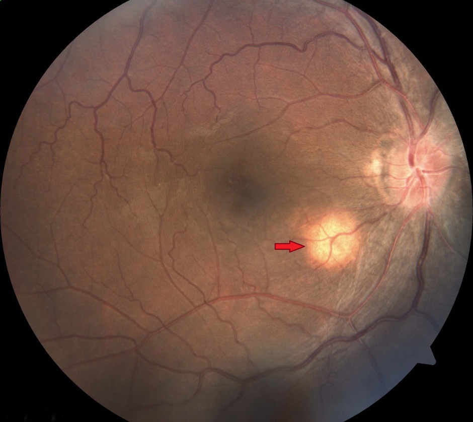
A 26 year old male was brought to Emergency department with history of altered sensorium of 1 day duration. He had a 2 week history of fever prior to admission. On examination, meningeal signs were present. Fundus examination showed evidence of papilloedema and a round pale yellow spot near the optic disc (Figure 1). CT scan of head did not reveal any abnormality.
Mantoux test and HIV ELISA were negative. CSF analysis showed:
- Glucose – 40mg/dl; Protein: 2gm/l;
- Cell Count: 1200cells/µl;
- Cell Type: 80% lymphocytes;
- CSF VDRL- negative;
- CSF Grams stain, India ink staining and Ziehl Neelsen staining were unremarkable.
What is the Fundus finding?
- Roth Spot
- Cotton Wool Spot
- Choroidal tubercle
- A-V malformation
Discussion:
Correct answer: 3) Choroidal Tubercle.
Intraocular tuberculosis is a rare event and occurs in 1% of all diagnosed cases of tuberculosis.1 It occurs by haematogenous spread of mycobacterial organism. Choroidal tuberculosis is the most common initial manifestation of intraocular tuberculosis. They may be seen in 1.4% to 60% of patients with different forms of tuberculosis and are highly specific for tuberculosis. 2, 3
Choroidal tubercles may be unilateral or bilateral and appear as polymorphic yellowish lesions with discrete borders. They are of 2 types, solitary tubercle or granuloma (seen in chronic tuberculosis) and choroidal miliary tubercles (seen in acute miliary tuberculosis). Their size varies from 0.4 to 5 mm and may be associated with retinal vasculitis, panuveitis, choroiditis and neuroretinitis.
When they involve macula they present with visual loss and any delay in appropriate treatment results in irreversible visual loss. Peripherally situated tubercles are asymptomatic. Definitive diagnosis can be daunting due to difficulty in getting ocular samples for histological evaluation, however when available reveal features of granulomatous inflammation. Fundus angiography exhibits hypofluorescence in early stages and hyperfluorescence in later stages.
On treatment they heal by varying degrees of scar formation and marginal pigmentation.4 Untreated tubercles grow into large tumour like mass called tuberculoma.
Roth spots are retinal haemorrhages with a pale centre and are associated with bacterial endocarditis. Cotton wool spots appear as fluffy white patches on the retina and are associated with diabetes. A-V malformations are developmental vascular anomalies and appear as marked arterial and venous dilation associated with a tortuous pattern of vessels. They may have an associated bruit or chemosis of the eye.
The presence of ocular tuberculosis may be subtle. A high index of suspicion is required for its diagnosis. Delay in treatment or misdiagnosis may lead to irreversible visual loss.
|
Acknowledgements The authors wish to thank Department of Ophthalmology, JSS Medical College, JSS University, Mysore for their contributions to the case. Competing Interests None declared Author Details M Suresh Babu, MBBS, MD, FCCP, FICP, Associate Professor of Internal Medicine, JSS Medical College, JSS University, Mysore, Karnataka, India. K.V.K.S.N.Murthy, MBBS, MD, JSS Medical College, JSS University, Mysore, Karnataka, India. CORRESPONDENCE: Dr. M SURESH BABU, Associate Professor of Internal Medicine, JSS Medical College, JSS University, Mysore, Karnataka, India. Email: drmsureshbabu@yahoo.co.in |
References
- Ocular tuberculosis. A prospective study in a general hospital. Medicine (Baltimore) 1997, 76:53–61. Bouza E, Merino P, Munoz P, et al.
- Biswas J, Badrinath SS. Ocular morbidity in patients with active systemic tuberculosis. Int Ophthalmol 1995- 1996;19:293-8.
- Illingworth RS, Lorber J. Tubercles of the choroid. Arch Dis Child 1956;31:467-9.
- Mehta S. Healing patterns of choroidal tubercles after antitubercular therapy: a photographic and OCT study. J.Ophthalmic Inflamm. Infect. 2(2), 95–97(2012).

The above article is licensed under a Creative Commons Attribution-NonCommercial-NoDerivatives 4.0 International License.