


ISSN 1757-8515
Gastrointestinal Tract Perforations Due to Ingested Foreign Bodies; A review of 21 cases
Arif Hussain Sarmast, Hakim Irfan Showkat, Asim Mushtaq Patloo, Fazl Q Parray, Rubina Lone and Khurshid Alam Wani
Cite this article as: BJMP 2012;5(3):a529
|
|
Abstract Aim: To study the etiology, presentation and complications of Gastrointestinal tract (GIT) perforations due to ingestion of foreign bodies. |
Introduction:
Foreign body ingestion is a common occurrence, especially in children, alcoholics, mentally handicapped and edentulous people wearing dentures. However, majority of the individuals pass these objects without any complications.1 Most foreign bodies pass readily into the stomach and travel the remainder of the gastrointestinal tract without difficulty; nevertheless, the experience is traumatic for the patient, the parents, and the physician, who must await the removal or the ultimate passage of the foreign body.2 The alimentary canal is remarkably resistant to perforation: 80% of ingested objects pass through the gastrointestinal tract without complications. 3 About 20% of ingested foreign bodies fail to pass through the entire gastrointestinal tract.4 Any foreign body that remains in the tract may cause obstruction, perforation or hemorrhage, and fistula formation. Less than 1% result in perforations from the mouth to the anus and those are mostly caused by sharp objects and erosions. 5, 18 Of these sharp objects, chicken bones and fish bones account for half of the reported perforations. The most common sites of perforation are the ileo-ceacal junction and sigmoid colon.3
Materials and Methods
This study, “Gastrointestinal tract perforations due to foreign bodies; a review of 21 casesover a ten year period” was carried out in the Department of General Surgery at the Sher-i-Kashmir Institute of Medical Sciences Srinagar (SKIMS), a tertiary care hospital in North India, from January 2002 to December 2011. A total of 21 consecutive patients who underwent surgery for an ingested foreign body perforation of the GI tract over a period of ten years were retrospectively reviewed. Computer database and extensive case note search of patient’s personal data including age, sex, residence, presenting complaints with special stress on clinical examination findings was done. The type and nature of the foreign objects, mode of entry into the gastrointestinal tract, preoperative diagnosis, perforation site, and treatment received were recorded. The complications arising due to perforation of GIT because of the foreign body ingestion and complications arising due to specific treatment received were noted. Important findings on various laboratory tests, including a complete blood count, erythrocyte sedimentation rate, [pre-op/post-op/follow up], blood cultures, and serum chemistry, chest and abdominal X-rays were penned down. Special efforts were made to identify the predisposing factors for ingestion of foreign bodies including edentulous patients with dentures, psychosis, extremes of age and hurried eating habits. Clinical, laboratory and radiological findings, treatment modalities, operative findings and therapeutic outcomes were summarized. Data collected as such was described as mean and percentage.
I/V Antibiotics ( Ceftriaxone + Metronidazole ) were given in the emergency room and changed to specific therapy as per the culture sensitivity postoperatively.
Results
The average follow up duration was 13 months (range 7 – 19 months). There were 14 male(66.66%) and 7 female (33.33%) patients ranging in age from 7 years to 82 years with a median age of 65 yrs at the time of diagnosis . The most frequently ingested objects were dietary foreign body (n = 17). Four patients had ingested objects like toothpicks (n =2) and metallic staples (n=2) {as shown in figure 1}. Among the dietary foreign bodies fish bone was found in 7(33.3%) and chicken bone in 10(47%) {as shown in figure 2} . All the patients described their ingestion as accidental and involuntary. A definitive preoperative history of foreign body ingestion was obtained in 4(19.04%) patients and an additional 9(42.8%) patients admitted ingestion of foreign body in the post operative period. Of these 13 patients the average duration between ingestion of foreign body and presentation was 9.3 days. Remaining 8 (38.09%) patients did not recall any history of foreign body ingestion; dietary or otherwise. In terms of impaction and perforation of ingested foreign body, ileum was the commonest site with 14(66.66%) patients showing perforation near the distal portions of the ileum followed by sigmoid colon in 5(23.8%). Jejunal perforation was seen in 2(9.5%) patients.
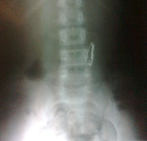
Fig 1: X ray abdomen AP view showing ingested metallic pin
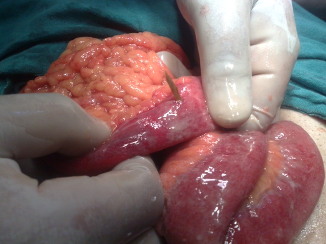
Fig 2: Intra operative picture showing perforation of small gut due to chicken bone
All our patients presented with acute abdomen and were admitted first in emergency department. Since majority of patients did not give any specific history of foreign body ingestion, they were managed as cases of acute abdomen with urgency and level of care varying according to the condition of patients. Eight patients presented with free air in the peritoneum and air under the right side of diaphragm. The most common preoperative diagnoses were acute abdomen of uncertain origin: 12 (57.14%); acute diverticulitis:5 (23.8%) and acute appendicitis: 4 (19.04%).
Table 1: Showing demographic profile, site of perforation, etiology, presentation and management.
| S No | Age | Sex | Site | Foreign Body | Presentation & Pre Op Diagnosis | Procedure Performed |
| 1 | 78 | Male | 40 cm from ileo-caecal valve | Fish bone | Acute abdomen, peritonitis | Removal of foreign body and repair |
| 2 | 65 | Female | 30 cm from ileo-caecal valve | Chicken bone | Acute abdomen, peritonitis | Resection of the perforated distal ileum and ileum stoma |
| 3 | 80 | Male | 30 cm from ileo-caecal valve | Chicken bone | Acute abdomen, peritonitis | Resection of the perforated distal ileum and ileum stoma |
| 4 | 43 | Male | Jejunum | Tooth pick | Acute abdomen, peritonitis | Removal of foreign body and repair |
| 5 | 10 | Male | 10 cm from ileo-caecal valve | Metallic staple | Acute abdomen, appendicitis | Removal of foreign body and repair |
| 6 | 72 | Female | Jejunum | Chicken bone | Acute abdomen, peritonitis | Resection of the perforated distal ileum and ileum stoma |
| 7 | 65 | Male | 20 cm from ileo-caecal valve | Fish bone | Acute abdomen, peritonitis | Resection of the perforated distal ileum and ileum stoma |
| 8 | 59 | Male | Sigmoid colon | Chicken bone | Acute abdomen, diverticulitis | Removal of foreign body and repair |
| 9 | 65 | Female | 30 cm from ileo-caecal valve | Chicken bone | Acute abdomen, peritonitis | Removal of foreign body and repair |
| 10 | 49 | Female | 40 cm from ileo-caecal valve | Chicken bone | Acute abdomen, peritonitis | Removal of foreign body and repair |
| 11 | 7 | Male | Sigmoid colon | Metallic staple | Acute abdomen, diverticulitis | Removal of foreign body and repair |
| 12 | 78 | Female | 15 cm from ileo-caecal valve | Fish bone | Acute abdomen, appendicitis | Resection of the perforated distal ileum and ileum stoma |
| 13 | 72 | Male | 15 cm from ileo-caecal valve | Fish bone | Acute abdomen, appendicitis | Resection of the perforated distal ileum and ileum stoma |
| 14 | 56 | Male | 20 cm from ileo-caecal valve | Tooth pick | Acute abdomen, appendicitis | Resection of the perforated distal ileum and ileum stoma |
| 15 | 65 | Male | Sigmoid colon | Fish bone | Acute abdomen, diverticulitis | Removal of foreign body and repair |
| 16 | 63 | Male | 30 cm from ileo-caecal valve | Chicken bone | Acute abdomen, peritonitis | Resection of the perforated distal ileum and ileum stoma |
| 17 | 82 | Female | 30 cm from ileo-caecal valve | Chicken bone | Acute abdomen, peritonitis | Removal of foreign body and repair |
| 18 | 55 | Female | Sigmoid colon | Fish bone | Hematochizia acute abdomen, diverticulitis | Removal of foreign body and repair |
| 19 | 56 | Male | 20 cm from ileo-caecal valve | Chicken bone | Acute abdomen, peritonitis | Resection of the perforated distal ileum and ileum stoma |
| 20 | 69 | Male | Sigmoid colon | Fish bone | Acute abdomen, diverticulitis | Removal of foreign body and repair |
| 21 | 71 | Male | 40 cm from ileo-caecal valve | Chicken bone | Acute abdomen, peritonitis | Resection of the perforated distal ileum and ileum stoma |
All the patients underwent an emergency celiotomy and confirmation of foreign body induced perforation was possible in all the 21 patients .Patients with a suspected appendicitis were explored via classical grid iron incision and rest via midline incision. Varying degrees of abdominal contamination was present in all the patients. Out of the 21 patients 11(52.38%) underwent removal of foreign body and primary repair of their perforations after minimal debridement. Intestinal resection with stoma formation (resection of the perforated ileum and ileum stoma) was done in 10 (47.6%) of the 21 patients as shown in Table 1. Take down of stoma was done at a later date. Three (14.28%) patients developed incisional superficial surgical site infection which responded to local treatment. Two (9.5%) patients died in the postoperative period due to sepsis. One patient (Patient no. 3 in table 1) who was a diabetic on Insulin, Chronic obstructive pulmonary disease and Hypertension died on 3rd postoperative day in surgical Intensive care unit due to severe sepsis. Another patient, (Patient no. 12 in table 1 ) an elderly female with no co-morbid illness developed severe sepsis due to Pseudomonas aeruginosa, died on 4th postoperative day. She was managed at a peripheral primary care center for first 3 days for her vague abdominal pain with minimal signs. All the other patients had an uneventful recovery and were discharged home between 6-14th postoperative day.
Discussion:
Foreign bodies such as dentures, fish bones, chicken bones, toothpicks and cocktail sticks have been known to cause bowel perforation6. Perforation commonly occurs at the point of acute angulation and narrowing. 7, 8 The risk of perforation is related to the length and the sharpness of the object.9 The length of the foreign body is also a risk factor for obstruction, particularly in children under 2 years of age because they have considerable difficulty in passing objects longer than 5 cm through the duodenal loop into the jejunum. In infants, foreign bodies 2 or 3 cm in length may also become impacted in the duodenum.10 The most common sites of perforation are the ileo-ceacal junction and sigmoid colon. Other potential sites are the duodeno-jejunal flexure, appendix, colonic flexure, diverticulae and the anal sphincter.3 Colonic diverticulitis or previously unsuspected colon carcinoma have been reported as secondary findings in cases of sigmoid perforation caused by chicken bones.11,12 Even colovesical or colorectal fistulas have been reported as being caused by ingested chicken bones. 13,14 .In our study ileum was the most common site with 14 patients showing perforation near the distal portions of the ileum followed by sigmoid colon. Jejunal perforation was seen in 2 patients.
The predisposing factors for ingestion and subsequent impaction are dentures causing defective tactile sensation of the palate, sensory defects due to cerebro-vascular accident, previous gastric surgery facilitating the passage of foreign bodies, achlorhydria where the foreign body passes unaltered from the stomach, previous bowel surgery causing stenosis and adhesions and diverticula predisposing to impaction.3 Overeating, rapid eating, or a voracious appetite may be contributing factors for ingesting chicken bones. The mean time from ingestion to perforation is 10.4 days.15 In cases when objects fail to pass the tract in 3 to 4 weeks, reactive fibrinous exudates due to the foreign body may cause adherence to the mucosa, and objects may migrate outside the intestinal lumen to unusual locations such as the hip joint, bladder, liver, and peritoneal cavity.16 The length of time between ingestion and presentation may vary from hours to months and in unusual cases to years, as in the case reported by Yamamoto of an 18 cm chopstick removed from the duodenum of a 71-year-old man, 60 years after ingestion.17 In our study the average duration between ingestion of foreign body and presentation was 9.3 days.
In a proportion of cases, definitive preoperative history of foreign body ingestion is uncertain.18 Small bowel perforations are rarely diagnosed preoperatively because clinical symptoms are usually non-specific and mimic other surgical conditions, such as appendicitis and caecal diverticulitis.19 In our study the most common preoperative diagnoses were acute abdomen of uncertain origin (n =12), acute diverticulitis (n = 5) and acute appendicitis (n = 4). Patients with foreign body perforations in the stomach, duodenum, and large intestine are significantly more likely to be febrile with chronic symptoms with a normal total white blood cell count compared to those with foreign body perforations in the jejunum and ileum.18 Plain radiographs of neck and chest in both anteroposterior and lateral views are required in all cases of suspect foreign body ingestion and perforations in addition to abdominal films. CT scans are more informative especially if radiographs are inconclusive.20 Computerised tomography (CT) scanning and ultrasonography can recognise radiolucent foreign bodies. An ultrasound scan can directly visualize foreign bodies and abscesses due to perforation. The ability to detect a foreign body depends on its constituent materials, dimensions, shape and position.21 Contrast studies with Gastrograffin may be required in excluding or locating the site of impaction of the foreign body as well as determining the level of a perforation. Using contrast is important in identifying and locating foreign bodies if intrinsically non-radiopaque substances, such as wooden checkers or fish and chicken bones are ingested.20 The high performance of computed tomography (CT) or multi-detector-row computed tomography (MDCT) scan of the abdomen in identifying intestinal perforation caused by foreign bodies has been well described by Coulier et al. 22 Although, in some cases imaging findings can be nonspecific, however, the identification of a foreign body with an associated mass or extraluminal collection of gas in patients with clinical signs of peritonitis, mechanical bowel obstruction, or pneumoperitoneum strongly suggests the diagnosis.8,20 Finally, endoscopic examination, especially in the upper gastrointestinal tract, can be useful in diagnosis and management of ingested foreign bodies.
Whenever a diagnosis of peritonitis subsequent to foreign body ingestion is made, an exploratory laparotomy is performed. However, laparoscopically assisted, or complete, laparoscopic approaches have been reported.17,23 The treatment usually involves resection of the bowel, although occasionally repair has been described.8 The most common treatment was simple suture of the defect. 24 Once the foreign body passes the esophagogastric junction into the stomach, it will usually pass through the pylorus25; however, surgical removal is indicated if the foreign body has sharp points or if it remains in one location for more than 4 to 5 days especially in the presence of symptoms. A decision should be based on the nature of the foreign body in those cases, as to whether a corrosive or toxic metal in ingested. 26 Occasionally, objects that reach the colon may be expelled after enema administration. However, stool softeners, cathartics and special diets are of no proven benefit in the management of foreign bodies.7
|
Competing Interests None declared Author Details ARIF HUSSAIN SARMAST, Postgraduate scholar, Dept of Surgery, SKIMS, India. HAKIM IRFAN SHOWKAT, Postgraduate scholar, Dept of Internal medicine, SKIMS, India. ASIM MUSHTAQ PATLOO, Senior Resident, Dept of General Surgery, SKIMS, India. FAZL Q PARRAY, Additional Professor, Dept of General Surgery, SKIMS, India. RUBINA LONE, Assistant Professor, Dept of Microbiology, SKIMS, India. KHURSHID ALAM WANI, Professor & Head, Dept of General Surgery, SKIMS, India. CORRESPONDENCE: HAKIM IRFAN SHOWKAT, Postgraduate scholar, Dept of Internal medicine, SKIMS, Srinagar, Kashmir, India. Email: docirfanshahi512@gmail.com |
References
- Kimbrell FT Jr, Tepas JJ 3d, Mullen JT. Chicken bone perforation of the sigmoid colon: a report of three cases. Am Surg 1975; 41(12): 814-7
- Eldridge WW, Jr. Foreign bodies in the gastrointestinal tract. JAMA 1961; 178: 665–7.
- Cleator IG, Christie J. An unusual case of swallowed dental plate and perforation of the sigmoid colon. Br J Surg 1973; 60 (2): 163-5
- Nandi P, Ong GB. Foreign body in oesophagus: review of 2394 cases. Br J Surg 1978; 65: 5–9.
- Perelman H. Tooth pick perforations of the gastrointestinal tract. J Abdom Surg 1965; 51–3.
- Akhtar S, Mcelvanna N, Gardiner KR, Irwin ST. Bowel perforation caused by swallowed chicken bones;a case series. Ulster Med J. 2007;76 (1): 37-8.
- McManus JE. Perforation of intestine by ingested foreign bodies: report of two cases and review of literature. Am J Surg. 1941;53(3):393–402.
- Singh RP, Gardner JA. Perforation of the sigmoid colon by swallowed chicken bone. Int Surg. 1981;66(2):181–3.
- Sarliève P, Delabrousse E, Michalakis D, Robert A, Guichard G, Kastler B: Multidetector CT diagnosis of jejunal perforation by a chicken bone. JBR-BTR 2004, 87:294-295.
- Erbes J, Babbitt DP. Foreign bodies in the alimentary tract of infants and children. Appl Ther 1965; 7: 1103–9.
- Gomez N, Roldos F, Andrade R. Intestinal perforation caused by chicken bone mimicking perforated colonic diverticulitus. Acta Gastroenterol Latinoam 1997;27:329–330
- Osler T, Stackhouse CL, Dietz PA, Guiney WB. Perforation of the colon by ingested chicken bone, leading to diagnosis of carcinoma of the sigmoid. Dis Colon Rectum 1985;28:177–179
- Khan MS, Bryson C, O’Brien A, Mackle EJ. Colovesical fistula caused by chronic chicken bone perforation. Ir J Med Sci 1996;165:51–52
- Read TE, Jacono F, Prakash C. Coloenteric fistula from chicken bone perforation of the sigmoid colon. Surgery 1999;125:354–356
- Rodríguez-Hermosa JI, Codina-Cazador A, Sirvent JM, Martín A, Gironès J, Garsot E: Surgically treated perforations of the gastrointestinal tract caused by ingested foreign bodies. Colorectal Disease 10(7):701-707.
- Carp L. Foreign bodies in the intestine. Ann Surg 1927; 85: 575–91.
- Yamamoto M, Mizuno H, Sugawara V. A chopstick is removed after 60 years in the duodenum. Gastrointest Endosc 1985; 31: 51–2.
- Goh BK, Chow PK, Quah HM, Ong HS, Eu KW, Ooi LL, Wong WK: Perforation of the gastrointestinal tract secondary to ingestion of foreign bodies. World J Surg 2006, 30(3):372-7.
- Yao CC, Yang CC, Liew SC, Lin CS: Small bowel perforation caused by a sharp bone: laparoscopic diagnosis and treatment. Surg Laparosc Endosc Percutan Tech 1999, 9(3):226-7.
- Maglinte DD, Taylor SD, Ng AC. Gastrointestinal perforation by chicken bones. Radiology 1979; 130: 597–599.
- Matricardi L , Lovati R. Intestinal perforation by a foreign body: diagnostic usefulness of ultrasonography. J Clin Ultrasound 1992; 20(3): 194-6
- Coulier B, Tancredi MH, Ramboux A. Spiral CT and multidetector- row CT diagnosis of perforation of the small intestine caused by ingested foreign bodies. Eur Radiol, 2004 Oct, 14 (10) :1918-25.
- Law WL, Lo CY. Fishbone perforation of the small bowel :laparoscopic diagnosis and laparoscopically assisted management. Surg Laparosc Endosc Percutan Tech, 2003 Dec, 13 (6) :392-3.
- Pinero Madrona A, Fernández Hernández JA, Carrasco Prats M, Riquelme Riquelme J, Parrila Paricio P: Intestinal perforation by foreign bodies. Eur J Surg 2000, 166(4):307-9.
- Henderson CT, Engel J, Schlesinger P. Foreign body ingestion: review and suggested guidelines of management. Endoscopy 1987; 19: 68–71
- Seo JK. Endoscopic management of gastrointestinal foreign bodies in children. Indian J Pediatr 1999; 66 (1 Suppl): S75–80.

The above article is licensed under a Creative Commons Attribution-NonCommercial-NoDerivatives 4.0 International License.