


ISSN 1757-8515
Retinitis Pigmentosa
M Suresh Babu, C R Venkatesh, P K Kiran, S Sunil Kumar and K Prabhath Kiran Reddy
Cite this article as: BJMP 2015;8(3):a825
|
|
Keywords: Retinitis pigmentosa Abbreviations: RP- Retinitis pigmentosa |
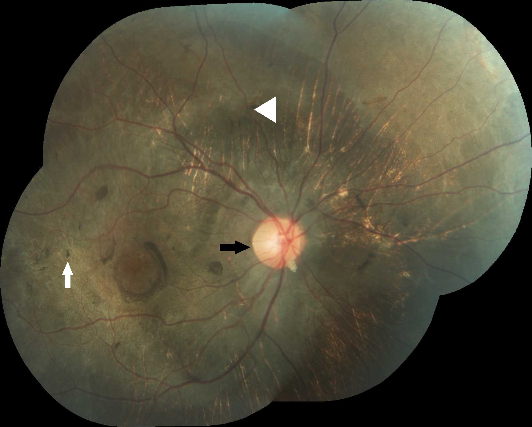
A 19 year old male presented with a history of recurrent respiratory tract infections and progressive diminution of vision. Fundoscopy was performed and showed the changes in image below.
What is the finding suggestive of?
1. Retinitis pigmentosa
2. Drug toxicity
3. Congenital rubella
4. Syphilis
Answer: Retinitis Pigmentosa
Retinitis pigmentosa (RP) is a bilateral inherited progressive retinal degeneration presenting in the first to second decades of life.1 The inheritance can be autosomal dominant, autosomal recessive or X-linked recessive. Hallmark symptoms of RP are nightblindness and visual field constriction. Fundus changes in retinitis pigmentosa include waxy pallor of optic disc (black arrow), arteriolar attenuation (white arrow head) and bony spicule pigmentation (white arrow) in the mid-peripheral fundus, which is predominantly populated by rods. Vessel attenuation is the earliest feature seen clinically. Although intraretinal pigmentary migration is relatively easy to observe, it requires years to develop, so early RP may only exhibit vessel attenuation without pigmentation (previously known as RP sine pigmento). Prognosis is variable and tends to be associated with the mode of inheritance.
Drug toxicity with chloroquine can result in visual disturbances. History of drug usage prior to vision disturbance can be present. Fundus examination shows a subtle bulls eye macular lesion characterized by a central foveolar island of pigment surrounded by a depigmented zone of RPE atrophy, which is itself encircled by a hyperpigmented ring.2 In congenital rubella, a history of maternal infection will be present. Fundus findings include salt and pepper pigmentary disturbance involving the periphery and posterior pole with normal vessels, RPE mottling and no intraretinal pigmentary migration. Syphilitic retinopathy may have sectorial or generalised pigmentation.3 The onset can be from adulthood to old age. History of genital ulcer may be present.
|
Acknowledgements Department of Ophthalmology,JSS Medical College&Hospital,JSS University, Mysore, India. Competing Interests None declared Author Details M SURESH BABU, Professor of Medicine, JSS Medical College, JSS University, Mysore, India. C R VENKATESH, Senior Resident, Dept of Medicine, JSS Medical College, JSS University, Mysore, India. P K KIRAN, Senior Resident,Dept of Medicine, JSS Medical College, JSS University, Mysore, India. S SUNIL KUMAR, Senior resident, Dept of Medicine, JSS Medical College, JSS University, Mysore, India. K ORABHATH KIRAN REDDY, Post-Graduate Trainee, Dept of Medicine, JSS Medical College, JSS University, Mysore, India. CORRESPONDENCE: Dr M SURESH BABU, Professor of Medicine, JSS Medical College, JSS University, Mysore, India. Email: drmsureshbabu@yahoo.co.in |
References
- Jack J Kanski Brad Bowling Clinical Opthalmology 7th Edition
- Parsons Diseases of the Eye 21st Edition
- The Sankara Nethralaya Atlas of Retinal Diseases 1st Edition

The above article is licensed under a Creative Commons Attribution-NonCommercial-NoDerivatives 4.0 International License.